ABSTRACT
Rhegmatogenous retinal detachment (RRD) remains a major cause of emergency assistance in our ophthalmology departments. We struggle for the improvement of visual outcome of our patients after RRD surgery by trying to optimize our surgical methods and techniques. The decision of which surgical procedure to use in the treatment of RRD still depends on a variety of factors and surgeon’s preference. Until date there was only one major randomized clinical trial that drew some guidelines for our clinical practice. This present work has the main purpose of reviewing the major articles published so far on RRD surgery with the goal of creating a rational for our day practice for the best functional and anatomic results. Although many variables are involved in RRD treatment, prospective, multicentric, randomized studies are needed.
Keywords: Rhegmatogenous Retinal Detachment; Pars Plana Vitrectomy; Scleral Buckling; Treatment; Outcome.
INTRODUCTION
Rhegmatogenous retinal detachment (RRD) remains a major cause of emergency assistance in our ophthalmology departments mainly because of the resulting severe and sudden visual impairment. With an incidence of 1/10000 patients a year, it also remains a disease with a severe functional outcome. (Haimann et al. 1982) Although in the latest years there has been great improvement and development of new techniques and equipment, only 42% of patients with a retinal detachment achieve a visual acuity of 20/40. (Mitry et al. 2010)
The pathologic events behind a RRD involve three concomitant factors: a physiologic posterior vitreous detachment, eventually a retinal tear or hole formation due
to traction forces over the retina and subretinal fluid mobilization. (Feltgen et al. 2014)
Prevention of a retinal tear/retinal detachment is challenging since the associated risk factors are not only not modifiable: myopia, ocular trauma, retinal degenerations; but also not acceptable as with the needs of our cataract patients and the resulting pseudophakic status after cataract removal. (Tielsch et al. 1996; Mitry et al. 2011)
Thereby we struggle to improve the visual outcome of our patients after RRD surgery by optimizing our surgical methods and techniques, handling them the best way for a particular patient in a particular clinical situation. Until recent past years the majority of surgeons were doing surgery based on their experience.
A study by Ga Eun Cho and co-workers analysed the trend in RRD surgery among the Korean Retina Society from 2001 to 2013. They concluded that for myopia, pseudophakia and media opacity, surgical interventions over the last decade have drastically shifted from scleral buckling and pneumatic retinopexy to vitrectomy. (Ga Eun Cho et al. 2014) A recent study by McLaughlin et al. using data from the US Medicare evaluated the vitreoretinal practice patterns from 2000 to 2014. For retinal detachment treatment there has been a switch from scleral buckling surgery toward vitrectomy with a distribution of 83% vitrectomy, 5% scleral buckling, and 12% pneumatic retinopexy in 2014. (McLaughlin et al 2017) Scleral buckling procedures have declined from 6502 in 2000 to 1260 in 2014 (P<0.01), whereas vitrectomy procedures increased from 13814 operations in 2008 to 19288 in 2014 (P<0.01). (McLaughlin et al 2017). A recent report from the Royal College of Ophthalmologistis’ National Database Study of Vitreoretinal Surgery evaluated a total of 5857 primary RRD operations performed by 117 surgeons, from 2000 to 2013, retrospectively extracted from 15 centers. In that period, RRD surgery comprised 79.7% vitrectomy operations, 13.9% SB and 6.4% combined PPV/SB procedures. (Sallam et al. 2017)
So, having a shift in our current practice on RRD surgery, this present work has the purpose of reviewing the major articles published so far on RRD surgery with the goal of creating a rational for our day practice to achieve the best anatomic and functional results for our patients.
MATERIALS AND METHODS
We compared reattachment and functional success rates after 3 commonly practiced surgical interventions for pseudophakic RRDs: PPV, SB, and the combined procedure (PPV/SB). Articles were retrieved from Medline and by cross-reference searches. Articles with sufficient data on preoperative evaluation, applied surgical technique, and anatomical and functional success rates were included in this analysis. Article analysis was made dividing them in two categories randomized control trials and non-randomized trials. Several clinical situations regarding RRD were included for a practical RRD treatment general approach.
Randomized control trials
A study by Heimann and co-workers (the Scleral Buckling versus Primary Vitrectomy in Rhegmatogenous Retinal Detachment study – SPR study) was one of the first multi-centric, prospective and randomized trials on surgical retina, with a level of evidence A1b which involved 25 European centres with trained surgeons. (Heimann et al. 2007), (Shekelle et al.1999) During the recruitment phase of this study two clinical concepts were well established: Localized retinal detachments - only 1 to 4 hours’ extension with an isolated tear - were treated with scleral buckling (SB); Complex RRD with proliferative vitreoretinopathy (PVR) grade B or C, giant retinal tears, or RRD associated with a macular hole were treated with pars plana vitrectomy (PPV). The great doubt was in the remaining spectre that included all the medium complexity retinal detachments which comprehend approximately 30% of the retinal detachments in our day practice. (Heimann et al. 2007)
The goal of the SPR study was to define the best surgical method for medium complexity RRD. A medium complexity RRD was defined as: tears with 1-2 hours’ extension, multiple tears, tears with a central extension, superior bullous retinal detachment and marked vitreous traction over a tear or an undetermined hole situation. Excluded from this study were all the RRD with: PVR grade B or C, those that could be treated with a single radial episcleral silastic sponge, associated severe ocular morbidity, high myopia (>7D) and age under 18 years-old. (Heimann et al. 2007)
A total of 681 patients were included with a minimum one-year follow-up. These patients were divided into two groups: phakic and aphakic/pseudophakic. A decision based in the fact that these groups represent retinal detachments with particular and unique clinical characteristics. In each group the patients were randomized to follow PPV or SB. The vitrectomy consisted on a standard 3-port gauge, laser retinopexy or criopexy, and tamponade with a mixture of air/SF6 (20%-40%). The patients submitted to PPV could have a SB procedure based on surgeon decision. The SB was performed using silicone sponges or an encircling band. The primary end point of this study was to determine in each group the procedure that would present a better functional result. Second end points were: PVR rate after surgery, primary and final anatomical success (primary anatomic success being defined has a treated RRD with no need of an additional procedures). On the phakic group cataract formation rate was also analysed.
On the phakic group they concluded that scleral buckling presented statistically significant better functional results
when compared with vitrectomy. (Heimann et al. 2007) The cataract formation rate was clearly superior on the vitrectomized patients and the rates of primary and final anatomic success, and PVR were similar between the two procedures. (Heimann et al. 2007) On the aphakic/pseudophakic group they realized that there was no difference on the functional results of a procedure over another. However, the primary anatomical success was better in those submitted to PPV. After one-year follow-up, both groups presented a final anatomical success rate of 95% independent from the chosen surgical procedure. (Heimann et al. 2007)
The SPR study group concluded that in medium complexity RRD phakic patients benefit with a SB procedure and aphakic/pseudophakic patients with PPV.
In this study sub-analysis of adding a scleral buckle on vitrectomized patients was also performed. Although subjected to bias due to the fact that this variable was not randomized, it suggested the use of scleral buckling in aphakic/pseudophakic patients. Phakic patients did not show benefit of adding a scleral buckling procedure to vitrectomy. (Heimann et al. 2007)
Brazitikos et al did a prospective, randomized clinical trial comparing PPV with SB for the treatment of pseudophakic retinal detachment. (Brazitikos et al. 2006) As in the SPR study, their findings suggested that PPV offered potential advantages over SB surgery in the treatment of pseudophakic RRD with less operating time, a better diagnosis of breaks location, higher reattachment rate with a single surgery and no postoperative axial length changes. Similar final anatomic success rates and final visual acuity were achieved between two groups. (Brazitikos et al. 2006)
Recently, a randomized study compared PPV and 360o endolaser therapy with PPV combined with SB (PPV/SB). (Falkner-Radler et al. 2015) Sixty eyes with primary RRD presenting with mild and/or grade A or grade B proliferative vitreoretinopathy (PVR) were included in this study. Although there were differences between groups regarding type of RRD, localization of tears and type of tamponade used, there were no significant differences on single surgical anatomic success and final visual acuity at 6 months follow-up. They suggested that PPV combined with 360o endolaser therapy seems to be as effective as PPV/SB, with possible benefits of an improved patient’s comfort and a more stable refractive status after surgery. (Falkner-Radler et al. 2015)
Non randomized control trials
A number of studies tried to answer some of the unanswered questions left by the SPR study as well to some particular RRD situations.
Although there is a growing popularity of vitrectomy in the treatment of RRD the benefits of performing a supplemental encircling SB are still on debate. Some authors show that additional buckling element could improve surgical results, while others report the opposite.
Isolated PPV versus combined PPV with scleral buckling for medium complexity RRD
Wheichel and co-workers evaluated 152 pseudophakic eyes submitted to PPV or PPV/SB for medium complexity RRD treatment. (Weichel et al. 2015) The case series included 68 patients who underwent PPV and 84 patients who underwent a PPV/SB. The PPV/SB group included a 360o encircling band. They found no statistically significant difference on the single and final anatomic success rates between the two groups. There was no statistically significant difference in complications rate between the two groups. Post operative visual acuity improvement was better in isolated PPV. However, as the authors stated, this visual improvement in the PPV group may be the result of some confounding factors. One to consider was the postoperative myopic shift that occurs after an encircling SB. They concluded that PPV for repair of noncomplex pseudophakic RRD was a noninferior technique compared with PPV/SB. (Weichel et al. 2015)
Mehta et al in also compared isolated PPV and PPV/SB in the treatment of medium complexity RRD. (Mehta et al. 2011) They reviewed 219 eyes; 85 submitted to PPV, and 134 submitted to PPV and SB. With a similar approach as in the SPR study, they divided the primary RRD in two groups; phakic and pseudophakic eyes. Different from the SPR study they found that phakic eyes had a significant primary anatomical success when treated with PPV/SB than with isolated PPV. Pseudophakic eyes had a better primary anatomical success with combined surgery (PPV/SB) although not statistically significant. Regarding final anatomical success and visual outcome there were no differences between the chosen procedures. The authors suggested that the better primary anatomical success observed on phakic eyes submitted to PPV/SB might be explained by the fact that a SB supporting the vitreous base could decrease traction forces and the probability of a secondary retinal tear. (Mehta et al. 2011) Difficult when shaving the vitreous base
when the lens is present; the incomplete removal of the anterior vitreous might to incarcerated vitreous on the scleral wound or post operative hyaloid contraction with vitreous retinal traction, were the causes pointed by the authors of this study. (Mehta et al. 2011) On pseudophakic eyes the addition of a SB might improve primary anatomical success, since the RRD on pseudophakic patients clinically tend to present with multiple and small anterior tears. However, the existing data in this study and as others presented suggest that the primary anatomical success is similar irrespective of isolated or combined PPV with SB. (Mehta et al. 2011)
In 2014, Orlin and co-workers reviewed 74 eyes with medium complexity primary RRD: 52 submitted to PPV and 22 submitted to PPV/SB. (Orlin et al. 2014) No differences were seen between two groups regarding preoperative macular status or BCVA. They found no difference between the two groups on final anatomical success, or visual improvement, with both groups showing good final anatomic and functional results. Different from the previous study they found no differences between groups suggesting that the addition of a scleral buckling procedure did not show an advantage over isolated vitrectomy. (Orlin et al. 2014)
Kinori and co-workers did a retrospective study reviewing 181 cases of primary RRD submitted to isolated PPV or PPV/SB. (Kinori et al. 2011) They concluded that independently from lens status the addition of SB did not improve the success rates being associated with longer surgical time and with the inconvenience of a more frequent use of general anaesthesia. The visual acuity improved significantly in both groups, with a trend towards better visual function in the PPV group. Postoperative complications rates were similar in both groups, with glaucoma development slightly higher in the PPV/SB group. (Kinori et al. 2011)
Kessner and Barack also evaluated 65 eyes of 63 patients with primary non-complex pseudophakic RRD. Their results did not find significant diferences in single surgery anatomic successs and visual acuity improvement between combined PPV/SB (360° encircling band) and vitrectomy alone for pseudophakic RRD. (Kessner et al. 2016) Complications were significantly more frequent in the combined PPV/SB group. (Kessner et al. 2016) From the total 53 complicating events observed after surgery, that included mainly transient intraocular pressure elevation and epiretinal membrane formation; only 4 events were directly related to the buckle in the PPV/SB group. (Kessner et al. 2016)
A recent meta-analysis of 1704 patitents by Totsuka and co workers found that supplemental SB procedure increases
the primary reattachment rate in PPV, although final reattachment rate is equally high with or wihtout SB (odds ratio [OR], 1.70; 95% confidence interval (CI), 1.21–2.39; P =0.002). (Totsuka et al. 2015)
A subcategory meta-analysis for the 419 pseudophakic eyes indicated that the advantage of the additional SB was not significant for primary reattachment in pseudophakic eyes. (odds ratio [OR], 1.28; 95% confidence interval (CI), 0.60–2.73; P = 0.52). (Totsuka et al. 2015) This results were concordant with a meta-analysis in 2006, of 2230 pseudophakic eyes undergoing RRD repair that found no statistically significant difference between PPV and PPV/SB groups regarding primary and final reattachment rates (OR, 2.09; 95% CI, 0.95-4.59, and OR, 1.48; 95% CI, 0.48-4.64, respectively). (Arya et al. 2006)
Isolated PPV versus combined PPV with scleral buckling for complex RRD
Storey and co-workers reviewed 678 patients with RRD, where 65 were identified with high risk for PVR. A RRD was considered of high risk if the detachment involved two or more quadrants, retinal tears with plus one clock hour extension, preoperative PVR or vitreous hemorrage. (Storey et al. 2014) They identified 65 RRD as having high risk for PVR. 36 eyes were submitted to PPV/SB and 29 eyes were treated with isolated PPV. They found statistically significant higher success rates of anatomical success with PPV/SB when compared with PPV alone. The strength of this study flaws by its retrospective nature. (Storey et al. 2014)
Lai et al recently also evaluated these two premises but in primary RRD complicated with grade C proliferative vitreoretinopathy. (Lai et al. 2015) Seventy-seven patients were identified for analysis. At the end of 12-month follow-up, 80.5% eyes in the PPV/SB group and 58.3% eyes in the PPV group achieved single surgery anatomical success. In a multiple logistic regression model, none of the baseline variables (age, gender, macula status, grade of PVR, extent of detachment, presence of vitreous haemorrhage, lens status, status of high myopia) nor types of retinal detachment surgery had significant effect on single surgical anatomical success and in the postoperative visual acuity. This study was the first to compare PPV/SB with PPV specifically for RRD with grade C PVR, and did not demonstrate a superiority of PPV/SB over PPV alone in achieving single surgery anatomical success for grade C PVR. (Lai et al. 2015)
RRD in high myopic
Bernheim and coworkers did a prospective comparative study where phakic and pseudophakics high myopic patients submitted to PPV were analysed. (Bernheim et al. 2015) One hundred and ninety-one eyes were included in the study. The minimal eye axial length for study inclusion was 26mm. They concluded that baseline characteristics of primary RRD in phakic and pseudophakic high myopic eyes are similar, suggesting that the main factor for RRD in pseudophakic eyes is related to high myopia itself. (Bernheim et al. 2015) The final anatomic success was similar from that seen in emmetropic eyes achieving approximately a 96% final reattachment rate. (Bernheim et al. 2015) Functional results were satisfactory and independent from lens status. (Bernheim et al. 2015)
Scleral Buckle Removal
Scleral buckles were originally intended to be temporary, but are commonly left in place because they are well tolerated, being removed only in the occurence of complications. (Tsui et al 2012) The removal of scleral buckle is a relatively infrequent and simple procedure to perform. Previously reported rates of scleral buckle removal ranged from 1% to 24% of cases. (Moisseiev et al. 2016) In an era where the use of sleral buckles is decreasing, a recent study from Moisseiev et al. found a rate of scleral buckle removal of 5.7%. Main indications for removal included extrusion, infection and strabismus. (Moisseiev et al. 2016). The removal of scleral buckle was associated with a 8.2% risk for redetachment and the authors suggest long-term follow up in these patients. (Moisseiev et al. 2016) Previously reported rates of redetachment after scleral buckle removal varied from 4% to 8%. (Hilton et al. 1978; Deutsch et al. 1992; Deokule et al. 2003)
When repairing a RRD, complications related with the scleral buckle must be bear in mind when performing them as a single or on a combined procedure.
Intersurgeon variations in primary RRD failure An ultimatelly raised question related to primary retinal detachment outcomes is related to the level of surgeon’s experience. A recent report from the Royal College of Ophthalmologists’ National Database Study Of Vitreoretinal Surgery concluded that the grades of surgeons and the technique of surgery were not associated with a significant difference in primary unadjusted RRD failure rates. (Sallam et al. 2017) The RRD reoperation rate at 6
months after primary surgery was 13.9% and did not differ significantly between consultants and trainees (P = 0.382). For surgeons contributing 50 cases, the mean (range) reoperation rates were 13.1% (6.7%–26.8%), 15.1% (11.3%–18.2%), and 15.3% (9.4%–22.1%) for consultants, independent nonconsultants, and trainee surgeons, respectively. (Sallam et al. 2017) The unadjusted for case-mix complexity failure rate similarity across differing grades of surgeons, suggests that there is an appropriate case selection and supervision of trainee surgeons. (Sallam et al. 2017)
Cost comparison of Scleral Buckle versus PPV for RRD
Seider an coworkers, used the data from SPR study for comparison of pharmacoeconomics of isolated SB and PPV. To calculate the total cost, they combined the cost of the initial surgery with the subsequent procedures according to the American insurance system (MEDICARE). Average total cost per-patient in phakic patients submitted to SB or PPV was $5461.66$ and $6116.80, respectively. (Seider et al. 2013) In pseudohakic/aphakic average total cost per-patient submitted to SB or PPV was $5117.40 and $4499.82, respectively. (Seider et al. 2013) Analysing total cost for the treatment of medium complexity RRD they found that SB is 10.7% ($655.14) less expensive than PPV in phakic eyes; PPV is 12% ($617.58) less expensive than SB in pseudophakic/aphakic because of the significantly reduced cost of postoperative procedures with PPV compared with SB in this group. (Seider et al. 2013) The major limitation of this study is that is only generalizable to the types of RRD included in the SPR study.
DISCUSSION
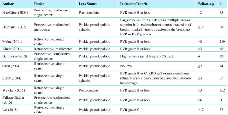
Table 1 summarizes the major studies until date on RRD surgery. Most of the non randomized trials evaluating primary RRD treatment show that PPV with or without SB is an acceptable approach in the management of noncomplex RRD, with good anatomic and functional results. The value of adding a SB to PPV is still controversial.
Limitations of the majority of these studies include the unavailability of high-quality RCT data, selection bias, and heterogeneous populations with regard to the characteristics of RRD.
Table 1 - Primary RRD treatment studies reviewed
Author Design Lens Status Inclusion Criteria Follow-up n
Brazikitos (2006) Prospective, randomized,
single center Pseudopahkic PVR grade B or less 12 75
Heimann (2007) Prospective, randomized, multicenter
Phakic, pseudopahkic, aphakic
Large breaks 1 to 2 clock hours, multiple breaks, superior bullous detachment, central extension of breaks, marked vitreous tracion on the break, no PVR or PVR grade A
≥12 681
Mehta (2011) Retrospective, single
center Phakic, pseudopahkic PVR grade B or less ≥3 219
Kinori (2011) Retrospective, multicenter Phakic, pseudopahkic PVR grade B or less ≥3 181
Bernheim (2013) Prospective, comparative,
single center Phakic, pseudopahkic High myopia (axial length > 26 mm) 6 191
Orlin (2014) Retrospective, single
center Phakic, pseudopahkic No PVR ≥2 74
Sorey (2014) Retrospective, single center
Phakic, pseudopahkic, aphakic
PVR grade B or C, RRD in 2 or more quadrants, retinal tears > 1 clock hour or associated vitreous hemorrhage
≥3 65
Weichel (2015) Retrospective, single
center Pseudopahkic PVR grade B or less ≥3 152
Falkner-Radler (2015)
Prospective, randomized,
single center Phakic, pseudopahkic PVR grade B or less ≥6 60
Lai (2015) Retrospective, single
center Phakic, pseudopahkic PVR grade C ≥12 77
The decision of which surgical procedure to use in the treatment of RRD still depends on a variety of factors and the surgeon’s preference. Until date there was only one major randomized clinical trial that drew some guidelines for our clinical practice. Although many variables are involved in RRD treatment prospective, multicentric, randomized studies are needed to determine the best anatomical and functional results.
REFERENCES
1. Arya AV, Emerson JW, Engelbert M, Hagedorn CL, Adelman RA. Surgical management of pseudophakic retinal
detachments: a meta-analysis. Ophthalmology
2006;113(10):1724– 1733.
2. Bernheim D, Rouberol F, Palombi K, Albrieux M, Romanet JP, Chiquet C. Comparative prospective study of thegmatogenous retinal detachments in phakic or pseudophakic with high myopia. Retina 2013 Nov-Dec; 33(10): 2039-48
3. Brazitikos PD, Androudi S, Christen WG, Stangos NT.
Primary pars plana vitrectomy versusscleral buckle surgery for the treatment of pseudophakic retinal detachment: a randomized clinical trial. Ophthalmology. 2006 Nov;113(11):2033-40.
4. Deokule S, Reginald A, Callear A. Scleral explant removal: the last decade. Eye (Lond) 2003;17:697–700.
5. Deutsch J, Aggarwal RK, Eagling EM. Removal of scleral explant elements: a 10-year retrospective study. Eye (Lond) 1992;6:570–573.
6. Falkner-Radler CI, Graf A, Binder S. Vitrectomy combined with endolaser or an encirclingscleral buckle in primary retinal detachment surgery: a pilot study. Acta Ophthalmol. 2015 Aug;93(5):464-9.
7. Feltgen N, Walter P. Rhegmatogenous Retinal Detachment—
an Ophthalmologic Emergency. Deutsches Ärzteblatt
International. 2014;111(1-2):12-22.
doi:10.3238/arztebl.2014.0012.
8. Ga Eun Cho, Seong Wook Kim, Se Woong Kang. Changing Trends in Surgery for Retinal Detachment in Korea Korean J Ophthalmol. 2014 Dec;28(6):451-9.
9. Haimann MH, Burton TC, Brown CK. Epidemiology of retinal detachment. Arch Ophthalmol. 1982;100:289–292.
10. Heimann H, Bartz-Schmidt KU, Bornfeld N, Weiss C, Hilgers RD, Foerster MH. Scleral buckling versus primary vitrectomy in rhegmatogenous retinal detachment: a prospective randomized multicenter clinical study. Ophthalmology. 2007 Dec; 114(12):2142-54
11. Hilton GF, Wallyn RH. The removal of scleral buckles. Arch Ophthalmol 1978;96:2061–2063.
12. Kessner R, Barak A. Pseudophakic rhegmatogenous retinal detachment: combined pars plana vitrectomy and scleral buckle versus pars plana vitrectomy alone. Graefes Arch Clin Exp Ophthalmol. 2016 Nov;254(11):2183-2189.
13. Kinori M, Moisseiev E, Shoshany N, Fabian ID, Skaat A, Barak A, Loewenstein A, Moisseiev J.Comparisonof Pars Plana Vitrectomy With and Without Scleral Buckle for the Repair of Primary Rhegmatogenous Retinal Detachment. Am J Ophthalmol.2011Aug;152(2):291-297.
14. Lai FH, Lo EC, Chan VC, Brelen M, Lo WL, Young AL.
Combined pars plana vitrectomy-scleral buckle versus pars plana vitrectomy for proliferative vitreoretinopathy. Int Ophthalmol 2015 Aug 11. doi:10.1007/s10792-015-0104-4
15. McLaughlin MD1, Hwang JC2. Trends in Vitreoretinal Procedures for Medicare Beneficiaries, 2000 to 2014. Ophthalmology. 2017 Mar 7. pii: S0161-6420(16)31843-7. doi: 10.1016/j.ophtha.2017.01.001.
16. Mehta S, Blinder KJ, Shah GK, Grand MG. Pars plana vitrectomy versus combined pars plana vitrectomy and scleral buckle for primary repair of rhegmatogenous retinal detachment. Can J Ophthalmol. 2011 Jun;46(3):237-41
17. Mitry D, Charteris DG, Fleck BW, Champbell H, Singh J. The epidemiology of rhegmatogenous retinal detachment: geographical variation and clinical associations. Br J Ophthalmol. 2010 Jun; 94(6):678-64
18. Mitry D, Singh J, Yorston D, Siddiqui MA, Wright A, Fleck BW, Campbell H, Charteris DG.
19. The predisposing pathology and clinical characteristics in the Scottish retinal detachment study. Ophthalmology. 2011 Jul;118(7):1429-34. doi: 10.1016/j.ophtha.2010.11.031.
20. Moisseiev E, Fogel M, Fabian ID, Barak A, Moisseiev J,
Alhalel A. Outcomes of Scleral Buckle Removal: Experience from the Last Decade. Curr Eye Res. 2016 Dec 2:1-5.
21. Orlin A, Hewing NJ, Nissen M, Lee S, Kiss S, DʼAmico DJ, Chan RV. Pars plana vitrectomycompared with pars plana vitrectomy combined with scleral Buckle in the primary managment of noncomplex rhegmatogenous retinal detachment. Retina. 2014 Jun;34(6):1069-1075
22. Sallam AB, Donachie PH, Yorston D, Steel DH, Williamson TH, Jackson TL, Sparrow JM, Johnston RL. ROYAL COLLEGE OF OPHTHALMOLOGISTS' NATIONAL DATABASE STUDY OF VITREORETINAL SURGERY: Report 7, Intersurgeon Variations in Primary Rhegmatogenous Retinal Detachment Failure. Retina. 2017 Feb 17. doi: 10.1097/IAE.0000000000001538. [Epub ahead of print]
23. Seider MI, Naiseri A, Stewart JM. Cost comparison of scleral buckle versus vitrectomy for rhegmatogenous retinal
detachment repair. Am J Ophthalmol. 2013 Oct;156(4):661-6
24. Shekelle PG, Woolf SH, Eccles M, Grimshaw J. Developing
clinical guidelines. Western Journal of Medicine.
1999;170(6):348-351.
25. Storey P, Alshareef R, Khuthaila M, London N, Leiby B, DeCroos C, Kaiser R; Wills PVR Study Group. Pars plana vitrectomy and scleral buckle versus pars plana vitrectomy alonefor patients with rhegmatogenous retinal detachment at high risk for proliferative vitreoretinopathy. Retina. 2014 Oct;34(10):1945-51.
26. Tielsch JM, Legro MW, Cassard SD, et al. Risk factors for retinal detachment after cataract surgery: a population-based case-control study. Ophthalmology 1996;103:1537–45.
27. Totsuka K, Inui H, Roggia MF, Hirasawa K, Noda Y, Ueta T.
SUPPLEMENTAL SCLERAL BUCKLE IN
VITRECTOMY FOR THE REPAIR OF
RHEGMATOGENOUS RETINAL DETACHMENT: A Systematic Review of Literature and Meta-Analysis. Retina. 2015 Nov;35(11):2423-31. doi: 10.1097/IAE.0000000000000797.
28. Tsui I. Scleral buckle removal: indications and outcomes. Surv Ophthalmol 2012;57:253–263.
29. Weichel ED, Martidis A, Fineman MS, McNamara JA, Park CH, Vander JF, Ho AC, Brown GC. Pars plana vitrectomy versus combined pars plana vitrectomy-scleral buckle for primary repair of pseudophakic retinal detachment.
Ophthalmic Surg Lasers Imaging Retina. 2015 Jul-Aug;46(7):702-7.
CONTACTO
Nuno Pinto Ferreira Av. Prof Egas Moniz 1649-035 Lisbon PortugalE-mail: [email protected]
This work was supported by the Department of Ophthalmology, Centro Hospitalar Lisboa Norte, Hospital de Santa Maria, Lisbon, Portugal.