Universidade de Lisboa
Probing the role of host lipids during hepatocyte infection
by malaria parasite
Investigando o papel dos lípidos da célula hospedeira durante
a infecção hepática pelo parasita da malária
Maurice Ayamba Itoe
Dissertation presented to the Faculty of Medicine of the University of Lisbon in fulfilment of the requirements for a Doctoral degree in Biomedical Sciences,
specialization in Bio-pathological Sciences
Dissertação apresentada à Faculdade de Medicina da Universidade de Lisboa, em cumprimento dos requisitos de um Doutoramento em Ciências Biomédicas
especialização emciências bio-patológicas
University of Lisbon 2012
Probing the role of host lipids during hepatocyte infection
by malaria parasite
Investigando o papel dos lípidos da célula hospedeira durante
a infecção hepática pelo parasita da malária
Maurice Ayamba Itoe
Dissertation presented to the Faculty of Medicine of the University of Lisbon in fulfilment of the requirements for a Doctoral degree in Biomedical Sciences,
specialization in Bio-pathological Sciences
Dissertação apresentada à Faculdade de Medicina da Universidade de Lisboa, em cumprimento dos requisitos de um Doutoramento em Ciências Biomédicas
especialização emciências bio-patológicas
Faculty of Medicine University of Lisbon
II
Cover image: Uptake of phosphatidylcholine (green) from host hepatocyte by P. berghei parasite (red)
III
This thesis was undertaken under an FP7-Marie Curie Actions Initial Training Networks- “Intervention strategies against malaria-InterMal Training” PhD program, reference PITN-GA-2008-215281, under the guidance of Dr. Maria M. Mota, Instituto de Medicina Molecular, Faculty of Medicine, University of Lisbon, Portugal and Dr. Friedrich Frischknecht, Hygiene Institute, Department of Parasitology, University of Heidelberg Medical School, Heidelberg, Germany.
Tese realizada sob o Programa de Doutoramento do 7º Programa Quadro - Rede Marie Curie "Intervention strategies against malaria-InterMalTraining", referência PITN-GA-2008-215281, sob a orientação da Dra. Maria M. Mota, Instituto de Medicina Molecular, Faculdade de Medicina da Universidade de Lisboa, Portugal e Dr. Friedrich Frischknecht, Departamento de Parasitologia da Universidade de Heidelberg, Heidelberg, Alemanha
IV
Acknowledgements
This thesis could not have been realized without the constant support from so many people to whom I will eternally be grateful and indebted to. My deepest appreciation goes to the following:
My supervisors; Maria and Freddy, for giving me the opportunity to be trained under their guidance in the InterMal Training PhD programme. Maria, thanks so much for allowing me to think freely and develop this project to the point it is now. I was deeply humbled by your constant availability to discuss problems that arose during the course of the project.
Freddy, thanks so much for the constant support and encouragement.
To the Marie Curie-funded InterMal Training PhD program, I will always be grateful to have been trained and supported by the network. A big thank you to all the students and coordinators of the InterMal Training program for haven made this program so successful in one way or another.
My thesis advisory committee members: Dr. Markus Meissner, Dr. Antonio Jacinto, Dr. Friedrich Frischknecht, and Dr. Maria Mota for their advice during my training.
To all present and former colleagues of UMA and associated groups, for making this environment an enjoyable place to work. It was a great opportunity for me to have worked with such a diverse and talented group of people. Ghislain and Eliana: thanks for your constant engagement in discussing the project. Your suggestions and discussions were always helpful. Ghislain, special appreciation for reading through the thesis and your suggestions. Eliana, thanks for helping me out with the translation. Kirsten, thanks so much for everything; for showing a lot of interest in discussing my data during or after lab meetings. Thanks also for reading this thesis and for making suggestions. Antonio and Vanessa, a big thank you for always willing to help. Amigo Peter: danke. Patricia I, Patricia M, Vanessa, Fernanda, Ana Roberto, Ana Parreira, Silvia, Ana Pena, Ines, Audrey, Joanas, Gunar, Celine, Claudia, Margarida, Miguel, Iset, Rita N, and Ana Pamplona. Thanks for all your support. Muito Obrigado!!!!!!!!!!!
A Carina: thanks for introducing me to malaria work and for your encouragement when things seemed doom. It was a wonderful opportunity to have worked with you.
To my all my dear friends for always being there for me.
To my mum and my entire family for their constant support over the years.
V
Abbreviations
ABHD5: αβ-hydrolase domain-containing protein 5 ACSL: Long chain acyl CoA synthetase
ATGL: Adipose triglyceride lipase CDS: Chanarin Dorfman Syndrome CE: Cholesterol Ester
Cer: Ceramide
CGI-58: Comparative gene identification-58 Chol: Cholesterol
Cho-P: Phosphocholine CoA: Coenzyme A
CSP: Circumsporozoite protein DAG: Diacylglycerol
DL-FBS: Delipidated fetal bovine serum EEF: Exo-erythrocytic forms
ESI-MS: Electron spray ionisation mass spectrometry FA: Fatty acid
FAS II: Fatty acid synthesis type II FAS II: Fatty acid synthesis type II FAS: Fatty acid synthase
FBS: Fetal bovine serum
GFP: Green Fluorescence Protein HGF: Hepatocyte Growth Factor
HPRT: Hypoxanthine guanine phosphoribosyltransferase HSPG: Heparan Sulphate Proteoglycan
HT: Hexose transporter LD: Lipid droplet
VI
L-FABP: Liver fatty acid binding protein LPA: Lysophosphatidic acid
LPC: Lysophosphatidylcholine mRNA: messenger ribonucleic acid MSP1: Merozoite Surface Protein 1
NBD-PC: 12-(7-nitro-2-1-3-benzoxadiazol-4-yl) amino Phosphatidylcholine NF-κB: Nuclear Factor κB
PA: Phosphatidic acid
PBS: Phosphate buffered Saline PC: Phosphatidylcholine
PC-TP: Phosphatidylcholine transfer protein
PCYT1α: CTP: Phosphocholine cytidylyltransferase 1 alpha PDH: Pyruvate dehydrogenase
PE: Phosphatidylethanolamine
PEMT: Phosphatidylethanolamine Methyltransferase Peri A: Perilipin A
PfSCD: P. falciparum Stearoyl-CoA Desaturase PG: Phosphatidylglycerol
PI: Phosphatidylinositol PS: Phosphatidylserine PUF: Pumilio protein
PV: Parasitophorous vacuole
PVM: Parasitophorous vacuole membrane RBC: Red Blood Cell
RBM: Roll Back Malaria RFP: Red Fluorescence Protein
VII
SCD: Stearoyl-CoA desaturase SiRNA: short interfering RNA SM: Sphingomyeline
SPECT: Sporozoite Microneme Protein Essential for Cell Traversal SR-B1: Scavenger receptor B type 1
TAG: Triacylglycerol
TRAP: Thrombospondin-Related Anonymous Protein UIS3/4: Up-regulated in infective sporozoites
VIII
Thesis outline
The work presented in this thesis was carried out from January 2009- February 2012 at the Institute of Molecular Medicine. The thesis is preceded by a summary in Portuguese and English and followed by six chapters that are organised as follows:
Chapter I: Starts off with a general introduction on malaria and the parasite´s life cycle, followed by an overview of lipid metabolism in hepatocytes and Plasmodium parasites. The significance of Plasmodium encoded lipid metabolism genes during liver stage infection is described.
Chapter II: This outlines the main aims that were undertaken in this study. Chapter III: Details the results from this study
Chapter IV: Contains the general discussion of the results Chapter V: Ends with the conclusions and future directions Chapter VI: Materials and Methods
References Appendix
IX
Table of contents
Acknowledgements IV Abbreviations V Thesis outline VIII
Sumario 1
Summary 4
1 Introduction 1.1 MALARIA DISEASE 8
1.1.1. Symptoms and diagnosis 10
1.1.2. Treatment and prevention 10
1.2 PLASMODIUM SPECIES, VECTORS, AND LIFE CYCLE 11
1.2.1. Erythrocytic infection 13
1.2.1.1 Host cell invasion and intra-erythrocytic development 13
1.2.1.2 Host cell egress 14
1.2.2 Mosquito stage: where the mix happens 15
1.2.3 Deposition of sporozoites in the skin and migration to the liver 16
1.2.4 Intra-hepatic development, merozoite formation, and release 20
1.3 LIPID METABOLISM 24
1.3.1 Lipid metabolism in hepatocytes 24
X
1.3.1.2 Fatty acid modification: role of stearoyl- CoA desaturase (SCD) 24
1.3.1.3 Lipogenesis and glycerophospholipid biosynthesis 25
1.3.1.4 The Kennedy pathway for PC and PE biosynthesis: Role of PCYT1α for PC synthesis 27
1.3.1.5 Catabolism of stored FAT: Role of ATGL and ABDH5 or CGI-58 28
1.3.2 Basic functions of lipids 30
1.3.3 Lipids and infection 31
1.3.4 Plasmodium infection and lipid metabolism 31
1.3.4.1 De novo lipid biosynthesis in Plasmodium and blood stage infection 32
1.3.4.1.1 Fatty acid synthesis and modification 32
1.3.4.1.2 Glycerophospholipid synthesis in Plasmodium 33
1.3.4.2 Lipids and Plasmodium liver stage infection 35
2. Aims 39
3. Results Plasmodium infection alters the lipid composition of infected cells 45
Plasmodium liver stage infection requires host cell de novo PC biosynthesis for intracellular survival of parasite 56
Host cell lipogenic and lipolytic processes are required during intracellular survival of Plasmodium parasites 66
XI
4. General discussion 79
5. Conclusions and future perspectives 90
6. Materials and Methods 93
References 104
1
Sumário
A malária, uma doença causada por protozoários do género Plasmodium, constitui ainda um sério problema de saúde no mundo, sendo a causa directa de mais de 650000 mortes todos os anos. Após transmissão através da picada de um mosquito Anopheles infectado, os parasitas migram da derme do hospedeiro até ao fígado através da circulação sanguínea e, uma vez no fígado, atravessam vários hepatócitos antes de infectarem aquele que se tornará a célula hospedeira. No interior da célula infectada, cada parasita irá originar dezenas de milhares de novos parasitas. A infecção do fígado do hospedeiro vertebrado precede a manifestação clínica da doença e é uma etapa obrigatória para o estabelecimento da infecção sanguínea. As interacções moleculares entre parasita e hospedeiro têm um papel determinante no estabelecimento da susceptibilidade à infecção e, portanto, a identificação de factores do hospedeiro relevantes para o desenrolar da infecção é essencial numa perspectiva de desenvolvimento de novas estratégias anti-maláricas.
O conhecimento actual sobre a natureza dos factores do hospedeiro que influenciam a infecção hepática é ainda rudimentar, apesar do interesse que esta fase da doença tem suscitado. Diversos estudos demonstraram que tanto o parasita como o hospedeiro alteram os seus perfis de expressão genética durante a fase hepática da infecção. Mais especificamente, a expressão de genes envolvidos no metabolismo de lípidos e na síntese de ácidos gordos encontra-se aumentada na célula hospedeira e parasita, respectivamente. Por esta razão, colocámos a hipótese de que a infecção hepática poderá alterar substancialmente o perfil lipídico da célula hospedeira.
De forma a testar esta hipótese, procedemos à extracção e caracterização por espectroscopia de massa do conteúdo lipídico de células infectadas e não-infectadas, previamente separadas com base na fluorescência da proteína “green fluorescence protein” expressa pelo parasita transgénico P. berghei. Esta análise permitiu identificar alterações consistentes com um aumento no metabolismo de armazenamento de lípidos (triglicéridos, esteres de colesterol) e síntese de lípidos estruturais (fosfatidilcolina, esfingolípidos, ceramida, esfingomielina) nas células infectadas. A fosfatidilcolina destaca-se nesta análise por ser o único fosfolípido que não se encontra sub-representado nas células infectadas. O perfil lipídico patente nas células
2
infectadas sugere que os processos de lipogénese, biosíntese de fosfatidilcolina e lipólise são essenciais durante a fase hepática da infecção por Plasmodium. Hipoteticamente, estas alterações poderão ser mediadas pela célula hospedeira ou pelo parasita, uma vez que este possui vias biosintéticas compatíveis com o perfil lipídico observado.
Numa tentativa de discriminar entre estas duas possibilidades, procedemos ao silenciamento da expressão de diversos genes necessários para os processos de lipogénese, síntese de fosfatidilcolina e catabolismo de lípidos na célula hospedeira. Esta análise demonstrou que a biosíntese de novo de fosfatidilcolina é essencial para o desenrolar da infecção hepática, uma vez que o silenciamento das enzimas “CTP:phosphocholine cytidylyltransferase 1 alpha (PCTY1a)” e “phosphatidylethanolamine N-methyltransferase (PEMT)” reduz drasticamente a infecção. Em paralelo, foi possível observar a transferência de análogos de colina e fosfatidilcolina da célula hospedeira para o parasita, o que sugere que o parasita utiliza fosfatidilcolina sintetizada pela célula hepática. O silenciamento da enzima lipolítica “adipose triglyceride lipase (ATGL)” e do seu co-activador “αβ-hydrolase domain containing protein 5 (ABDH5/CGI-5)”, bem como da enzima “stearoyl-coenzyme A desaturase 1 (SCD-1)”, necessária para o processo de lipogénese, tem como consequência uma redução da infecção hepática por P. berghei.
Este estudo caracteriza pela primeira vez o repertório lipídico da célula hospedeira durante a fase hepática da infecção por Plasmodium e estabelece uma correlação entre diversas vias do metabolismo hepático de lípidos e a viabilidade do parasita na célula hospedeira.
4
Summary
Malaria, a vector-borne disease caused by members of the apicomplexa parasites of the genus
Plasmodium, continues to pose serious threat to global health whereby it is responsible for
the death of over 650000 people from about 216 million infections each year (WHO World Malaria Report 2011). Upon transmission to the mammalian host by an infected Anopheles mosquito, Plasmodium sporozoites exit the dermis of the skin into the blood stream and travel to the liver where they traverse several hepatocytes before eventually invading a final one. Within the hepatocytes, the parasite undergoes an exponential replication generating tens of thousands of blood stage-infectious merozoites. Though clinically silent, a successful liver stage infection is the first obligatory step for the onset of a blood stage infection which is associated with the clinical manifestations of the disease. A better understanding of the molecular mechanisms involved in the establishment of liver stage infection is crucial for rational design of vaccines and drugs against the disease.
Despite intense interest over several decades in studying Plasmodium liver stage infection, our knowledge of the role of host factors during this stage of the parasite´s life cycle is rudimentary. Previous studies using rodent malaria parasites have revealed that both parasite and host genes are transcriptionally modulated during intracellular replication of Plasmodium species in hepatocytes (Albuquerque et al., 2009, Tarun et al., 2008). Specifically, a subset of genes involved in lipid metabolism in the host (Albuquerque et al, 2009) or fatty acid synthesis enzymes of the parasite (Tarun et al, 2008) are up-regulated during infection. Taken together, we hypothesized that the lipid repertoire of Plasmodium infected hepatocytes would change during infection.
To address this, total cellular lipids was extracted from Green Fluorescence Protein (GFP)-expressing P. berghei-infected and non-infected cells that were sorted by Fluorescence Activated Cell Sorting (FACS) at different time points after infection. The lipids were subjected to shotgun mass spectrometry to identify or monitor which lipid classes change as infection progressed. Our analysis revealed enrichment in infected cells of lipid classes that represent a typical signature of storage and structural membrane lipid biosynthesis. This includes triglycerides (TAG), cholesterol esters (CE), phosphatidylcholine (PC) and the sphingolipids; ceramide (Cer) and sphingomyeline (SM). Concomitant with enrichment in PC, the levels of all the other major phospholipids are decreased at all time points examined.
5
The lipidomic profile of infected cells is suggestive of an engagement of key elements of lipid metabolism such as lipogenesis, PC biosynthesis, and lipolysis. These changes could be mediated by the host or P. berghei parasites which do express several enzymes involved in lipid biosynthesis.
In order to decipher the functional relevance of the host cell lipid biosynthetic machinery, as indicated by our lipidomic data, to the establishment of a successful infection, we performed targeted silencing of host genes involved in lipogenesis, PC synthesis, and lipolytic breakdown of stored fat in Huh7 cells. Interestingly, our data shows that upon knockdown of
host CTP: phosphocholine cytidylyltransferase 1 alpha (PCTY1α) and
phosphatidylethanolamine methyltransferase (PEMT), key enzymes in de novo PC synthesis via the Kennedy pathway and trimethylation of PE to PC, respectively, severely abrogates infection. In addition, a possible utilisation of host-derived PC during infection was demonstrated using an analogue of choline or PC. Also, knockdown of host lipolytic enzyme adipose triglyceride lipase (ATGL) and its co-activator αβ-hydrolase domain-containing protein 5 (ABHD5) or comparative gene identification 5 (CGI-5), and lipogenic enzyme stearoyl-CoA desaturase 1 (SCD1) greatly impacts P. berghei infection in Huh7 cells.
This study provides not only the first global picture of the lipid repertoire of Plasmodium infected cells but also novel evidence for a connection between different arms of host cell lipid metabolic pathways and the establishment of a successful liver stage infection.
7
8 1. Malaria disease
Malaria continues to be a leading cause of illness and death globally. In humans, malaria is caused by four Plasmodium species; Plasmodium falciparum, P. vivax, P. malariae and P.
ovale. More recent reports indicate that, P. knowlesi, a species that was known to infect only
monkeys is now known to infect human populations in South East Asia (Cox-Singh et al. 2008, Jongwutiwes et al. 2011, Putaporntip et al. 2009). Majority of malaria infections are caused by P. falciparum and P. vivax. P. falciparum infections are responsible for most severe cases and deaths due to malaria and are localised in tropical regions such as Sub- Saharan Africa, South East Asia, and around the Amazon rainforest in South America (Gething et al. 2011, Hay et al. 2009) (Figure 1).
On the other hand, infections due to P. vivax are common in south East Asia and most endemic countries in the Americas. P. malariae infections usually occur in areas endemic to
P. falciparum but with very low incidence. (RPM report 2011). Globally, an estimated 3.3
billion people, spanning about 106 countries, were at risk of malaria in 2010, with about 2.1 billion living in regions of low risk and 1.2 billion at high risk. Of these, an estimated 216 million episodes of malaria were recorded during this period globally with the Africa region bearing the brunt of the disease; of the over 650000 deaths reported annually worldwide, about 91% occur in the Africa region, with most of the cases occurring in children under the age of 5 (WHO world malaria report, 2011). However, these reports have been challenged very recently by a study conducted by Murray and colleagues, 2012, suggesting about a
two-Figure 1. Global spatial distribution of P falciparum malaria endemicity. P. falciparum annual mean parasite rate
(PfPR)-proportion of individuals infected at a given time, within stable transmission. Endemicity is confined mainly to Sub-saharan Africa. Adapted from Gething et al, 2011.
9
fold increase in the number of deaths due to malaria globally and a similar factor of increase in those registered in Africa. Their report also stated that adults and children above 5 years are also badly affected by the disease (Murray et al. 2012).
Despite being home to the bulk of stable transmission, there exist huge disparities in epidemiological settings and control strategies among African countries, thus resulting in different stable transmission dynamics across the continent; low stable transmission in the east to high stable transmission in west and central regions. Noteworthy is the fact that most countries in the West Africa sub-region have the highest malaria transmission rates in the Africa region (Figure 1).
As an example to illustrate malaria endemicity and impact of the disease in west/central Africa sub-region, a recent report released in 2011 for 2010 by Cameroon´s National Malaria Control Programme (NMCP).
Malaria was responsible for 36% of medical consultations with 62.2% of malaria cases being
uncomplicated, with 38% morbidity among pregnant women and 24% of death recorded in hospitals, 67% of these occurring in children under the age of 5. This was recently echoed by WHO world malaria report for 2011, indicating that of a total population of around 20 million people, the entire country is at risk of malaria, with nearly three quarters at high stable transmission risk and the remaining at low stable transmission risk with Plasmodium
falciparum as the main parasite (Figure 2).
Figure 2. Malaria transmission pattern in Cameroon. More than
three-third of the population are at high stable risk of transmission. Infections occur all-year round. Adapted from WHO World
10 1.1 Symptoms and diagnosis
The main common symptoms associated with malaria involve periodic fever and chills, cerebral convulsions due to cerebral malaria, severe anemia, loss of foetus due to stillbirths (Miller et al. 1994).
The standard malaria diagnostic test involves staining of blood films with diluted Giemsa solution. In addition, other microscopy techniques utilise fluorescence dyes such as acridine orange (AO) and benzothiocarboxypurine (BCP), both of which have high affinity for nucleic acid in the parasite nucleus and would emit fluorescence upon excitation at 490nm. However, in recent years, other non-microscope techniques have been developed such as PCR-based amplification of parasite antigen, or immunochromatographic rapid diagnostic tests (RDT) to detect abundant parasite proteins from blood (reviewed in (Moody 2002).
1.2 Treatment and prevention
Following the emergence and spread of chloroquine-resistant P. falciparum in the eighties and nineties, the WHO recommended first-line of treatment for uncomplicated malaria are Artesunate plus amodiaquine and Artemether or Quinine are used for severe malaria. In most endemic regions of, of the entire population that are risk to Plasmodium infections, only about 40% that get sick are potentially treated with Artemisinin-based combination therapy (ACT) alone, and the majority with other antimalarial medicines.
The main intervention strategies in controlling the disease involve the use of insecticide treated bed nets (ITN)/Long Lasting Insecticide Treated Nets (LLINs), Indoor Residual Spraying (IRS), and Intermittent Preventive Treatment for pregnancy (IPTp). It is important to emphasize here that, the major strides made in reducing the incidence of malaria in endemic regions has been hugely attributed to the implementation of one or a combination of these control strategies, thanks to enormous financial support from donor countries and other international funding agencies.
In addition, other preventive measures in place involve the use of chemoprophylaxis to interfere with blood and liver stages of Plasmodium infections. The main prophylaxes include atovaquone-proguanil, chloroquine, mefloquine, doxycycline, and primaquine. Of these, only
11
primaquine and atovaquone-proguanil acts on hepatic schizonts of all human malaria species. In addition, primaquine is uniquely active in killing dormant forms (hypnozoites) of P. ovale and P. vivax, which can relapse and cause onset of second round of clinical malaria. Together with mefloquine, doxycycline, and chloroquine, atovaquone-proguanil also acts on the blood stage schizonts of all species, thus preventing clinical onset of disease (Freedman 2008). Despite the discovery of the causative agent of malaria over a hundred years ago, dreams for an efficacious vaccine against this deadly disease have only been recently rekindled after a recent report of an efficacy of over 45% protection against severe malaria from phase 3 trials of a recombinant vaccine, RTS,S/AS01, in Africa children (Agnandji et al. 2011).
In the human host, the life cycle of Plasmodium species, the causative agent of malaria is characterised by two stages: an intra-hepatocytic stage in the liver and blood stage in red blood cells (RBC). All the clinical signs and symptoms associated with the disease occur in the blood stage while the liver stage is asymptomatic. A better understanding of the molecular mechanisms of pathogenesis is necessary for the development of drugs and vaccines against this disease. Below, I present an overview of the peculiarities of the
Plasmodium life cycle which involves vertebrate and invertebrate hosts.
2. Plasmodium species, vectors, and their life cycle
Apicomplexan parasites belonging to the genus Plasmodium are of great medical significance as they are known to pose serious threat to public health systems globally (Greenwood et al. 2005). Of the over 5000 Plasmodium species that exist, only five are known to cause infections in humans (see section 1.1). However, our knowledge on the basic biology of the disease has been obtained mainly from the use of rodent malaria parasites- P. berghei, P.
chabaudi and P. yoelii- and avian- P gallinaecium, whose entire life cycles can be
recapitulated in the laboratory.
Malaria parasites are transmitted from one individual to another by arthropods belonging to the genus Anopheles. Of the over 45 Anopheles species that exist across Africa and many other malaria endemic regions, 14 of them are known to be capable of transmitting malaria,
12
with the most common and efficient vector species being Anopheles gambiae, Anopheles
arabiensis, Anopheles funestus, Anopheles nili and Anopheles moucheti (Fontenille and
Simard 2004). Because of disparities in eco-systems across the region, differences may exist in the type of species that are predominant in transmitting the disease in different localities (Antonio-Nkondjio et al. 2005, Bigoga et al. 2007, Tanga et al. 2010, Wanji et al. 2003).
Plasmodium parasites exhibit a characteristic life cycle involving vertebrate and mosquito
hosts, each harbouring very distinct transformation stages crucial in the pathogenesis and transmission of the disease, respectively. In the mammalian host, the life cycle is composed of an exo-erythrocytic liver stage and an erythrocytic stage occurring in RBCs (Figure 3).
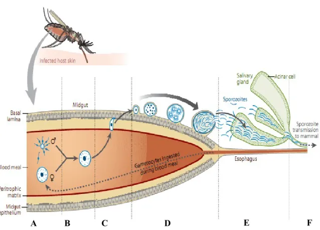
Figure 3. Schematic representation of Plasmodium life cycle. (A): Deposition of salivary gland sporozoites into the dermis. (B): sporozoites exit the dermis into the blood stream and travel to the liver sinusoid. (C): Sporozote traverses several hepatocytes before arresting in a final host cell. (D): Parasite differentiates into tens of thousands of merozoites. (E): merozoites are released into the blood stream as merozomes. (F): merozoites infect red blood cells and replicate to generate 10-30 daughter merozoites. (G): some merozoites differentiate into sexual forms, gametes. (H): Sexual forms are ingested by a mosquito during a blood meal and undergo fertilisation in the midgut. (I): after sporulation, sporozoites are ready to be ingested into a new host during the next blood meal.
B
E
D
C
I
A
G
H
F
13
Though clinically silent, a successful liver stage infection is a pre-requisite for the establishment of symptomatic blood stage infection (Prudencio et al. 2006). Following injection of Plasmodium sporozoites into mammalian hosts by an infected female Anopheles mosquito during a blood meal, the sporozoites travel through the blood stream to the liver where they traverse several hepatocytes before eventually invading the final one with the formation of a parasitophorous vacuole (PV) (Mota et al. 2001). Within the target cell, the parasite undergoes extensive replication; producing tens of thousands of infectious merozoites, which when released from the host cell will initiate a blood stage infection. Upon invasion of an erythrocyte by a merozoite, the parasite undergoes replication producing 10-30 merozoites, which upon rupture of the infected cell, are released into the blood stream. These, go on to re-invade other RBC. After several cycles of replication in RBCs, some merozoites can differentiate and undergo gamtogenesis to produce sexual forms. These are released into the blood circulation upon rupture of infected cells and can be ingested by a female
Anopheline mosquito during a blood meal. The remaining segment of the parasite´s life cycle
occurs within the mosquito host; fertilisation of gametes and subsequent developments within the mosquito results in the production of infectious sporozoites which when present in the salivary glands are ready for injection/transmission to an individual during the mosquito´s subsequent blood meal (Figure 3).
2.1 Erythrocytic infection
2.1.1 Host cell invasion and intracellular development
Upon completion of liver stage infection, merozoites are released into the blood stream to initiate RBC infection. Infection of RBCs by Plasmodium merozoites occurs through a tightly regulated process common to members of the phylum Apicomplexan. Binding of merozoites to erythrocyte is mediated by a wide range of ligands on the merozoite surface, mainly proteins belonging to Duffy Binding-like (DBL) and reticulocyte binding homolog (Rh) families, with several proteins on RBCs, reviewed in (Cowman and Crabb 2006, Kappe et al. 2010). Upon binding, invasion of host cell is mediated by proteins that are stored in apical organelles, hence the name Apicomplexan. The process proceeds as follows: (1) attachment of merozoites to the surface of RBCs via parasite surface protein, MSPs, in a low-affinity reversible binding, (2) re-orientation and apical attachment with the erythrocyte membrane
14
through the involvement of a micronemal protein, AMA1 (Triglia et al. 2000), (3) apical discharge of secretory organelles and tight junction formation between the parasite and host cell plasma membrane. Once formed, the tight junction moves from the apical to the posterior end through actin-myosin machinery. This movement propels the parasite forward into host cell leading to the formation of a parasitophorous vacuole (PV), (reviewed in (Carruthers and Blackman 2005, Cowman and Crabb 2006).
Upon entry into the cell, in order to develop, the parasite initiates an extensive remodelling of the host cell, which is void of any protein synthesis machinery, into a convenient environment suitable for survival and replication, culminating in the generation of 10-30 merozoites at the end of schizogony over a very short time. Asexual development in erythrocytes involves several stages: rings, trophozoites, and schizonts. A key feature of host cell remodelling includes the formation of Maurer´s cleft. This connects the parasitophorous vacuole/parasitophorous vacuole membrane to the erythrocyte surface and plays a major role in protein trafficking and the appearance of tiny protrusions called knobs, which mediate adherence of infected cells to the vascular endothelium (Crabb et al. 1997). Infected cells-vascular endothelium interaction in the brain is the hallmark of cerebral malaria, which is the leading cause of death in children. The main proteins in Knobs are knobs associated histidine-rich proteins (KHARP) and erythrocyte membrane protein 1 (EMP1) with the later exhibiting high antigenic variability (Crabb et al. 1997) and highly implicated in the pathogenesis of the disease. Also, an important characteristic of asexual development in erythrocytes is the catabolism of haemoglobin in the food vacuole of the parasite in order to generate amino acids which its uses for protein synthesis, with a resultant release of a toxic metabolic product, heme (reviewed in (Tilley et al. 2011)). Heme is converted into an inert crystalline product called hemozoin which gets sequestered in the food vacuole (reviewed in (Hanscheid et al. 2007).
2.1.2 Host cell Egress
An intra-erythrocytic merozoite undergoes several rounds of mitotic replication to generate 10-30 merozoites per infected erythrocyte. For progression of blood stage infection to occur, mature merozoites must be released from infected cells to continue the cell. Earlier studies by (Wickham et al. 2003), showed that rupture of infected cells proceeds through a two step
15
process, starting with the rupture of the PVM and followed by the rupture of the host cell plasma membrane. More specifically, (Yeoh et al. 2007), further showed that a parasite-encoded serine protease called PfSUB1 (Plasmodium falciparum subtilisin 1) was highly expressed at late blood schizogony and was released into the PV just prior to PV and erythrocyte membrane rupture. PfSUB1-mediated PV and PVM rupture occurred via activation of SERA proteins (Yeoh et al. 2007). Upon release from host cells, the merozoites re-invade other red blood cells. After several rounds of replication in erythrocytes, some of the merozoites differentiate into sexual forms called gametocytes.
2.2 Mosquito stages: where the mix happens
During a blood meal from an infected individual, gametocytes are ingested alongside blood into the mosquito midgut lumen where they are activated to form male and female gametes (Figure 4A) which undergo fertilisation to produce zygotes (Figure 4B). The resultant zygote undergoes further transformation to form ookinete (Figure 4C). This is the first motile and invasive stage of the parasite in the mosquito, with invasion and migratory capabilities akin to those observed during cell transmigration in liver stage infection with sporozoites. The ookinete traverses several epithelial cells and eventually forms an oocyst (Figure 4D) which becomes immobile in the extracellular lumen. This then undergoes extensive replication within the lumen, usually 10-14 days, leading to the production of thousands of sporozoites. Upon egress from the oocyst, the sporozoites are transported in the hemolymph to the salivary glands where they attach and invade through the basal lining of the salivary glands (Figure 4E). Upon arrival at the salivary gland, sporozoites reside in salivary cavities where a good proportion of them have been shown to display three patterns of locomotion; back-and-forth, without change in displacement, circular manner, and straight (Frischknecht et al. 2004). Upon colonization of the salivary duct during salivating, a great proportion of the sporozoites also display characteristic motility patterns (Frischknecht et al. 2004), and are ready to be injected to the mammalian host (reviewed in (Aly et al. 2009). While in the salivary glands, very recent data has shown that Plasmodium sporozoites are held in a developmentally quiescent state by Pumilio-2(Puf2), a member of the RNA binding protein family PUF. In the absence of Puf2, salivary glands sporozoites undergo morphological and transcriptional changes akin to early intra-hepatic EEFs (Gomes-Santos et al. 2011).
16 2.3 Deposition in the skin and migration to the liver
Transmission of malaria to vertebrate host is initiated at the skin during a blood meal of an infected Anopheline mosquito which contains sporozoites in the salivary glands (Ponnudurai
et al, 1991). Several decades ago, it was only known that upon injection of sporozoites into
the skin, the sporozoites would travel through the blood circulation to the liver where they infect hepatocytes and differentiate into blood-infectious form. Recent advances in imaging technologies and availability of a suitable rodent malaria models, have aided greatly in the description of events that occur after inoculation of sporozoites into the dermis. It has been previously demonstrated that sporozoites injected into the skin remain at the injection site for at least 5 minutes before migrating to the blood vessels to enter the blood stream and journey to the liver (Matsuoka et al. 2002, Sidjanski and Vanderberg 1997). More recent intravital
Figure 4. Fertilisation and differentiation of malaria parasites in the mosquito host. Gametes (A) ingested with a
blood meal from an infected individual undergo fertilisation in the midgut to form zygotes (B). The zygotes undergo meiosis and form ookinetes (C). The Ookinetes go through the peritrophic matrix of the midgut, enter the apical end of the epithelium and traverses several epithelia cells before arriving at the basal epithelial surface. Here, they form oocysts (D) which undergo sporogony to form thousands of sporozoites, which upon egress from the sporoplast travel in the hemolymph to reach the salivary gland. (E): Sporozoites attach to acinar cells and traverse into the salivary gland ducts where they´re ready to be transmitted to an individual during a subsequent blood meal (F). Adapted from Aly, AS et al, 2009.
17
microscopy studies elegantly demonstrated that sporozoites injected into the dermis can glide and migrate through the dermis (Vanderberg and Frevert 2004), and can also traverse skin cells (Amino et al. 2008), before exiting into the blood stream, with a significant number of them leaving after 30 minutes of inoculation (Figure 5).
Detailed quantification revealed that of the bulk of sporozoites that are inoculated into the dermis, about half of them remain in the bite-site, one-third eventually reaches the liver to undergo exo-erythrocytic developments in hepatocytes, and roughly about 15% end up in the lymph nodes where they are eliminated (Amino et al. 2006). Furthermore, of the residual sporozoites at the bite-site, some can invade skin cells and undergo complete development into merozoites as it occurs in the liver (Gueirard et al. 2010). Still, these parasites do not seem to be able to initiate malaria infection (Voza et al. 2012). Visualisation of EEFs in the skin of infected mice seems to be re-echoing the consideration of a `skin stage`in the malaria
life cycle, as was previously suggested by Matsuoka and colleagues, 2002.
Once sporozoites enter the peripheral blood circulation at the inoculation site, they travel within a few minutes to reach the liver where they invade their target cells, hepatocytes (Shin et al. 1982) and replicate into tens of thousands of blood-stage infectious merozoites. The liver architecture as shown in Figure 6 is composed of a sinusoidal cell layer which is made up of endothelial and Kupffer cells and is separated from the hepatocytes by the Space of Disse. Following the report by Shin and colleagues (Shin et al. 1982), it was generally believed that Plasmodium sporozoites directly infect liver hepatocytes once they reach the liver. However, subsequent studies over the last two decades have elegantly demonstrated that is not the case. In fact, upon arrival at the liver, the sporozoites are initially arrested at the
Figure 5. Deposition of sporozoites into the dermis of the vertebrate host. During a blood
meal, an infected Anopheline mosquito injects sporozoites (in green) into the dermis of the skin. The sporozoites migrate in the dermis and exit into the blood capillaries, wherein they travel to the liver sinusoids. Adapted from Prudencio et al, 2006.
18
sinusoids via specific interactions of parasite surface proteins; circumsporozoite protein (CSP) and thrombospondin-related adhesive proteins (TRAP) with heparin sulfate proteoglycans (HSPG) of the liver cells (Coppi et al. 2007, Frevert et al. 1993, Robson et al. 1995), with differences in the degree of binding to kupffer cells, stellate cells and hepatocytes (Pradel et al. 2002). The degree of binding of TRAP and CS is highest for hepatocytes because they are enriched in HSPG than the other cell types in the liver. Binding of CS is mediated via specific interaction of a conserved a domain on the carboxyl terminal named region II-plus with sulfated residues on HSPGs (Sinnis et al. 1994). Subsequent studies by Coppi and colleagues have demonstrated that the binding of HPSGs on hepatocytes to
Plasmodium sporozoites signals the parasite to switch from a `migratory mode` to an
`invasive mode`. Switching involves proteolytic cleavage of CSP and exposure of a thromspondin-spanning region (TSR) (Coppi et al. 2005, Coppi et al. 2007, Coppi et al. 2011) and induction of parasite calcium-dependent protein kinase signalling. Once arrested at the liver sinusoids the sporozoite glide over and transmigrate through Kupffer cells (stationary liver professional macrophages), sometimes with the formation of a non-fusogenic vacuole (Pradel and Frevert 2001) before invading a final target hepatocyte (Figure 6).
2.4 Invasion of hepatocytes Figure 6. Attachment and traversal of sinusoidal layer by sporozoites. Sporozoites travel in the blood and arrive at liver sinusoid. They attach to heparin sulphate proteoglycans (HSPG) extending from hepatocytes and stellate cells, and transmigrate through kupffer cells in the Space of Disc before invading hepatocytes. Adapted from Prudencio and Mota, 2007.
19
Upon exit from Kupffer cells and the Space of Disse, Plasmodium sporozoites traverse on average 4-5 hepatocytes, sometimes resulting in damage and leakage of the plasma membrane of traversed cells (Mota et al. 2001). Subsequent studies by Mota and colleagues (2002), suggested that cell-traversal activates sporozoites, thus preparing them for final host cell invasion. However, this hypothesis was later challenged by studies conducted by Ishino and colleagues (Ishino et al. 2004) using P. berghei sporozoites deficient in a micronemal protein named Sporozoites microneme protein essential for cell traversal (SPECT). SPECT-deficient sporozoites lack cell traversal ability but still invade hepatocytes normally in vitro like the wild-type. In addition, SPECT-mutants exhibit greatly reduced infectivity in vivo. However, this defect could be reverted to wild type levels upon depletion of kupffer cells, thus suggesting that traversal is required for passage through Kupffer cells in liver stage infection by Plasmodium species. In addition to SPECT mutants, Kariu and colleagues (Kariu et al. 2006), have also described another micronemal protein, cell traversal protein of
Plasmodium ookinetes and sporozoites (CelTOS), to be essential for liver stage infectivity
and ookinete migration through the midgut epithelium. That notwithstanding, very recent studies by Coppi and colleagues (Coppi et al. 2011), supports the notion of activation of sporozoites by migration but alludes this activation to processing of CSP by a protease whose activity exposes the TSR domain of CSP thereby causing sporozoites to stop migrating and invade a target cell.
Invasion of hepatocytes by Plasmodium sporozoites occurs via invagination of host plasma membrane, which results in the formation of a parasitophorous vacuole whose membrane, the PVM, is host cell-derived (Lingelbach and Joiner 1998). Previous studies by (Carrolo et al. 2003, Leiriao et al. 2005), showed that migration of sporozoites through hepatocytes induces the release of hepatocyte growth factor (HGF) from traversed cells which renders neighbouring cells susceptible to infection and protects them from undergoing apoptosis; through activation of a HGF/MET signalling cascade in the host cells. However, a recent follow-up study conducted by Kaushansky and Kappe (Kaushansky and Kappe 2011) cautions against generalisation of the effect of HGF/MET signalling on liver stage infection across Plasmodium species; HGF/MET signalling was observed to be crucial only for P.
berghei infection but not for P. yoelii nor P. falciparum infections. In addition, infected cells
are also reported to over-express heme oxygenase 1(HO-1), an anti-inflammatory enzyme, which serves to protect them from inflammatory insults (Epiphanio et al. 2008). On the other
20
hand, Torgler and colleagues (Torgler et al. 2008), showed that cytosolic factors that are released from traversed cells activate NF-κB expression in neighbouring cells and controls infection via apoptosis. NF-κB is the main host regulator of inflammatory response controlling the transcription of several genes involved in processes such as apoptosis and survival. However, like other protozoan parasites such as Trypanosoma cruzi, the causative agent of chegas disease (Monteiro et al. 2001), P. berghei parasites express a cysteine protease inhibitor named PbICP (P. berghei inhibitor of cystein protease) (Rennenberg et al. 2010). PbICP is released by sporozoite during invasion of hepatocytes, accumulates within the PV, and is released during late liver stage at which point its acts as a potent inhibitor of cathepsin-like cysteine proteases of the host and parasite, thus preventing premature apoptotic programmed host cell death (Rennenberg et al. 2010).
Efforts during the last decade have identified two key molecules on the surface of hepatocytes, namely tetraspanin `cluster of differentiation` 81 (CD81) and scavenger receptor B1 (SR-B1), that regulate permissiveness of hepatocytes to Plasmodium sporozoites, though no direct binding between these molecules and sporozoites has been yet observed (Rodrigues et al. 2008, Silvie et al. 2003, Yalaoui et al. 2008). While SR-B1 mediates cellular uptake of cholesterol ester from high-density lipoproteins, CD81 aggregates into proteo-lipidic membrane micro-domains that associates with cholesterol to enhance the function of surface proteins. Thus, SR-B1 availability indirectly affects CD81 levels on the surface of hepatocytes (Yalaoui et al. 2008).
2.4. Intra-hepatocytic development, merozoite formation, and release.
Upon residence in host hepatocytes, Plasmodium parasites undergo extensive replication generating tens of thousands of RBC-infective merozoites from a single salivary gland infective sporozoite (reviewed in (Prudencio et al. 2006). This multiplication step stakes 2-16 days, depending on the Plasmodium species, before appearing in blood circulation. For example, in the case of rodent malaria, this pre-patent period takes 2 days after inoculation of sporozoites.
Intra-hepatocytic development is accompanied by huge morphological transformations (reviewed in (Graewe et al. 2012) (Figure 7) as follows: Stage A: trophozoite: 0- 20 hours
21
after infection, the parasite maintains a single nucleus, with a single apicoplast and mitochondrion. Stage B: schizont: soon after the trophozoite stage, the parasite undergoes extensive rounds of nuclei division generating tens of thousands of merozoites with a concomitant rapid increase in size, paralleled by highly intertwined branched structures of the apicoplast and the mitochondrion. Stage C: cytomere: at the end of nuclei division, the plasma membrane of the parasite invaginates partitioning the cytoplasm into several spherical units. Stage D: at the end of apicoplast and mitochondria fission, merozoites are formed and are released into the host cell upon disintegration of the PVM. Towards the end of PVM breakdown, the parasite induces host cell death and detachment (Figure 7).
1hpi 20hpi 36hpi 44hpi 46-50hpi 40hpi Organelle formation and segregation Sporozoite invasion and development
Figure 7. Intracellular development and organelle differentiation of sporozoites in hepatocytes. Upon host cell
invasion, Plasmodium parasites differentiate to generate tens of thousands of merozoites from a single infected cell. Main stages of differentiation include A: Trophozoite (0-20hpi), B: Schizont (20-36hpi), C: Cytomere (40-44hpi), D: PVM breakdown and release of merozoites into the cytoplasm. Sequence of morphological changes: A: parasite possesses a single apicoplast and mitochondria, B: extensive replication of apicoplast and mitochondria, C: Apicoplast and mitochondria disintergration and partitioning to individual merozoites. Adapted from Graewe et al, 2011.
A
B
C
22
PVM breakdown, subsequent release of merozoites into the host cell cytoplasm, and eventual induction of host cell death and detachment from the substratum or neighboring cells is mediated by the action of cystein proteases. In fact, this sequence of events in infected cells can be inhibited by treatment with a well characterised cystein protease inhibitor, E64 (Sturm et al. 2006). At the end of EEF development, the merozoites are packaged into membrane-bound structures called merosomes. These bud off from the detached cell into the liver sinusoid cell layer where they remain intact for a while before entering the blood circulation (Baer et al. 2007, Sturm et al. 2006). The merosomal membrane disintegrates upon arrival at the lungs before the release of merozoites which go on to initiate the asexual blood stage life cycle of Plasmodium parasites (Baer et al. 2007).
For some time now, there has been renewed interest in unravelling parasite or host factors necessary for successful liver stage development by Plasmodium species. Two parasite proteins, p36p and p52, involved in PVM formation after invasion, have been shown to be critical for infective sporozoites to successfully establish in hepatocytes. P36p and p52 are glycosylphosphatidylinositol (GPI)-anchored proteins on the sporozoite membrane. Genetic disruption of p36p in P berghei led to the generation of sporozoites that abort infection abruptly in hepatocytes, though exhibit same levels of invasion, cell traversal, gliding motility, ookinete production like the wild type sporozoites (Ishino et al. 2005, van Dijk et al. 2005). Similarly, disruption of an ortholog of p36p in P. falciparum, p52, attenuates liver stage infection in human primary hepatocytes (van Schaijk et al. 2008). In addition, double disruption of p36p and p52 (p36p/p52) in P. yoelii yielded sporozoites that are completely unable to form PV in vitro and in vivo in hepatocytes. Like the P. berghei p36p-deficient sporozoites, p36p/p52 parasites progress through blood and mosquito stages normally, have same gliding motility, cell traversal, and cell invasion abilities like wild type sporozoites (Labaied et al. 2007). Also, studies by Mueller colleagues (Mueller et al. 2005a, Mueller et al. 2005b) showed that UIS3 and UIS4 (upregulated in infective sporozoites gene 3 and 4), PVM-resident proteins, are essential for early liver stage development (Mueller et al. 2005a, Mueller et al. 2005b). UIS3 and UIS4 knockout sporozoites invade hepatocytes normally but are unable to progress beyond trophozoite stage nor cause blood stage infection. Subsequent studies showed that UIS3 interacts with liver fatty acid binding protein (L-FABP) and phospholipid, phosphatidylcholine (PE), thus suggesting that UIS3 might be serving as a conduit for uptake and transport of lipids from host cell cytoplasm into the EEF (Mikolajczak
23
et al. 2007, Sharma et al. 2008). Further elucidation of the significance of L-FABP-UIS3 interaction showed that knockdown of host cell liver fatty acid binding protein impaired liver stage infection (Mikolajczak et al. 2007). Furthermore, targeted disruption of asparagine-rich proteins; SLARP (sporozoite and liver asparagine-rich protein) and SAP1 (sporozoite asparagine-rich protein 1) in P. berghei and P. yoelii, respectively, generated mutants that were defective in initiating a complete liver stage development in vitro and in vivo (Aly et al. 2008, Silvie et al. 2008). This also correlates with decreased abundance of UIS genes such as UIS3, UIS4, and p52; thus suggesting their post-transcriptional regulation by SLARP/SAP1 (Aly et al. 2008, Silvie et al. 2008).
On the other hand, several Plasmodium knockout parasites have been described to show normal liver stage development, as opposed to early stage arrest for those described above, but are impaired at very late developmental stages essential for liver-blood stage transition. Indeed, disruption of fatty acid synthesis enzymes of the FASII system of the parasite, FabI, FabB/Z and FabF, generated parasites that develop normally but are impaired in late liver-blood stage transition step (Vaughan et al. 2009, Yu et al. 2008). Similarly, pyruvate dehydrogenase (PDH)-deficient parasites failed to form blood stage infective merozoites, even though they showed normal early stage development and no apparent effect on blood and mosquito stage development (Pei et al. 2010). In addition, other parasite lines with disruption of proteins that are normally expressed exclusively or abundantly during late liver stage infection have been reported to exhibit impairment in late liver stage development or transition to blood stage. This includes liver-specific protein 1 (LISP1) and Plasmodium-specific apicoplast protein for liver merozoite formation (PALM) (Haussig et al. 2011, Ishino et al. 2009). While LISP1-deficient parasites are defective in PVM rupture, PALM-deficient parasites seem to be defective in merozoite segregation, even though host membrane-bound structures (merosomes) prepared from both lines are still able to cause blood stage infection (Haussig et al. 2011, Ishino et al. 2009). Also, P. berghei sporozoites deficient in cGMP-dependent protein kinase were reported to be blocked at late liver stage development and failed to release merozoites into the medium (Falae et al. 2010). Liver specific antigen 1 (LSA-1)-deficient P. falciparum parasites are also severely impaired in differentiating into mature merozoites at late liver stages (Mikolajczak et al. 2011).
24 3. Lipid metabolism
3.1 Lipid metabolism in hepatocytes 3.1.1 Fatty acid synthesis
Fatty acid biosynthesis in adipose and non-adipose tissues in mammals is mediated by a fatty acid synthase I (FASI) system. FASI system shares a lot of similarities in terms of the reactions and the enzymes thereof with the FASII system (reviewed below) found in
Plasmodium, plants, and bacteria but differ in a number of ways : (1) FASII system has a
multifunctional polypeptide performing the same enzymatic reactions and using the same intermediates as in FASII. In fact, it is believed that FASI systems have evolved from FASII system through gene fusion processes that led to the formation of multifunction protein complex. (II). FabD in FASI system has both Malonyl and Acetyl-CoA transacylase acyl carrier protein (ACP) function for loading units on to ACP unlike the FASII system where these processes are performed by FabH and FabD (Figure 9). (III). FASI systems use a dedicated set of thioesterases that cleave acyl chains from ACP and liberates them freely into the cytosol as free FA. On the contrary, FASII systems use specific enzymes that transfer acyl chains from ACP and incorporate directly into biological membranes (reviewed in (Chan and Vogel 2010). After several rounds of elongations, the main products from FASI systems are saturated fatty acids with 16 or 18 carbons, palmitic and stearic acids, respectively, which are then modified according to the need of the cell and for maintenance of homeostasis.
3.1.2 Fatty acid modification: Role of Stearoyl-CoA desaturases (SCD).
The main fatty acids derived from diet or from de novo fatty acid synthesis by FASI are palmitic and oleic acid, C16:0 and C18:1, respectively. Once synthesized or taken up by the cell, the fatty acids are immediately activated by long chain acyl-CoA synthetases to generate the corresponding saturated or unsaturated acyl-CoA chains, palmitoyl-CoA and oleoyl-CoA (Li L. O. et al. 2010). A key family of enzymes that regulate cellular levels of saturated and unsaturated acyl-CoA is stearoyl-CoA desaturases (SCD). There are four SCD isoforms in mouse (SCD1, SCD2, SCD3 and SCD4) and two in humans; SCD1 and SCD5. While SCD1 is the main isoform that is ubiquitously expressed in all tissues, SCD5 is a pseuodgene whose expression is restricted to the brain (reviewed in (Paton and Ntambi 2009). Stearoyl-CoA
25
desaturases catalyse the rate-limiting step converting long chain saturated acyl-CoAs to monounsaturated acyl-CoAs through the introduction of ∆9-cis double bond in acyl chains ((Enoch et al. 1976). The preferred substrates for SCD are palmitoyl-CoA (C16:0) and stearoyl-CoA (C18:0), to yield palmitoleoyl-CoA (C16:1) and Oleoyl-CoA (C18:1), respectively. Palmitoleate and oleate are highly preferred substrates for cholesterol esters (CE), triglycerides (TG), and phosphatidylcholine (PC) biosyntheses, (reviewed in (Paton and Ntambi 2009).
The existence of mice with a natural mutation in SCD-1 expression (asebia mice) or the generation of SCD-/- knockout mice have aided greatly in enhancing our understanding of the role of SCD-1 in lipid homeostasis. SCD-/- knockout mice are deficient in triglycerides, choleseterol esters, and wax esters synthesis. They are also characterised by decreased levels of palmitoleate and oleate acid in plasma and tissue lipids and increased palmitate and oleate (Miyazaki et al. 2001, Miyazaki et al. 2000). More importantly, hepatic-specific disruption of SCD-1 expression in mice greatly impaired levels of palmitoleate and oleate acids in TG (Miyazaki et al. 2007). In addition, the ratio of SAFA/MUFAs in membrane phospholipids must be tightly regulated; disruption of this balance can have consequences on membrane fluidity and cellular function (Ariyama et al. 2010, Collins et al. 2010, Miyazaki et al. 2001). Thus, SCD1 seems to regulate cellular homeostasis of cholesterol esters (CE), triglycerides (TG) and phosphosphatidylcholine (PC).
Generally, SCD-1 expression can be modulated by a number of factors such as sterol regulatory element binding proteins 1c (SREBP-1c) transcription factors. Upon maturation, SREBP-1c can translocate into the nucleus and induce SCD1 expression by binding to the sterol regulatory element (SRE) transcription binding site on SCD1 promoter region. In addition, SCD-1 expression can also be regulated by two nuclear receptor transcription factors LXRs, PPARs and the hormonal nuclear estrogen receptor (reviewed in (Paton and Ntambi 2009).
3.1.3 Lipogenesis and Glycerophospholipid biosynthesis
Following fatty acid synthesis and modification, and depending on the cellular and homeostatic requirements of the cell, both saturated and monounsaturated or polyunsaturated
fatty ac reaction (DAG) the mon action position fatty ac through Being phospho conjuga (Figure Figure 8 acid syn (FA-CoA cholester CEs are phospha DGAT: Monoacy GPAT: acyltrans
cids are cha ns in the g or triacylg noacylglyce of monoac n of the gly cid. In add h an esterific at the bran olipid bios ated to the p 8). 8. Schematic re nthase 1 (FAS1 A). FA-CoA en
rol esters (CEs hydrophobic atodylcholine, P diacylglycero ylglycerol acyl Glycerol-3-pho sferase, FASI: F annelled into glycerol-3-p lycerol (TA erol pathway cylglycerol ycerol backb dition, fatty cation react nch point o synthesis e products of epresentation ) system or can nter the glycer ). DAG is inter molecules and PS: phosphatidy olacyltransferas transferase, AT osphate acyltra Fatty acid synth
o lipid synt phosphate p AG), respec y, which in acyyltransf bone of TA acyl-CoA tion to form of glycerol either via the Kenned of lipid synthe n be uptaken fr rolipid pathway rmediate substr are stored in ylserine, PI: ph e.Dgat: diacy TGL: Adipose t ansferase, PAP hase 1, SCD1: S 26 thesis throu pathway to ctively (Figu nvolves acyl ferase (MG AG and DA chains can m cholesterol lipid synthe a CDP-DA dy pathway esis in hepatocy rom extracellul y to generate d rate for triacylg lipid droplets hosphatidylinos lglycerol acyl triglyceride lipa P: Phosphatidic Stearoyl-CoA d ugh two or t generate th ure 8). DAG lation of mo GAT) (Shi AG is usuall n also be c l esters (Ch esis, DAG AG-depend for choline
ytes. Fatty acid
lar medium and diacylglycerol ( glycerol (TAG) (yellow circle) sitol, PG: phos ltransferase, M ase, AGPAT: A c acid phospah desaturase, FAT three seque he glycerol G can also onoacylglyc and Cheng ly occupied conjugated hang et al, 1 also plays dent pathw and ethano ds (FA) can be s d become activ (DAG) or can ) or phospholip ). PE: phospha sphatidyglycero MOAG: Mono Acylglycerol ph htase, ACAT: TPs: fatty acid t ential transa lipid diacyl be generat cerol (MAG g 2009). T d by an uns to free cho 997). s a central way or is olamine met synthesized by vated to fatty ac be utilised to pid synthesis. T atidylethanolam ol. FFA: free fa oacylglycerol, hosphate acyltra Acyl-CoA ch transfer proteins acylation glycerol ted from G) by the The sn-2 saturated olesterol role in directly tabolism the Fatty cyl-CoAs generate TAGs and mine, PC: atty acid. MGAT: ansferase, holesterol s
Both T intracel phospho monola anchora (ATGL (DGAT How th 3.1.4 Th It is wo homeos respecti choline Etn Figure from th phospho is the ra enzyme TAG: t ethanola choline, TAG and C llular lipid olipid mon ayer protect age of seve L), compara T) (reviewed his fat is mob
he Kenned orth noting stasis. Chol ively, via th e n 9. Kennedy pa he medium an ocholine (cho-P ate limiting enz for PE biosyn triacylglycerols amine, PSS1/2 E are high storage org olayer mad ts the core eral lipid-dr ative gene d in (Guo e bilised for s dy Pathway
that the liv line and et he classical athway for PC nd phosphoryl P) and phospho yme in PC syn nthesis. PE can s, DAG: diac : phosphatidyls hly hydroph ganelles term de mainly o from the a roplet asso identifica et al. 2009), subsequent y for PC an ver serves thanolamine Kennedy pa C and PE synt lated by chol ethanolamine ( nthesis. PCYT2 n be converted ylglycerol, PA serine syntheta 27 hobic lipids med lipid d of PC (Tau aqueous en ociated prot ation 58 (C , (Kuerschn metabolic n d PE biosy as the majo e serve as athway (Ke thesis in hepat line kinase an (Etn-P). PCYT1 : CTP: phospho d to PC by PE A: phosphatidi ase 1, Ser: ser
s and thus droplets (LD uchi-Sato et nvironment teins such CGI-58), d ner et al. 20 needs of the ynthesis: Ro or site for substrates ennedy and W tocytes. Cholin nd ethanolami 1α (CCTa): CT oethanolamine EMT: Phosphat ic acid, ATGL rine, PSD: Pho are stored Ds). LDs a t al. 2002). and serves as adipose diacylglycer 08, Zimmer e cell will be ole of CCT phosphilipi for PC and Weiss 1956 ne and ethanola ine kinase res TP: phosphocho cytidylyltransfe idylethanolami L: adipose tr osphatidylserine in highly d are envelop . The phosp s as a platf triglycerid rol acyltra rmann et al e discussed α id biosynthe d PE biosy 6) (Figure 9)
amine (Etn) are spectively, to oline cytidylyltr ferase is the rate
ne N-methyltra riglyceride lipa e decarboxylat dynamic ped by a pholipid form for de lipase ansferase l. 2009). later. esis and ynthesis, ). e uptaken generate ransferase e limiting ansferase. ase. Etn: tion, cho:
28
Once taken up by hepatocytes, they are phosphorylated by choline kinase and ethanolamine kinase to yield choline-phosphate and ethanolamine-phosphate respectively. Subsequently, choline phosphate and ethanolamine phosphate are activated by CTP:phosphocholine cytylydyltransferase (CCT) and CTP:phosphoethanolamine cytylydyltransferase (ECT) to yield CDP-Choline and CDP-ethanolamine, respectively. CCT and ECT are the rate limiting enzymes in the synthesis of these phospholipids because they determine or regulate the amount of their product that would be available for use in the last step of the Kennedy pathway (reviewed in (Gibellini and Smith 2010) (Figure 9).
In humans CCT exists as two isoforms: CCTα (PCYT1α) and CCTβ (PCYT1β). PCYT1α is exclusively expressed in the liver where it is responsible for the bulk of PC that is synthesized in this tissue. It activity can be regulated by three main factors (1) the cellular level of PC, (2) calmodulin, (3) diacylgylcerol, and (4) fatty acids (Chen and Mallampalli 2007, Pelech and Vance 1982, Utal et al. 1991). In addition PC can also be synthesized from the trimethylation of phosphatidylethanolamine (PE) by phosphatidylethanolamine methytransferase (PEMT) (Ridgway and Vance 1987). PEMT is mainly expressed in the liver where it helps stabilise levels of PC. It contributes to about 30% of total PC synthesis in the liver. In addition, PE can be synthesized from the decarboxylation of phosphatidylserine (PS) by phosphatidylserine decarboxylase (PSD) or undergo a base exchange of ethanolamine for serine (reviewed in (Vance and Vance 2009). PC and PE constitute the major phospholipid of biological membranes and as such are classified as structural phospholipids. Maintenance of a proper PC/PE ratio on the plasma membranes is essential for membrane integrity and function (Li Z. et al. 2006). Though present on membranes, the other phospholipids play minor roles.
3.1.5 Catabolism of Fat: Role of Adipose triglyceride lipase (ATGL) and Comparative gene identification protein 58 (CGI-58)/ab-hydrolase domain-containing protein 5 (ABHD5).
During periods of increased lipogenesis, excess fatty acids are stored in lipid droplets (LDs) in the form of triglycerides (TAG) and cholesterol esters (CE). While the storage of fatty acid in LDs is considered to protect the cell from lipotoxicity (Listenberger et al. 2003), catabolic breakdown of these molecules serves as an essential source of ATP through β-oxidation of FA, membrane phospholipid biosynthesis precursors, or signalling molecules. During periods