Multidrug resistance 1 gene polymorphisms may
determine Crohn’s disease behavior in patients from
Rio de Janeiro
Ana Teresa P. Carvalho,I Renata S. B. Fro´es,IBarbara C. Esberard,IJuliana C. V. C. Santos,III Davy C. M. Rapozo,IIIAna B. Grinman,ITatiana A. Sima˜o,II,IIIPedro Nicolau Neto,IIRonir R. Luiz,IV Antonio Jose´ V. Carneiro,V Heitor S. P. de Souza,VLuis Felipe Ribeiro-PintoII,III
IUniversidade do Estado do Rio de Janeiro (UERJ), Disciplina de Gastroenterologia e Endoscopia Digestiva, Rio de Janeiro/RJ, Brazil.IIUniversidade do
Estado do Rio de Janeiro (UERJ), Laborato´rio de Toxicologia e Biologia Molecular, Rio de Janeiro/RJ, Brazil.IIIUniversidade do Estado do Rio de Janeiro (UERJ), Instituto Nacional de Caˆncer, Programa de Carcinogeˆnese Molecular, Rio de Janeiro/RJ, Brazil.IVUniversidade Federal do Rio de Janeiro (UFRJ), Instituto de Epidemiologia e Sau´de Coletiva, Rio de Janeiro/RJ, Brazil.VUniversidade Federal do Rio de Janeiro (UFRJ), Departamento de Clı´nica Me´dica, Servic¸o de Gastroenterologia, Rio de Janeiro/RJ, Brazil.
OBJECTIVES:Conflicting data from studies on the potential role of multidrug resistance 1 gene polymorphisms in inflammatory bowel disease may result from the analysis of genetically and geographically distinct populations. Here, we investigated whether multidrug resistance 1 gene polymorphisms are associated with inflammatory bowel diseases in patients from Rio de Janeiro.
METHODS: We analyzed 123 Crohn’s disease patients and 83 ulcerative colitis patients to determine the presence of the multidrug resistance 1 gene polymorphisms C1236T, G2677T and C3435T. In particular, the genotype frequencies of Crohn’s disease and ulcerative colitis patients were analyzed. Genotype-phenotype associations with major clinical characteristics were established, and estimated risks were calculated for the mutations.
RESULTS: No significant difference was observed in the genotype frequencies of the multidrug resistance 1 G2677T/A and C3435T polymorphisms between Crohn’s disease and ulcerative colitis patients. In contrast, the C1236T polymorphism was significantly more common in Crohn’s disease than in ulcerative colitis (p= 0.047). A significant association was also found between the multidrug resistance 1 C3435T polymorphism and the stricturing form of Crohn’s disease (OR: 4.13;p= 0.009), whereas no association was found with penetrating behavior (OR: 0.33;p= 0.094). In Crohn’s disease, a positive association was also found between the C3435T polymorphism and corticosteroid resistance/refractoriness (OR: 4.14; p= 0.010). However, no significant association was found between multidrug resistance 1 gene polymorphisms and UC subphenotypic categories. CONCLUSION: The multidrug resistance 1 gene polymorphism C3435T is associated with the stricturing phenotype and an inappropriate response to therapy in Crohn’s disease. This association with Crohn’s disease may support additional pathogenic roles for the multidrug resistance 1 gene in regulating gut-microbiota interactions and in mediating fibrosis. Understanding the effects of several drugs associated with multidrug resistance 1 gene variants may aid in the selection of customized therapeutic regimens.
KEYWORDS: Multidrug Resistance 1 Gene; Inflammatory Bowel Disease; Crohn’s Disease; Brazilian Population; Genotype-Phenotype Associations.
Carvalho AT, Fro´es RS, Esberard BC, Santos JC, Rapozo DC, Grinman AB, et al. Multidrug resistance 1 gene polymorphisms may determine Crohn’s disease behavior in patients from Rio de Janeiro. Clinics. 2014;69(5):327-334.
Received for publication onSeptember 3, 2013;First review completed onSeptember 19, 2013;Accepted for publication onSeptember 19, 2013
E-mail: [email protected] / [email protected]
Tel.: 55 21 2562-2669
& INTRODUCTION
Inflammatory bowel diseases (IBDs) comprise Crohn’s disease (CD) and ulcerative colitis (UC) and are character-ized by chronic and relapsing intestinal inflammation due to an inappropriate immune response to the intestinal micro-biota in a genetically predisposed individual (1). The results obtained from genome-wide association studies have con-tributed to the identification of distinct genetic loci Copyrightß2014CLINICS– This is an Open Access article distributed under
the terms of the Creative Commons Attribution Non-Commercial License (http:// creativecommons.org/licenses/by-nc/3.0/) which permits unrestricted non-commercial use, distribution, and reproduction in any medium, provided the original work is properly cited.
No potential conflict of interest was reported.
implicated in IBD susceptibility, including pathways involved in pro-inflammatory cell activation (2) and autophagy (3). However, after the discovery of nucleotide-binding oligomerization domain 2/caspase recruitment domain-containing protein 15 (NOD2/CARD15) as the first susceptibility gene for CD (4,5), research on IBD has progressively shifted toward investigation of the innate immune system and the integrity of the epithelial barrier.
Among the genes regulating innate immunity is a member of the adenosine triphosphate-binding cassette superfamily, ABCB1, which is also known as multidrug resistance 1 (MDR1). MDR1 is located in an IBD suscept-ibility locus on chromosome 7q21 (6,7) and is also involved in epithelial integrity (8); thus, this gene has emerged as an interesting candidate for the study of IBD pathogenesis. Moreover, because the encoded product of theMDR1gene, P-glycoprotein 170 (P-gp), is highly expressed on epithelial surfaces such as the brush borders of enterocytes (9), it has been suggested that this transmembrane efflux pump could participate in the function of the intestinal barrier, prevent-ing the accumulation of toxins (10). In addition, proper P-gp function appears to contribute to the prevention of colon inflammation because mdr1a-knockout mice develop spon-taneous colitis under specific pathogen-free conditions (11). Several drugs routinely used in IBD therapy, including corticosteroids (12,13) and immunosuppressants, such as methotrexate (14) and cyclosporin A (15), are also substrates of P-gp. This glycoprotein functions by transporting molecules from the inner to the outer leaflet of the cell membrane. High expression of the P-gp protein was demonstrated in the peripheral blood lymphocytes and the enterocytes of patients with CD and UC who required surgical treatment after the failure of medical therapy. This result prompted the investigators to hypothesize that the lack of a response to steroids in IBD could be explained by constitutively high MDR1 expression (16). An increased efflux of steroids, mediated by P-gp, would then result in decreased concentrations of cytoplasmic steroids in enter-ocytes, reducing the drugs’ pharmacological effectiveness (17). Nevertheless, the biological functions of these gene variants and the question of whether they can modulate the IBD phenotype are still unclear.
To date, studies onMDR1gene polymorphisms and their potential association with IBD have provided conflicting results. Furthermore, no studies on MDR1 alleles, MDR1 genotypes and their respective frequencies have been performed in Brazilian patients with IBD. Therefore, in view of the conflicting data and the potential relevance of
MDR1 gene polymorphisms to determining specific IBD behaviors, we examined the contributions of the MDR1
polymorphisms C1236T, C3435T and G2677T/A in a southeastern Brazilian population. Additionally, we inves-tigated the relationship between genotype and clinical phenotype in IBD patients from Rio de Janeiro.
& MATERIALS AND METHODS
Study population
A total of 206 patients with IBD (comprising 123 patients with CD and 83 patients with UC) were enrolled in this study from February 2009 to January 2011. The patients were regularly followed up at the Outpatient Unit for Intestinal Diseases of the Disciplina de Gastreonterologia e Endoscopia Digestiva of the Hospital Universita´rio Pedro
Ernesto, Universidade do Estado do Rio de Janeiro (HUPE/ UERJ), and of the Servic¸o de Gastreonterologia of the Hospital Universita´rio Clementino Fraga Filho, Universidade Federal do Rio de Janeiro (HUCCF/UFRJ). The diagnosis of IBD was based on established diagnostic criteria, including clinical, imaging, endoscopic and histological parameters (18). Clinicopathological data were collected from all patients, including gender, ethnicity, age, age at diagnosis, disease activity, their history of surgery related to IBD, chronic steroid use (including steroid-dependent or steroid-refractory dis-ease) and the presence of side effects of medical treatment. For the patients with CD, the disease location was character-ized as the terminal ileum (L1), colon (L2), ileocolon (L3) or upper gastrointestinal tract (L4), and the predominant disease behavior was defined as non-stricturing, non-penetrating (B1); stricturing (B2); or perforating (B3) according to the Montreal classification (19). Perianal disease was considered separately as an additional feature. CD activity assessment was based on the Harvey-Bradshaw index (20). For the patients with UC, disease extension was characterized based on the Montreal classification, using modified criteria combining ulcerative proctitis and left-sided UC (E1+E2) and considering extensive UC separately (pancolitis; E3). Disease activity was also assessed using the Clinical Colitis Activity Index (21).
DNA extraction and genotyping
Peripheral blood samples were obtained from all of the participants by venipuncture and collected in EDTA tubes. Genomic DNA was isolated from peripheral blood leuko-cytes by proteinase-K/sodium dodecyl sulfate digestion and phenol-chloroform extraction, as described elsewhere (22). TheMDR1polymorphisms most commonly described in the literature, C1236T, G2677T and C3435T, were detected by real-time polymerase chain reaction (PCR) followed by direct sequencing. Specific primers were used for each region of interest (corresponding to exons 12, 21 and 26 of theMDR1gene). The primers used were as follows: C1236T sense, 59 CCTATATCCTGTGTCTGTG 39; C1236T anti-sense, 59CTGTGGGGTCATAGAGCCTC 39; G2677T sense, 59AGCAGGAGTTGTTGAAATGAA 39; G2677T anti-sense, 59 AGAGCATAGTAAGCAGTAGG 39; C3435T sense, 59 CGAGCACACCTGGGCATC 39; and C3435T anti-sense, 59 GAGGCTGCCACATGCTCCCA 39. The genotype frequen-cies of theMDR1polymorphisms were specifically analyzed in the study population of CD and UC patients.
Briefly, PCR was performed using the Rotor-Gene Q 2plex HRM (Qiagen, Limburg, Netherlands) with two channels (green and yellow). The reactions were performed in a buffer containing 0.75 mM MgCl2, 0.2 mM dNTPs and 1.0 U
of Platinum Taq DNA polymerase (all from Invitrogen, Life Technologies, Carlsbad, CA, USA); 20 pmol of each primer; 200 ng of genomic DNA; and sterile, ultra-pure water, to a final volume of 50mL. For amplification, the DNA was first
The sequencing reactions were performed using the ET Dye Terminator Cycle Sequencing Kit (GE Healthcare) according to the manufacturer’s protocol. The primers used were the same as those employed in the PCR (Table 1). For each product, eight sequencing reactions were performed: four with sense oligonucleotides and four with anti-sense oligonucleotides. The sequencing reactions were then analyzed using the MegaBACE 1000 automatic sequencer (GE Healthcare), and the sequences were analyzed using
Chromas software (http://www.technelysium.com.au/ chromas.html, accessed on March 19th, 2011) (Figure 1).
Statistical analyses
Tests for Hardy-Weinberg equilibrium were performed using Genepop software (Genepop web version 3.1). The 5% significance level for one degree of freedom is 3.84, and because the qui-square value was less than this, the null hypothesis that the population was in Hardy-Weinberg equilibrium was not rejected.
For all other data evaluation, we used SPSS 17.0 software (SPSS Inc., Chicago, IL, USA). The distribution of individual characteristics was evaluated by simple descriptive statistics. Differences among the distributions of selected variables were evaluated using the chi-square test for categorical data. All tests were two-tailed, andp-values of less than 0.05 were considered statistically significant. Genotype-phenotype associations were assessed using odds ratios (ORs) calculated for the minor allele at each single-nucleotide polymorphism (SNP). Multiple logistic regression models were also used to explore the effect of genotype on the phenotypic variables, with phenotype status as the dependent variable.
Ethical considerations
This study was approved by the Ethical Committees of the University Hospital Pedro Ernesto, Universidade do
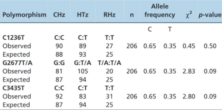
Table 1 -MDR1C1236T, G2677T/A and C3435T genotypes and allele frequencies.
Polymorphism CHz HTz RHz n
Allele
frequency x2 p-value
C T
C1236T C:C C:T T:T
Observed 90 89 27 206 0.65 0.35 0.45 0.50
Expected 88 93 25
G2677T/A G:G G:T/A T/A:T/A
Observed 81 105 20 206 0.65 0.35 2.83 0.09
Expected 87 94 25
C3435T C:C C:T T:T
Observed 92 83 31 206 0.65 0.35 2.80 0.09
Expected 87 94 25
CHz, common homozygote; HTz, heterozygote; RHz, rare homozygote.
Figure 1 -Electropherogram 1236. The wild-type sequence agggCc (left) and the polymorphism in which cytosine is exchanged for
thymine (agggTc) (right) (A). Electropherogram showing a polymorphism (C1236T) with overlapping cytosine and thymine curves,
Estado do Rio de Janeiro, and of the University Hospital Clementino Fraga Filho, Universidade Federal do Rio de Janeiro. Informed consent was obtained from all subjects. The study protocol was in accordance with the ethical principles for medical research involving human subjects described in the Declaration of Helsinki.
& RESULTS
The CD group consisted of 49 men and 74 women with a mean age of 39.8 years (range: 11–80 years). In total, 51 were
classified as white, whereas 72 were classified as non-white. The mean duration of CD was 8.8 years (range: 0.5–41 years). The UC group consisted of 40 men and 43 women with a mean age of 45.6 years (range: 21–73 years). Of these individuals, 31 were classified as white, and 52 were classified as as non-white. The mean duration of UC was 7.5 years (range: 0.2–27 years).
The distributions of the selectedMDR1gene polymorph-isms (C1236T, G2677T/A and C3435T) are shown in Table 1 and demonstrate that the respective allele frequencies were in Hardy-Weinberg equilibrium in the study population.
Subsequently, we investigated the distribution of each polymorphic genotype in the CD and UC patient groups (Table 2). The MDR1 G2677T/A and C3435T genotypes were similar between CD and UC patients (p = 0.477 and
p= 0.712, respectively). However, the homozygous C1236T genotype was significantly more prevalent among CD patients compared with UC patients (p= 0.047).
The association of the different polymorphic genotypes with the phenotypic characteristics of CD and UC was also investigated. Tables 3, 4 and 5 show the genotype frequencies of the SNPs in different subgroups of patients with CD. A significant positive association was found between the MDR1 homozygous C3435T polymorphism and the stricturing form of CD (p= 0.009). Interestingly, a tendency toward a negative association with the penetrating form of the disease was identified for the same SNP (p= 0.094). Among the 35 patients who had stricturing
Table 2 -Analysis ofMDR1gene polymorphisms for the differential diagnosis of inflammatory bowel disease.
MDR1SNP CHz HTz RHz p-value
C1236T C:C C:T T:T
CD n = 123) 50 51 22 0.047
UC (n = 83) 40 38 5
G2677T/A G:G G:T/A T/A:T/A
CD n = 123) 50 59 14 0.477
UC (n = 83) 31 46 6
C3435T C:C C:T T:T
CD n = 123) 52 52 19 0.712
UC (n = 83) 40 31 12
MDR1, multidrug resistance protein 1; SNP, single-nucleotide
polymorphism; CD, Crohn’s disease; UC, ulcerative colitis; CHz, common homozygous; HTz, heterozygous; RHz, rare homozygous. The data were analyzed using the Pearson chi-square test.
Table 3 -Genotype-phenotype associations of theMDR1SNP C1236T in patients with Crohn’s disease.
MDR1SNP C1236T CHz HTz OR p-value RHz OR p-value
Gender
Male (n = 49, 39.8%) 20 19 0.89 0.777 10 1.25 0.665
Female (n = 74, 60.2%) 30 32 12
Ethnicity
White (n = 51, 41.5%) 20 22 1.14 0.749 9 1.04 0.942
Non-white (n = 72, 58.5%) 30 29 13
Age at diagnosis
,40 (A1) (n = 62, 50.4%) 24 31 1.68 0.197 7 0.51 0.201
$40 (A2) (n = 61, 49.6%) 26 20 15
Disease location
Terminal ileum (L1) (n = 22, 17.9%) 10 11 1.10 0.846 1 0.19 0.093
Colon (L2) (n = 58, 47.1%) 23 21 0.82 0.625 14 2.05 0.168
Ileocolon (L3) (n = 38, 30.9%) 14 18 1.40 0.431 6 0.96 0.949
Upper GI (L4) (n = 5, 4.1%) 3 1 0.31 0.298 1 0.75 0.804
Disease behavior
NS/NP (B1) (n = 44, 35.8%) 17 19 1.15 0.732 8 1.11 0.846
Stricturing (B2) (n = 35, 28.4%) 15 14 0.88 0.777 6 0.88 0.814
Penetrating (B3) (n = 44, 35.8%) 18 18 0.97 0.941 8 1.02 0.976
Disease activity
Moderate/severe (n = 24, 19.5%) 8 12 1.62 0.342 4 1.17 0.819
Mild/remission (n = 99, 80.5%) 42 39 18
Surgery due to CD
Yes(n = 45, 36.6%) 23 14 0.44 0.053 8 0.67 0.447
No(n = 78, 63.4%) 27 37 14
Perianal disease
Yes(n = 37, 30.1%) 17 15 0.81 0.620 5 0.57 0.338
No(n = 86, 69.9%) 33 36 17
Chronic steroid use
Yes(n = 33, 26.8%) 10 15 1.67 0.273 8 2.29 0.139
No(n = 90, 73.2%) 40 36 14
Side effects of medication
Yes(n = 39, 31.7%) 15 18 1.27 0.570 6 0.88 0.814
No(n = 84, 68.3%) 35 33 16
disease, 11 had the homozygous C3435T polymorphism, whereas among the 44 patients with penetrating disease, only three presented this genotype. Furthermore, the homozygous C3435T polymorphism was associated with chronic steroid use (steroid-dependent/refractory) (p= 0.010) (Table 5). A significant association with disease behavior was also found between theMDR1heterozygous G2677T/A polymorphism and the stricturing, non-penetrating form of CD (p= 0.033; Table 4). In contrast, no significant associations were found between the MDR1
C1236T polymorphism and specific CD subphenotypes (Table 3). Furthermore, no significant association was found between theMDR1gene polymorphisms and UC subphe-notypic categories (Supplementary Tables S1, S2 and S3).
& DISCUSSION
In this study, we present information on the genotype frequencies of theMDR1 C1236T, G2677T/A and C3435T polymorphisms in patients with CD or UC as well as potential determination of the phenotypic features of IBD, in a southeastern Brazilian population from Rio de Janeiro. In particular, we found that the C1236T polymorphism was significantly more common in CD patients than in UC patients. Regarding CD phenotypes, a significant associa-tion was detected between theMDR1 C3435T polymorph-ism and the stricturing form of the disease. In addition, a positive association between the C3435T polymorphism and
the chronic use of steroids was identified in CD patients. We also found positive and negative trends regarding specific phenotypes and both heterozygous and homozygous poly-morphisms, suggesting that MDR1 gene variants may determine both susceptibility to and protection against CD. VariousMDR1gene polymorphisms have been reported thus far, and the C3435T polymorphism has been the most well studied in IBD. Similar to the results of the current study, associations between MDR1 gene polymorphisms have been reported in refractory CD and, to a lesser extent, in UC in a Slovenian population (23). In another study performed in a large cohort in the United States, investiga-tors observed a significant association between CD and a missense polymorphism in exon 21 (G2677T/C; Ala893Ser/ Thr), which was thought to be related to altered transporter and/or gene expression activity (24). In a case-control study in Spain, a significant association between the MDR1
C3435T polymorphism and CD was characterized, in addition to the identification of the CD susceptibility haplotype 2677T/C3435 (25). Moreover, in an Italian study, investigators found a significant association between the C3435T SNP and the ileocolonic localization of CD, whereas the same polymorphism appeared to be negatively asso-ciated with a positive family history and arthritis in CD patients (26). In a large case-control cohort study in the United Kingdom, investigators associated theMDR1SNPs C3435T and G2677T/A with an increased risk of developing UC, including an influence on disease behavior (27). In
Table 4 -Genotype-phenotype associations of theMDR1SNP G2677T/A in patients with Crohn’s disease.
MDR1SNP G2677T/A CHz HTz OR p-value RHz OR p-value
Gender
Male (n = 49, 39.8%) 17 26 1.53 0.284 6 1.46 0.541
Female (n = 74, 60.2%) 33 33 8
Ethnicity
White (n = 51, 41.5%) 19 26 1.29 0.521 6 1.22 0.742
Non-white (n = 72, 58.5%) 31 33 8
Age at diagnosis
,40 (A1) (n = 62, 50.4%) 24 31 1.20 0.636 7 1.08 0.894
$40 (A2) (n = 61, 49.6%) 26 28 7
Disease location
Terminal ileum (L1) (n = 22, 17.9%) 10 10 0.82 0.682 2 0.67 0.628
Colon (L2) (n = 58, 47.1%) 27 26 0.67 0.301 5 0.47 0.226
Ileocolon (L3) (n = 38, 30.9%) 11 21 1.96 0.120 6 2.66 0.118
Upper GI (L4) (n = 5, 4.1%) 2 2 0.84 0.865 1 1.85 0.623
Disease behavior
NS/NP (B1) (n = 44, 35.8%) 13 27 2.40 0.033* 4 1.14 0.847
Stricturing (B2) (n = 35, 28.4%) 16 13 0.60 0.240 6 1.59 0.449
Penetrating (B3) (n = 44, 35.8%) 21 19 0.66 0.290 4 0.55 0.362
Disease activity
Moderate/severe (n = 24, 19.5%) 8 12 1.34 0.559 4 2.10 0.286
Mild/remission (n = 99, 80.5%) 42 47 10
Surgery due to CD
Yes(n = 45, 36.6%) 23 17 0.48 0.063 5 0.65 0.493
No(n = 78, 63.4%) 27 42 9
Perianal disease
Yes (n = 37, 30.1%) 15 17 0.94 0.892 5 1.30 0.683
No(n = 86, 69.9%) 35 42 9
Chronic steroid use
Yes(n = 33, 26.8%) 12 14 0.99 0.973 7 3.17 0.060
No(n = 90, 73.2%) 38 45 7
Side effects of medication
Yes(n = 39, 31.7%) 17 20 1.00 0.991 2 0.32 0.153
No(n = 84, 68.3%) 33 39 12
CHz, common homozygous (G:G); HTz, heterozygous (G:T/A); RHz, rare homozygous (T/A:T/A); NS/NP, non-stricturing, non-penetrating; OR, odds ratio. All comparisons were performed in relation to the CHz group.
addition, in contrast to the results of the current study, case-control studies in European cohorts of patients from Germany (28) and Scotland (29) also suggested a potential association between the MDR1 SNP C3435T and UC. Nevertheless, in another Italian study, the investigated polymorphisms in theMDR1gene had no significant role in disease susceptibility or the response to medical therapy in IBD (30).
The discrepancies among these studies may be attributed to not only different study designs, sample sizes and selection of controls but also the distinct patient popula-tions. In fact, meta-analyses have been performed to attempt to overcome the heterogeneity of studies involving MDR1
SNPs. Of note, certain studies have revealed that the allele frequencies of the three main variants differ considerably among distinct populations. A significant association of the 3435T allele with UC was confirmed in a meta-analysis (27), and in another study, the 3435T allele and the 3435TT genotype were demonstrated to be significantly associated with UC, but not with CD (31). Differences in the C3435T SNP allele frequency have also been detected, with an increased frequency of the C allele (wild type) in African populations compared with Caucasian and Asian popula-tions (32). In an Asiatic study, investigators found distinct haplotype profiles and linkage disequilibrium at theMDR1
gene locus in all three ethnic groups enrolled in the study (33). In a recent study that was also performed in Rio de
Janeiro, Brazil, matching the recruitment area and ethnicity of our IBD patients, 278 healthy individuals were analyzed regarding the genotype and allele frequencies of MDR1
gene polymorphisms. The investigators found a peculiar variant distribution, with significant differences between C1236C and C3435T and also between C1236C and C3435C, which differed from the results obtained for several other ethnic groups (34). In this context, it must be emphasized that data from potential source populations, such as Europeans or Africans, cannot be deemed representative of the Brazilian genotype and allele frequencies due to the marked heterogeneity and the extensive admixture of the Brazilian population (35,36). Taken together with the results of our study, these observations highlight the critical importance of analyzing MDR1 gene polymorphisms in Brazilians, and particularly in Brazilian IBD patients.
Potential genotype-phenotype associations related to the
MDR1 gene have been investigated, with contradictory results. In the present study, we report positive associations between the C3435T polymorphism and both the chronic use of steroids and disease activity in CD patients. Similar to our results, an association between MDR1 variants and corticosteroid dependence was also reported in children with CD in Canadian tertiary centers (37). In a British study on IBD, the 2677T allele was increased in UC cases, and the TT genotype was strongly associated with disease severity and the use of steroids in UC (27). In contrast, in a large
Table 5 -Genotype-phenotype associations of theMDR1SNP C3435T in patients with Crohn’s disease.
MDR1SNP C3435T CHz HTz OR p-value RHz OR p-value
Gender
Male (n = 49, 39.8%) 22 18 0.72 0.420 9 1.23 0.703
Female (n = 74, 60.2%) 30 34 10
Ethnicity
White (n = 51, 41.5%) 22 21 0.92 0.842 8 0.99 0.988
Non-white (n = 72, 58.5%) 30 31 11
Age at diagnosis
,40 (A1) (n = 62, 50.4%) 25 27 1.17 0.695 10 1.20 0.734
$40 (A2) (n = 61, 49.6%) 27 25 9
Disease location
Terminal ileum (L1) (n = 22, 17.9%) 8 10 1.31 0.604 4 1.47 0.572
Colon (L2) (n = 58, 47.1%) 25 25 1.00 1.000 8 0.79 0.655
Ileocolon (L3) (n = 38, 30.9%) 16 16 1.00 1.000 6 1.04 0.948
Upper GI (L4) (n = 5, 4.1%) 3 1 0.32 0.307 1 0.91 0.934
Disease behavior
NS/NP (B1) (n = 44, 35.8%) 20 19 0.92 0.839 5 0.57 0.343
Stricturing (B2) (n = 35, 28.4%) 13 11 0.80 0.641 11 4.13 0.009*
Penetrating (B3) (n = 44, 35.8%) 19 22 1.27 0.547 3 0.33 0.094
Disease activity
Moderate/severe (n = 24, 19.5%) 7 11 1.72 0.299 6 2.97 0.080
Mild/remission (n = 99, 80.5%) 45 41 13
Surgery due to CD
Yes(n = 45, 36.6%) 20 20 1.00 1.000 5 0.57 0.342
No(n = 78, 63.4%) 32 32 14
Perianal disease
Yes(n = 37, 30.1%) 17 16 0.92 0.833 4 0.55 0.341
No(n = 86, 69.9%) 35 36 15
Chronic steroid use
Yes(n = 33, 26.8%) 11 12 1.12 0.813 10 4.14 0.010*
No(n = 90, 73.2%) 41 40 9
Side effects of medication
Yes(n = 39, 31.7%) 13 22 2.20 0.062 4 0.80 0.730
No(n = 84, 68.3%) 39 30 15
CHz, common homozygous (C:C); HTz, heterozygous (C:T); RHz, rare homozygous (T:T); NS/NP, non-stricturing, non-penetrating; OR, odds ratio. All comparisons were performed in relation to the CHz group.
*Indicates a significant difference,p
cohort of IBD patients using steroids, both the C3435T and the G2677T/A polymorphisms were evaluated, but no significant differences were found within subgroups or among subgroups. Additionally,MDR1genotypes were not found to influence the response to therapy (30). In another study, the expression and function ofMDR1in intraepithe-lial, lamina propria and peripheral blood lymphocytes were decreased in UC patients compared with CD patients and healthy controls (38). In accordance with this finding, the tissue expression of a number of detoxification genes and ABC transporters, including MDR1, was shown to be markedly downregulated in UC patients, supporting the notion that a defective mucosal detoxification system could predispose a patient to intestinal inflammation (39). Indeed, the effects of corticosteroids and other medications used in IBD onMDR1expression have not been fully established, and the question of whether the difference in P-gp expression reflects a primary defect or occurs secondary to therapy, influencing the response to treatment, remains to be clarified.
The stricturing form of CD, also known as the fibroste-notic form of CD, has been previously associated with
NOD2 variants and small bowel involvement in patients with CD (40). However, another study demonstrated that the fibrostenotic phenotype of CD was significantly asso-ciated withNOD2gene variants and also with a high titer of antibodies against oligomannan, OmpC, I2 and Cbir, regardless of disease location (41). These results support the notion that altered innate immunity may synergize with a loss of tolerance to microbial antigens and with the adaptive immune response, thus favoring a specific CD phenotype. In contrast to studies reported thus far onMDR1
gene polymorphisms in IBD, another novel genotype-phenotype association found in our study was the sig-nificant positive association between the C3435T SNP and the stricturing form of CD. Although the mechanism by which anMDR1 SNP can determine a specific phenotype has yet to be determined, certain evidence indicates that
MDR1 participates in a complex biological network with multiple physiologically relevant mediators and pathways, including pro-inflammatory cytokines (42), endotoxin-induced inflammation (43), transcription factors such as NF-kB (44,45) and cyclooxygenases (46), which can mod-ulate MDR1 expression and activity at different levels. Hence, these intricate mechanisms reinforce a key role for P-gp in drug bioavailability and epithelial homeostasis in the context of inflammation and infection. However, the contribution of theMDR1gene to the response to medica-tions and possibly to IBD susceptibility or phenotypes, including the stricturing form of CD, needs further clarification.
In conclusion, the results of this study indicate that the rare homozygous MDR1 gene polymorphism C3435T is associated not only with the stricturing phenotype but also with an inappropriate response to therapy in a population of CD patients from Rio de Janeiro. The relationship with the CD phenotype supports the existence of additional roles forMDR1in specific mechanisms underlying CD pathogen-esis, such as the control of gut-microbiota interactions and the regulation of fibrosis. Furthermore, understanding the effects of several drugs associated with these MDR1
variants may aid in the selection of customized therapeutic regimens.
& ACKNOWLEDGMENTS
The authors wish to thank the Brazilian research foundations CNPq and FAPERJ for their financial support. All authors approved the final version of this article.
& AUTHOR CONTRIBUTIONS
Carvalho AT, Froes R, Esberard B and Carneiro AJ participated in study design, patient selection and follow-up, data collection and interpretation and manuscript preparation. Grinman AB, Neto PN, Santos J, Raposo D and Simao T performed the laboratory experiments, technical trouble-shooting and data collection and interpretation. Luiz RR performed statistical analysis and data interpretation. Pinto LF and Souza HS interpreted the data, obtained funding, performed statistical analysis, wrote the manuscript and critically reviewed the manuscript.
& REFERENCES
1. Xavier RJ, Podolsky DK. Unravelling the pathogenesis of inflammatory bowel disease. Nature. 2007;448(7152):427-34, http://dx.doi.org/10.1038/ nature06005.
2. Duerr RH, Taylor KD, Brant SR, Rioux JD, Silverberg MS, Daly MJ, et al. A genome-wide association study identifies IL23R as an inflammatory bowel disease gene. Science. 2006;314(5804):1461-3, http://dx.doi.org/ 10.1126/science.1135245.
3. Hampe J, Franke A, Rosenstiel P, Till A, Teuber M, Huse K, et al. A genome-wide association scan of nonsynonymous SNPs identifies a susceptibility variant for Crohn disease in ATG16L1. Nat Genet. 2007;39(2):207-11, http://dx.doi.org/10.1038/ng1954.
4. Hugot JP, Chamaillard M, Zouali H, Lesage S, Cezard JP, Belaiche J, et al. Association of NOD2 leucine-rich repeat variants with susceptibility to Crohn’s disease. Nature. 2001;411(6837):599-603, http://dx.doi.org/10. 1038/35079107.
5. Ogura Y, Bonen DK, Inohara N, Nicolae DL, Chen FF, Ramos R, et al. A frameshift mutation in NOD2 associated with susceptibility to Crohn’s disease. Nature. 2001;411(6837):603-6, http://dx.doi.org/10.1038/35079114. 6. Satsangi J, Parkes M, Louis E, Hashimoto L, Kato N, Welsh K, et al. Two stage genome-wide search in inflammatory bowel disease provides evidence for susceptibility loci on chromosomes 3, 7 and 12. Nat Genet. 1996;14(2):199-202, http://dx.doi.org/10.1038/ng1096-199.
7. van Heel DA, Fisher SA, Kirby A, Daly MJ, Rioux JD, Lewis CM. Inflammatory bowel disease susceptibility loci defined by genome scan meta-analysis of 1952 affected relative pairs. Hum Mol Genet. 2004;13(7):763-70, http://dx.doi.org/10.1093/hmg/ddh090.
8. Van Limbergen J, Russell RK, Nimmo ER, Ho GT, Arnott ID, Wilson DC, et al. Genetics of the innate immune response in inflammatory bowel disease. Inflamm Bowel Dis. 2007;13(3):338-55, http://dx.doi.org/10. 1002/ibd.20096.
9. Thiebaut F, Tsuruo T, Hamada H, Gottesman MM, Pastan I, Willingham MC. Cellular localization of the multidrug-resistance gene product P-glycoprotein in normal human tissues. Proc Natl Acad Sci U S A. 1987;84(21):7735-8, http://dx.doi.org/10.1073/pnas.84.21.7735. 10. Marzolini C, Paus E, Buclin T, Kim RB. Polymorphisms in human MDR1
(P-glycoprotein): recent advances and clinical relevance. Clin Pharmacol Ther. 2004;75(1):13-33, http://dx.doi.org/10.1016/j.clpt.2003.09.012. 11. Panwala CM, Jones JC, Viney JL. A novel model of inflammatory bowel
disease: mice deficient for the multiple drug resistance gene, mdr1a, spontaneously develop colitis. J Immunol. 1998;161(10):5733-44. 12. Bourgeois S, Gruol DJ, Newby RF, Rajah FM. Expression of an mdr gene
is associated with a new form of resistance to dexamethasone-induced apoptosis. Mol Endocrinol. 1993;7(7):840-51.
13. Ueda K, Okamura N, Hirai M, Tanigawara Y, Saeki T, Kioka N, et al. Human P-glycoprotein transports cortisol, aldosterone, and dexametha-sone, but not progesterone. J Biol Chem. 1992;267(34):24248-52. 14. Norris MD, De Graaf D, Haber M, Kavallaris M, Madafiglio J, Gilbert J,
et al. Involvement of MDR1 P-glycoprotein in multifactorial resistance to methotrexate. Int J Cancer. 1996;65(5):613-9.
15. Chaudhary PM, Mechetner EB, Roninson IB. Expression and activity of the multidrug resistance P-glycoprotein in human peripheral blood lymphocytes. Blood. 1992;80(11):2735-9.
16. Farrell RJ, Murphy A, Long A, Donnelly S, Cherikuri A, O’Toole D, et al. High multidrug resistance (P-glycoprotein 170) expression in inflamma-tory bowel disease patients who fail medical therapy. Gastroenterology. 2000;118(2):279-88, http://dx.doi.org/10.1016/S0016-5085(00)70210-1. 17. Farrell RJ, Kelleher D. Glucocorticoid resistance in inflammatory bowel
disease. J Endocrinol. 2003;178(3):339-46, http://dx.doi.org/10.1677/joe. 0.1780339.
and management of Crohn’s disease: Definitions and diagnosis. J Crohns Colitis. 2010;4(1):7-27, http://dx.doi.org/10.1016/j.crohns.2009.12.003. 19. Satsangi J, Silverberg MS, Vermeire S, Colombel JF. The Montreal
classification of inflammatory bowel disease: controversies, consensus, and implications. Gut. 2006;55(6):749-53, http://dx.doi.org/10.1136/gut. 2005.082909.
20. Harvey RF, Bradshaw JM. A simple index of Crohn’s-disease activity. Lancet. 1980;1(8167):514, http://dx.doi.org/10.1016/S0140-6736(80)92767-1.
21. Walmsley RS, Ayres RC, Pounder RE, Allan RN. A simple clinical colitis activity index. Gut. 1998;43(1):29-32, http://dx.doi.org/10.1136/gut.43.1. 29.
22. Miller CA, 3rd, Martinat MA, Hyman LE. Assessment of aryl hydro-carbon receptor complex interactions using pBEVY plasmids: expres-sionvectors with bi-directional promoters for use in Saccharomyces cerevisiae. Nucleic Acids Res. 1998;26(15):3577-83, http://dx.doi.org/10. 1093/nar/26.15.3577.
23. Potocnik U, Ferkolj I, Glavac D, Dean M. Polymorphisms in multidrug resistance 1 (MDR1) gene are associated with refractory Crohn disease and ulcerative colitis. Genes Immun. 2004;5(7):530-9, http://dx.doi.org/ 10.1038/sj.gene.6364123.
24. Brant SR, Panhuysen CI, Nicolae D, Reddy DM, Bonen DK, Karaliukas R, et al. MDR1 Ala893 polymorphism is associated with inflammatory bowel disease. Am J Hum Genet. 2003;73(6):1282-92.
25. Urcelay E, Mendoza JL, Martin MC, Mas A, Martinez A, Taxonera C, et al. MDR1 gene: susceptibility in Spanish Crohn’s disease and ulcerative colitis patients. Inflamm Bowel Dis. 2006;12(1):33-7, http:// dx.doi.org/10.1097/01.MIB.0000194184.92671.78.
26. Ardizzone S, Maconi G, Bianchi V, Russo A, Colombo E, Cassinotti A, et al. Multidrug resistance 1 gene polymorphism and susceptibility to inflammatory bowel disease. Inflamm Bowel Dis. 2007;13(5):516-23, http://dx.doi.org/10.1002/ibd.20108.
27. Onnie CM, Fisher SA, Pattni R, Sanderson J, Forbes A, Lewis CM, et al. Associations of allelic variants of the multidrug resistance gene (ABCB1 or MDR1) and inflammatory bowel disease and their effects on disease behavior: a case-control and meta-analysis study. Inflamm Bowel Dis. 2006;12(4):263-71, http://dx.doi.org/10.1097/01.MIB.0000209791.98866. ba.
28. Schwab M, Schaeffeler E, Marx C, Fromm MF, Kaskas B, Metzler J, et al. Association between the C3435T MDR1 gene polymorphism and susceptibility for ulcerative colitis. Gastroenterology. 2003;124(1):26-33, http://dx.doi.org/10.1053/gast.2003.50010.
29. Ho GT, Nimmo ER, Tenesa A, Fennell J, Drummond H, Mowat C, et al. Allelic variations of the multidrug resistance gene determine suscept-ibility and disease behavior in ulcerative colitis. Gastroenterology. 2005;128(2):288-96, http://dx.doi.org/10.1053/j.gastro.2004.11.019. 30. Palmieri O, Latiano A, Valvano R, D’Inca R, Vecchi M, Sturniolo GC, et al.
Multidrug resistance 1 gene polymorphisms are not associated with inflammatory bowel disease and response to therapy in Italian patients. Aliment Pharmacol Ther. 2005;22(11-12):1129-38, http://dx.doi.org/10. 1111/j.1365-2036.2005.02701.x.
31. Annese V, Valvano MR, Palmieri O, Latiano A, Bossa F, Andriulli A. Multidrug resistance 1 gene in inflammatory bowel disease: a meta-analysis. World J Gastroenterol. 2006;12(23):3636-44.
32. Schaeffeler E, Eichelbaum M, Brinkmann U, Penger A, Asante-Poku S, Zanger UM, et al. Frequency of C3435T polymorphism of MDR1 gene in
African people. Lancet. 2001;358(9279):383-4, http://dx.doi.org/10. 1016/S0140-6736(01)05579-9.
33. Tang K, Ngoi SM, Gwee PC, Chua JM, Lee EJ, Chong SS, et al. Distinct haplotype profiles and strong linkage disequilibrium at the MDR1 multidrug transporter gene locus in three ethnic Asian populations. Pharmacogenetics. 2002;12(6):437-50, http://dx.doi.org/10.1097/00008571-200208000-00004.
34. Scheiner MA, Damasceno AM, Maia RC. ABCB1 single nucleotide polymorphisms in the Brazilian population. Mol Biol Rep. 2009;37 (1):111-8.
35. Alves-Silva J, da Silva Santos M, Guimaraes PE, Ferreira AC, Bandelt HJ, Pena SD, et al. The ancestry of Brazilian mtDNA lineages. Am J Hum Genet. 2000;67(2):444-61.
36. Parra FC, Amado RC, Lambertucci JR, Rocha J, Antunes CM, Pena SD. Color and genomic ancestry in Brazilians. Proc Natl Acad Sci U S A. 2003;100(1):177-82, http://dx.doi.org/10.1073/pnas.0126614100. 37. Krupoves A, Mack D, Seidman E, Deslandres C, Amre D. Associations
between variants in the ABCB1 (MDR1) gene and corticosteroid dependence in children with Crohn’s disease. Inflamm Bowel Dis. 2011;17(11):2308-17, http://dx.doi.org/10.1002/ibd.21608.
38. Yacyshyn B, Maksymowych W, Bowen-Yacyshyn MB. Differences in P-glycoprotein-170 expression and activity between Crohn’s disease and ulcerative colitis. Hum Immunol. 1999;60(8):677-87, http://dx.doi.org/ 10.1016/S0198-8859(99)00036-1.
39. Langmann T, Moehle C, Mauerer R, Scharl M, Liebisch G, Zahn A, et al. Loss of detoxification in inflammatory bowel disease: dysregulation of pregnane X receptor target genes. Gastroenterology. 2004;127(1):26-40, http://dx.doi.org/10.1053/j.gastro.2004.04.019.
40. Abreu MT, Taylor KD, Lin YC, Hang T, Gaiennie J, Landers CJ, et al. Mutations in NOD2 are associated with fibrostenosing disease in patients with Crohn’s disease. Gastroenterology. 2002;123(3):679-88, http://dx.doi.org/10.1053/gast.2002.35393.
41. Ippoliti A, Devlin S, Mei L, Yang H, Papadakis KA, Vasiliauskas EA, et al. Combination of innate and adaptive immune alterations increased the likelihood of fibrostenosis in Crohn’s disease. Inflamm Bowel Dis. 2010;16(8):1279-85, http://dx.doi.org/10.1002/ibd.21196.
42. Evseenko DA, Paxton JW, Keelan JA. Independent regulation of apical and basolateral drug transporter expression and function in placental trophoblasts by cytokines, steroids, and growth factors. Drug Metab Dispos. 2007;35(4):595-601, http://dx.doi.org/10.1124/dmd.106.011478. 43. Tomita M, Takizawa Y, Kanbayashi A, Murata H, Tanaka A, Nakaike M,
et al. Suppression of efflux transporters in the intestines of endotoxin-treated rats. Int J Pharm. 2012;428(1-2):33-8.
44. Kuo MT, Liu Z, Wei Y, Lin-Lee YC, Tatebe S, Mills GB, et al. Induction of human MDR1 gene expression by 2-acetylaminofluorene is mediated by effectors of the phosphoinositide 3-kinase pathway that activate NF-kappaB signaling. Oncogene. 2002;21(13):1945-54, http://dx.doi.org/10. 1038/sj.onc.1205117.
45. Inoue S, Nakase H, Matsuura M, Mikami S, Ueno S, Uza N, et al. The effect of proteasome inhibitor MG132 on experimental inflammatory bowel disease. Clin Exp Immunol. 2009;156(1):172-82, http://dx.doi. org/10.1111/j.1365-2249.2008.03872.x.