265 265 265 265 265 Mem Inst Oswaldo Cruz, Rio de Janeiro, Vol. 95(2): 265-270, Mar./Apr. 2000
Ultrastructural Study of the TG180 Murine Sarcoma Cell
Invasion by
Toxoplasma gondii
: Comparison between
in
Vivo
and
in Vitro
Cell Cultures
Hugo Marcelo Ribeiro Barbosa, Marcos Silva*, Eloisa Amália Vieira Ferro*,
José Roberto Mineo/
+Laboratório de Imunologia, Departamento de Patologia *Laboratório de Histologia, Departamento de Morfologia, Universidade Federal de Uberlândia, 38400-902 Uberlândia, MG, Brasil
Infection of non-adherent TG180 murine sarcoma cells with Toxoplasma gondii was compared, at the ultrastructural level, in both in vivo and in vitro conditions. Suspensions of 3.0 x 10⁶ TG180 cells
infected in vitro with 1.0 x 10⁶ parasites of the RH strain were harvested between the first and 6th day
post-infection and processed for transmission electron microscopy. In vivo infection was made by intra-peritoneal inoculation in mice of 1.0 x 10⁶ TG180 cells, that were co-inoculated with a parasite suspen-sion at the same cell concentration. Cells were harvested 10, 20, 30 min and 24, 48 h post-inoculation and processed for transmission electron microscopy at the same conditions of the in vitro culture. It was observed TG180 murine sarcoma cells with intense and equivalent intracellular parasitism in both conditions. Host cells with parasitophorous vacuoles containing up to 16 parasites, as well as parasites undergoing mitoses or presenting a bradyzoite-like morphology, were frequently seen in both culture methods.
Key words: Toxoplasma gondii - TG180 murine sarcoma cells - in vitro culture - transmission electron
microscopy
Toxoplasma gondii is an intracellular parasite of a wide range of vertebrate hosts, including the human host. The infection caused by this parasite may be asymptomatic in healthy adults, but it can be an important illness to non-immune pregnant women by inducing embryopathy. In immuno-compromised hosts, as in patients suffering of Aids or submitted to transplants, this parasite can origi-nate encephalitis.
The most effective treatment currently used is a combination of pyrimethamine and sulphadiaz-ine, which does not eliminate the encysted form of the parasite (Bunetel et al. 1995). Alternatively, it can be used spiramycin, a macrolide antibiotic, which shows no evidence of harmful effects on foetal development or toxicity (Desmonts & Couvreur 1974, Bunetel et al. 1995). There is a low number of toxoplasmicidic drugs on clinical use, but several new drugs have been proposed. New methods are necessary to test these drugs.
According to Bunetel et al. (1995), the infection of TG180 murine sarcoma cells in vitro allows an easy, rapid, accurate and economical method to determine the effects of drugs against T. gondii.
It has been described in the literature the study of the interaction of T. gondii and host cells by in vivo or in vitro models (Chang et al. 1972, Shepp et al. 1985, Pavesio et al. 1992, Saffer et al. 1992, Speer et al. 1997).
The major aim of the present investigation was to study the infection of TG180 cells by T. gondii by comparing cultures submitted to in vivo and in vitro conditions. In addition, it was verified the adequacy of this method, emphasizing the char-acteristics of the in vitro infection, by using the transmission electron microscopy to evaluate the results.
MATERIALS AND METHODS
T. gondii - The RH strain used was obtained from animal facilities of our laboratory. The tachyzoites were cultured by intraperitoneal inocu-lation of female Balb/C mice. The parasites were harvested from mice by rinsing of the peritoneal cavity with 5 ml of Eagle modified minimum es-sential medium (MEM).
TG180 murine sarcoma cell cultures - The TG180 cell line was derived from the ATCC (CCRFS-180 II) sarcoma 180 in 1958 (Sartorelli
This work was supported by CNPq, Fapemig and Capes.
+Corresponding author. Fax: +55-34-218.2333. E-mail:
266 266 266 266
266 Invasion of TG180 Cells by T. gondii Hugo Marcelo Ribeiro Barbosa et al.
& Booth 1961). A sub-population of this cell line adapted to in vitro culture (Couatarmanach et al. 1991) was used in our experiments. For the in vitro experiments the cells were grown in MEM supple-mented with 10% fetal calf serum, 40 µg/ml gentamycin and 25 µg/ml amphotericin B. The cultures were incubated at 37oC in an atmosphere of 95% air and 5% CO2 in 5 ml-tissue culture flasks. The cells were observed daily by inverted light microscopy, growing in suspension as non-adherent polymorphic cells.
Then, 3.0x10⁶ TG180 cells were inoculated into flasks containing 5 ml of MEM culture medium and used after 8-14 passages. Cell countings were made with the use of “Trypan blue”.
For in vivo experiments, TG180 sarcoma mu-rine cells were grown as ascitic tumors in adult female Balb/C mice in our animal facilities. For the experiments, mice were intraperitoneally in-oculated with 1.0x10⁶ TG180 cells.
In vitro infection - Six culture flasks were in-fected with 1.0x10⁶ tachyzoites each. This 3:1 TG180:T. gondii ratio was used as it has been al-ready shown to yield large numbers of intracellu-lar parasites (Couatarmanach et al. 1991). Cell sus-pensions were collected from the flasks daily from the first to the sixth day post-infection by centrifu-gation at 1,000g for 10 min. To avoid cell damage due to medium acidification, MEM culture medium was added on the second and third day post-infec-tion. Cell suspensions were fixed overnight with 2.5% glutaraldehyde/2% paraformaldehyde (Karnovsky 1965) diluted in 0.1M phosphate buffer, pH 7.2, washed three times in 5 ml of phos-phate buffered saline (PBS) pH 7.4 and processed for transmission electron microscopy as described below. On day 6, culture flasks of uninfected cells were also fixed and used as controls.
In vivo infection - Female Balb/C mice were intraperitoneally inoculated with a suspension of 1.0 x 10⁶ TG180 cells. Subsequently, these ani-mals were also inoculated with another cell sus-pension containing 1.0 x 10⁶ T. gondii by the same route. Mice were sacrificed after 10, 20, 30 min and 24 and 48 h post-infection and their ascitic fluids were collected. This material was fixed with 2.5% glutaraldehyde/2% paraformaldehyde, washed three times in PBS and processed for trans-mission electron microscopy as described below.
Transmission electron microscopy - Briefly, the material was post-fixed for 1 h in 1% OsO4, dehy-drated in increasing concentrations of ethanol and embedded in Epon. Ultrathin sections were cut with a diamond knife, stained with uranyl acetate and lead citrate and examined in a Zeiss EM 109 trans-mission electron microscope.
RESULTS
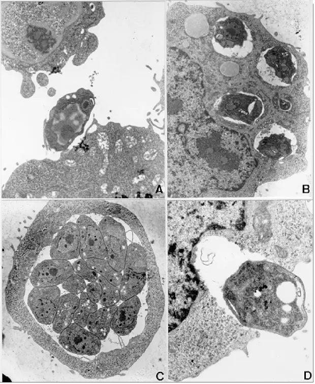
Infection of TG180 cells in vivo - The TG180 murine sarcoma cells present a spherical morphol-ogy, with finger-like extensions at their periphery after 24 h of infection. In the cytoplasm, it was observed a large amount of free ribosomes and poorly developed rough endoplasmic reticulum. Mitochondria, lipid droplets and multivesicular bodies were also present. The centrally located nucleus is frequently spherical and with chroma-tin distributed in groups of electron-dense masses. One or two irregularly shaped nucleoli could be observed. After 24 h, T. gondii could be found both in contact with the TG180 cell surfaces and start-ing the penetration process through invaginations of the host cell plasma membrane (Figs 1a, d). The parasites were also found inside parasitophorous vacuoles after 24 h (Fig. 1b). Vacuoles, free ribo-somes, Golgi complex, dense granules, rhoptries and mitochondria of several sizes were present in the intracellular parasites (Fig. 1c). It was also ob-served TG180 cells with vacuoles containing up to sixteen newly-divided T. gondii in the material collected after 48 h of infection (Fig. 1c).
267 267267 267267 Mem Inst Oswaldo Cruz, Rio de Janeiro, Vol. 95(2), Mar./Apr. 2000
Fig. 1: TG180 sarcoma murine cells after in vivo infection byRH strain of Toxoplasma gondii. A: TG180 host cell with parasite attached to its plasma membrane, after 24 h of infection, 4,400X; B: host cells presenting four parasitophorous vacuoles with parasites in mitotic process after 48 h of infection, 7,000X; C: cell presenting at least sixteen parasites inside a parasitophorous vacuole, after 48 h of infection, 7,000X; D: cell being invaded by tachyzoite 24 h after infection, 12,000X.
DISCUSSION
In vitro assays are very important to evaluate the effects of new drugs against intracellular micro-organisms, particularly when there are some asso-ciated in vivo factors, such as the host immune sys-tem, which can interfere in the effects at the
un-268 268 268 268
268 Invasion of TG180 Cells by T. gondii Hugo Marcelo Ribeiro Barbosa et al.
der in vitro conditions tumor cells are the most suit-able lines for the growth and multiplication of T. gondii. The TG180 murine sarcoma cells are com-monly utilized to produce T. gondii by co-inocula-tion in the peritoneal cavity of mice. Couatarmanach et al. (1991) demonstrated that the culture of TG180
in vitro could produce large amounts of T. gondii, up to 259 parasites/cell. Therefore, this method of culture can be very useful to evaluate the effect of new drugs against T. gondii.
We observed by transmission electron micros-copy that TG180 murine sarcoma cells are very
269 269269 269269 Mem Inst Oswaldo Cruz, Rio de Janeiro, Vol. 95(2), Mar./Apr. 2000
susceptible to T. gondii infection. Basically, the same parasitism degree was observed in both in vivo and in vitro cultures. We found parasites in the intracellular environments, forming large parasitophorous vacuoles containing up to 16 para-sites. Furthermore, extracellular parasites were
found close to cellular debris, probably arising from heavily infected cells that released these parasites to the extracellular environment.
Ultrastructural analysis of TG180 sarcoma cells showed that these cells, when submitted to in vitro culture, have basically the same cytoarchitectural
270 270 270 270
270 Invasion of TG180 Cells by T. gondii Hugo Marcelo Ribeiro Barbosa et al.
aspects of the in vivo cultured cells. As a cellular substrate for T. gondii infection, the TG180 can be utilized for diagnostic purpose, or to test new drugs with antitoxoplasmic activity. The TG180 cells can be a good option, as this cell line presents fast growth, is economically viable and is very suscep-tible to the T. gondii infection.
Lindsay et al. (1993) observed in culture of human foreskin fibroblast cells (Hs 68) the pres-ence of host cell mitochondria surrounding the T. gondii tissue cyst wall and suggested that this may contribute directly or indirectly to the formation of the vacuole membrane. Interestingly, we ob-served frequently the presence of uncountable mi-tochondria neighboring the parasitophorous vacu-oles, some of them completely enclosed by a net of hypertrophied mitochondria, and similar to those described by Lindsay et al. (1993).
Another remarkable observation was the pres-ence of parasites in the extracellular environment, some of them harboring an electron-dense sub-stance in their anterior portion that resembled bod-ies of amilopectine. These parasites presented morphology similar to bradyzoite forms. However, it will be necessary to search for bradyzoite spe-cific antigens in these preparations to confirm these findings. One of the major challenges nowadays is the study of new drugs that can cross the toxoplas-mic cyst wall and show toxic effect against para-sites located inside these structures. The TG180 murine sarcoma cells may be useful to test new drugs that have their toxoplasmicidic effect just against the bradyzoites.
Our results demonstrated that the TG180 mu-rine sarcoma is a suitable cellular substrate to T. gondii culture in both in vivo and in vitro condi-tions. Thus, this cell type might be used as a po-tential tool to isolate this parasite with diagnostic aims or as an alternative approach to test new toxoplasmicidic drugs in vitro against both tachyzoites and bradyzoites.
ACKNOWLEDGMENTS
To Dr Deise Aparecida Oliveira Silva and Dr Maria Aparecida de Souza for their suggestions during the preparation of this manuscript.
REFERENCES
Butenel L, Guerin J, Andre P, Robert R, Deunff J 1995. Calibration of an in vitro assay system using a
non-adherent cell line to evaluate the effect of a drug on
Toxoplasma gondii. Intern J Parasitol25: 699-704.
Chang CH, Stulberg C, Bollinger RO, Walker R, Brough AJ 1972. Isolation of Toxoplasma gondii in tissue
culture. J Ped 81: 790-794.
Couatarmanach A, Andre P, Le Minous D, Martin L, Robert R, Deunff J 1991. In vitro culture and
clon-ing of. Toxoplasma gondii in a newly established
cell line derived from TG180. Intern J Parasitol 21:
129-132.
Desmonts G, Couvreur J 1974. Congenital toxoplas-mosis. A prospective study of 378 pregnancies. New England J Med 290: 1110-1116.
Karnovsky MJ 1965. A formaldehyde-glutaraldehyde fixative of high osmolality for use in electron mi-croscopy. J Cell Biol 27: 137A-138A.
Lindsay DS, Mitschler RR, Toivio-Kinnucan MA, Upton SJ, Dubey JP, Blagburn BL 1993. Association of host cell mitochondria with developing Toxoplasma gondii tissue cysts. Am J Vet Res 54: 1663-1667.
Park BK, Moon HR, Yu JR, Kook J, Chai JY, Lee SH 1993. Comparative susceptibility of different cell lines for culture of Toxoplasma gondiiin vitro. Kisaengchunghak-chapchi 31: 215-222.
Pavesio CE, Chiappino ML, Setzer PY, Nichols BA 1992. Toxoplasma gondii: differentiation and death
of bradyzoites. Parasitol Res78: 1 1-19.
Saffer LD, Mercereau-Puijalon O, Dubremetz JF, Schwartzman JD 1992. Localization of a Toxoplasma gondii rhoptry protein by immunoelectron
micros-copy during and after host cell penetration. J Protozool 39: 526-530.
Sartorelli A C, Booth B A 1961. Studies on mechanisms of therapeutic synergy by combinations of asazerine and purine analogs. Fed Proceed 20: 156.
Shepp DH, Hackman RC, Conley FK, Anderson JB, Meyers JD 1985. Toxoplasma gondii reactivation
identified by detection of parasitemia in tissue cul-ture. Annals Intern Med 103: 218-221.