Intensive Care Med
DOI 10.1007/s00134-015-4206-2
SEVEN-DAY PROFILE PUBLICATION
The Intensive Care Global Study
on Severe Acute Respiratory Infection
(IC-GLOSSARI): a multicenter, multinational,
14-day inception cohort study
Yasser Sakr
1*, Ricard Ferrer
2, Konrad Reinhart
1, Richard Beale
3, Andrew Rhodes
4, Rui Moreno
5,
Jean Francois Timsit
6, Laurent Brochard
7,8, B. Taylor Thompson
9,10, Ederlon Rezende
11and Jean Daniel Chiche
12,13,14© 2016 Springer-Verlag Berlin Heidelberg and ESICM
Abstract
Purpose: In this prospective, multicenter, 14-day inception cohort study, we investigated the epidemiology, patterns of infections, and outcome in patients admitted to the intensive care unit (ICU) as a result of severe acute respiratory infections (SARIs).
Methods: All patients admitted to one of 206 participating ICUs during two study weeks, one in November 2013 and the other in January 2014, were screened. SARI was defined as possible, probable, or microbiologically confirmed respiratory tract infection with recent onset dyspnea and/or fever. The primary outcome parameter was in-hospital mortality within 60 days of admission to the ICU.
Results: Among the 5550 patients admitted during the study periods, 663 (11.9 %) had SARI. On admission to the ICU, Gram-positive and Gram-negative bacteria were found in 29.6 and 26.2 % of SARI patients but rarely atypical bac-teria (1.0 %); viruses were present in 7.7 % of patients. Organ failure occurred in 74.7 % of patients in the ICU, mostly respiratory (53.8 %), cardiovascular (44.5 %), and renal (44.6 %). ICU and in-hospital mortality rates in patients with SARI were 20.2 and 27.2 %, respectively. In multivariable analysis, older age, greater severity scores at ICU admission, and hematologic malignancy or liver disease were independently associated with an increased risk of in-hospital death, whereas influenza vaccination prior to ICU admission and adequate antibiotic administration on ICU admission were associated with a lower risk.
*Correspondence: [email protected]
1 Department of Anesthesiology and Intensive Care, Uniklinikum Jena,
Erlanger Allee 103, 07743 Jena, Germany
Full list of author information is available at the end of the article
Take-home message: Admission to the ICU for SARI is common and
associated with high morbidity and mortality rates. We identified several risk factors for in-hospital death that may be useful for risk stratification in these patients.
On behalf of the IC-GLOSSARI Investigators and the Trials Group of the European Society of Intensive Care (ESICM), Rue Belliard, 19, 1040 Brussels, Belgium. Email: [email protected].
Conclusions: Admission to the ICU for SARI is common and associated with high morbidity and mortality rates. We identified several risk factors for in-hospital death that may be useful for risk stratification in these patients.
Keywords: Severe acute respiratory infections, Pneumonia, Outcome, Intensive care Introduction
Recent outbreaks of severe acute respiratory syndrome (SARS) and H1N1 influenza infection [1–4] have stimu-lated an interest in the surveillance of patients with severe acute respiratory infection (SARI), defined by the World Health Organization as an acute respiratory illness of recent onset (within 7 days) that includes fever (≥38 °C), cough, and dyspnea requiring overnight hospitalization [5]. Whereas this definition of SARI may prove practi-cal for surveillance systems, it provides little informa-tion for intensive care physicians regarding the nature of infections, the spectrum of severity, or the diagnostic and treatment strategies used for the subset of these patients that ultimately requires intensive care unit (ICU) admis-sion. Indeed, only a subset of the patients with SARI will require ICU admission for oxygen therapy or mechanical ventilation; a smaller subset will develop acute respira-tory distress syndrome and sepsis and an even smaller proportion will require adjunctive treatments [6–8].
Obtaining accurate information on the epidemiology of critically ill patients with SARI and how these patients are currently diagnosed and treated should help inten-sive care practitioners to understand the factors associ-ated with progression from acute respiratory infection to more severe critical illness. Such information may also help identify high-risk populations and guide health-care providers in resource allocation. It is also required to identify patients for future interventional studies and provide a framework to facilitate a rapid response of the ICU community to emerging epidemics.
We conducted a prospective, observational, inception cohort study to investigate the epidemiology and micro-biological profiles of ICU-SARI, to document commonly used treatment and monitoring strategies, to measure current outcomes, and to identify risk factors for poor outcome in these patients.
Methods
The Intensive Care Global Study on Severe Acute Res-piratory Infection (IC-GLOSSARI) study group was an initiative of the Trials Group of the European Society of Intensive Care Medicine (ESICM-TG). Participation was entirely voluntary, with no financial incentive. Institu-tional review board approval was obtained by the partici-pating institutions according to local ethical regulations. Informed consent was not required because of the obser-vational and anonymous nature of the data collection. A steering committee was nominated by the ESICM-TG
and was responsible for the scientific conduct and con-sistency of the project. A list of contributing centers is provided in the “Appendix”.
Study design
The study was performed over 4 weeks, from 3 to 17 November 2013 and from 13 to 26 January 2014. Each center included patients for two 1-week periods, one in November and the other in January. These dates were arbitrarily chosen to capture the expected peaks of admissions due to SARI. The same centers participated in both periods. All adult (≥18 years) patients admit-ted to the participating ICUs during the study period (2 weeks per center) were screened. Patients admitted to the ICU due to SARI, defined as possible, probable, or microbiologically confirmed respiratory tract infec-tion together with recent onset (within 7 days prior to ICU admission) dyspnea and/or fever (≥38 °C), were included in the study. Patients were excluded if they (1) were less than 18 years of age; (2) had had symptoms for more than 7 days; (3) were receiving invasive mechanical ventilation prior to admission for any reason other than general anesthesia for surgical procedures; and (4) had previously been included in the study during the same study period. As a result of the lack of data from simi-lar studies on this specific population, it was not possible to estimate the sample size prior to inclusion. The study was purely observational and there was no study protocol for patient diagnosis or therapy. Patients were followed up for 60 days after admission to the ICU for in-hospital mortality.
Definitions
Infection was defined according to the definitions of the International Sepsis Forum (Table E1) [9]. Organ fail-ure was defined as a sequential organ failfail-ure assessment (SOFA) score >2 for the organ in question. Hospital-acquired SARI was defined as the development of SARI 48 h or more after hospital admission [10]. The definition of healthcare-related infections is provided in Table E1 [10]. Patients were classified as having community-acquired SARI if they did not fit the criteria for health-care-related or hospital-acquired SARI.
Antibiotic therapy was considered adequate if the results of microbiology confirmed in vitro sensitivity and/or if clinical improvement occurred with no need to change or escalate the antibiotic regimen within 7 days of the onset of therapy.
Data collection
Data were collected in individual centers on paper case record forms (CRFs) and were then transcribed by local investigators into a secure Internet-based platform. A minimal data set was recorded for all adult patients admitted to the participating ICUs during the two study weeks. This included the type of admission (surgical or not), use of mechanical ventilation on admission to the ICU and during the ICU stay, the ICU length of stay, and the ICU mortality. In patients with SARI, data collec-tion on admission included demographic data, comorbid diseases, and the presenting signs and symptoms. Clini-cal and laboratory data to Clini-calculate the SAPS II score [11] were recorded as the worst values within 24 h after admission. Microbiologic and clinical infections were recorded daily as were the antibiotics administered. Organ function was evaluated daily using the SOFA score [12]. The daily data collection was performed for 28 days following admission to the ICU or until ICU discharge or death.
Data management and quality control
Detailed instructions were available for all participants before starting data collection and throughout the study period. Plausibility checks were performed for each vari-able and between varivari-ables. Data were further reviewed by the primary investigator (YS) for plausibility and availability of outcome parameters, and any doubts were clarified with the center in question. Any file with more than 20 % missing data was excluded from the study. The reliability of data collection was further analyzed using Kappa statistics on a randomly selected 2 % of the col-lected comorbidity and mortality data. Discordance was clarified through direct contact with the investigators and corrected in the final database. The Kappa coefficient ranged from 0.84 to 1.00 denoting very good agreement between collected data and the randomly recollected sample.
For single missing values of the SOFA score, the mean value of the results on either side of the absent result was imputed. When first or last values were missing, the nearest value was carried backward or forward, respec-tively. When more than one consecutive result was miss-ing, it was considered as a missing value in the analysis. Missing data represented 6.7 % of the collected data and 1.5 % were replaced.
Outcome parameters
The primary outcome parameter in patients with SARI was in-hospital mortality within 60 days of admission to the ICU. Secondary outcome parameters included death in the ICU, ICU and hospital lengths of stay, and organ failure as assessed by the SOFA score.
Statistical analyses
Data were analyzed using IBM® SPSS® Statistics soft-ware, version 22 for Windows, R softsoft-ware, version 3.2.2 (CRAN project), and MLwiN v.2.28. The Kol-mogorov–Smirnov test was used to verify the normality assumption of continuous variables. Difference testing between groups was performed using analysis of vari-ance (ANOVA), Student’s t test, Mann–Whitney test, Chi square test, or Fisher’s exact test, as appropriate.
To identify the independent risk factors for in-hos-pital death in patients with SARI we used a three-level multilevel technique with in-hospital outcome as the dependent variable (Table E2). The explanatory variables considered in the model were
• Individual-level factors: age, sex, ethnicity, SAPS II and SOFA scores on admission to the ICU, source of admission, comorbidities, mode of acquisition of SARI, prior vaccination against pneumococcus or influenza, antibiotic administration, adequacy and timing (in days) of the antibiotic therapy initiated on admission to the ICU, need for mechanical ventila-tion during the ICU stay, number of days in the hos-pital prior to ICU admission, and microorganisms retrieved at any time during the ICU stay
• Hospital-level factors: type of hospital, level of care, number of staffed ICU beds
• Country-level factors: for this level, a random inter-cept model was considered
Data are given as means with standard deviation, medi-ans, and interquartile ranges (IQR), or numbers and percentages (n, %). All statistics were two-tailed and a p value less than 0.05 was considered as significant.
Results
Characteristics of study cohort
A total of 206 ICUs from 42 countries contributed to the study, mostly located in Western Europe (66.5 %,
n = 137) (Table E3). During the study periods, 5550
patients were admitted to these units (Fig. 1). A total of 663 (11.9 %) patients were admitted because of SARI; 364 in the first and 299 in the second inclusion periods. The epidemiology of SARI in the different geographic regions is presented in Table E4.
SARI admissions were more likely to be non-surgical and to require mechanical ventilation on admission to the ICU and at any time during the ICU stay than those who were admitted for reasons other than SARI (Table E5).
The characteristics of patients with SARI on admission to the ICU are presented in Table 1; these admissions were most commonly community-acquired (68.8 %). The most common comorbidities in the these patients were
systemic hypertension (51.3 %) and chronic obstructive pulmonary disease (COPD, 31.1 %) (Table E6).
Clinical manifestations, patterns of infection, and antimicrobial therapy in SARI patients
The first symptoms occurred at a median of 2 days (IQR 0–4) before admission to the ICU, most commonly dysp-nea (92.8 %), cough (75.3 %), and fever (64.7 %). Less common symptoms included headache (18.3 %), nausea and vomiting (17.4 %), and diarrhea (7.3 %). A history of recent foreign travel was reported by 27 patients (4.1 %).
Prior antimicrobial therapy was initiated in 378 patients (57.0 %) (Table E7) and in 611 patients (96.5 %) a new antimicrobial agent was started on admission to the ICU, mostly empirically (88.6 %). The initial antimi-crobial therapy was adequate in 80.6 % of cases. The anti-microbial regimen was changed in 209 patients (31.5 %) because of clinical deterioration (45.2 %), culture results (34.8 %), or in the context of de-escalation of therapy (19.9 %). Concomitant antimicrobials were used in 130 patients (19.6) for indications other than treatment of SARI.
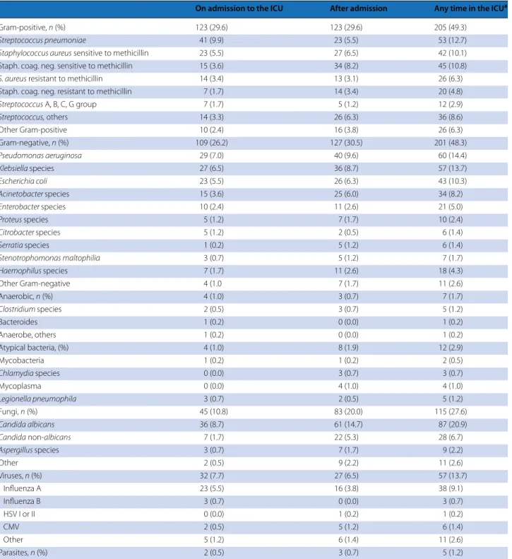
Specimens were obtained for microbiological examina-tion in 596 (89.9) patients and were positive for microor-ganisms in 62.7 % of patients (Table E8). Microbiological investigation on admission to the ICU revealed predomi-nantly Gram-positive (29.6 %) or Gram-negative bacte-ria (26.2 %) but rarely atypical bactebacte-ria (1.0 %); 7.7 % of the microorganisms isolated on admission were viruses (Table 2). Overall, the most frequently isolated Gram-positive bacteria was Streptococcus pneumoniae (12.7 %); the most frequently isolated Gram-negative microorgan-ism was Pseudomonas aeruginosa (14.4 %). Viruses were
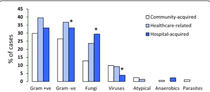
isolated in 13.7 % of positive cultures, atypical bacteria in 2.9 %, and fungi in 27.6 % of cases. In patients with hospital-acquired SARI, Gram-negative microorganisms (33.3 vs. 26.5 %, p = 0.002) and fungi (29.5 vs. 12.8 %,
p = 0.001) were more frequently and viral infections
less frequently (3.9 vs. 9.9 %, p = 0.006) isolated than in patients with community-acquired SARI (Fig. 2).
Morbidity and mortality
Organ failure was present on admission to the ICU in 427 (64.4 %) of the patients with SARI and occurred during the entire ICU stay in 495 (74.7 %). Respiratory, cardio-vascular, and renal failures were the most common organ failures (Table 3). The ICU and hospital mortality rates were 20.2 and 27.2 %, respectively.
The overall ICU mortality in all patients admitted to the contributing centers (n = 5550) was 13.1 % and the median ICU stay was 3 (IQR 2–8) days. Patients with SARI had higher ICU mortality (20.2 vs. 12.2 %, p < 0.001) and longer ICU lengths of stay [5 (2–12) vs. 3 (2–8)] days,
p < 0.001) than those without SARI (Table E5).
Survivors were younger, more likely to be female, less likely to be admitted to the ICU from a hospital ward, had lower severity scores on admission to the ICU, and had a lower incidence of organ failure during the ICU stay (Tables E9, E10) than non-survivors. Survivors were more likely to have received pneumococcal or influenza vaccines than non-survivors. Arrhythmias, chronic renal failure, cancer, asthma, hematologic malignancies, organ or bone marrow transplantation, liver disease, immuno-suppression, or chemotherapy prior to ICU admission were more frequent in non-survivors than in survivors (Table E7). The most common primary causes of in-hos-pital death were multiorgan failure (43.6 %) and pneumo-nia (31.9 %) (Table E11).
In multivariable analysis, older age, greater severity scores on admission to the ICU, hematologic malignancy, and liver disease were independently associated with an increased risk of in-hospital death. Influenza vaccination prior to ICU admission, and adequate antibiotic admin-istration on admission to the ICU were associated with a lower risk of in-hospital death (Table E2).
Discussion
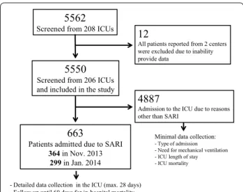
The main findings of our study were that (1) SARI was a common cause of admission to the ICU and was asso-ciated with considerable morbidity and high mortal-ity rates; (2) these infections were predominantly due to Gram-positive or Gram-negative bacteria and rarely due to atypical bacteria or viruses; and (3) older age, greater severity scores, and comorbid conditions were independently associated with an increased risk of in-hospital death in these patients, whereas prior influenza 5562
Screened from 208 ICUs 12
All patients reported from 2 centers were excluded due to inability provide data
5550
Screened from 206 ICUs and included in the study
663
Patients admitted due to SARI
364 in Nov. 2013 299 in Jan. 2014
4887
Admission to the ICU due to reasons other than SARI
Minimal data collection:
- Type of admission - Need for mechanical ventilation - ICU length of stay - ICU mortality
- Detailed data collection in the ICU (max. 28 days) - Follow up until 60 days for in-hospital mortality
Fig. 1 Flowchart of recruitment to and inclusion in the study. ICU intensive care unit, SARI severe acute respiratory infection
vaccination and adequate initial antibiotic administration were associated with a lower risk.
Repeated outbreaks of SARI-related epidemics repre-sent a major healthcare problem [1–4]. Several studies have investigated the epidemiology and clinical charac-teristics of SARI, with a special emphasis on viral etiology [13–17] or on the subset of patients admitted to the ICU with severe community-acquired pneumonia [17–22]. To the best of our knowledge, our study is the first to investi-gate this issue in a large prospective, multinational cohort
of critically ill patients with SARI, providing a global view of this condition worldwide.
One in ten patients with SARI was treated empiri-cally without any microbiologic sampling. Increased awareness of sepsis should be expected to improve compliance with early microbiologic sampling [23]. Nevertheless, the low index of suspicion in the absence of an ongoing epidemic may explain the relatively low rate of investigations aimed at detecting a viral etiol-ogy. In agreement with previous large epidemiologic
Table 1 Characteristics of patients with severe acute respiratory infections (SARI) on admission to the ICU, stratified according to the inclusion periods
Missing values n = 2–12 (max 1.8 %), valid percentages are presented after exclusion of missing values
ICU intensive care unit, SAPS simplified acute physiology score, IQR interquartile range, SOFA sequential organ failure assessment score
a India, Pakistan, and Sri Lanka
All patients Inclusion period p value
November 2013 January 2014
N 663 364 299
Age, years, mean ± SD 63.9 ± 16 64.5 ± 15.9 63.1 ± 16.2 0.226
Male, n (%) 386 (58.7) 216 (59.8) 170 (57.2) 0.501 Healthcare worker, n (%) 11 (1.7) 5 (1.4) 6 (2.0) 0.798 Ethnicity, n (%) White/Caucasian 441 (67.7) 246 (69.5) 195 (71.3) 0.392 Asian 49 (7.5) 22 (6.2) 27 (9.1) Latin American 46 (6.9) 24 (6.8) 22 (7.4) South Asiana 41 (6.2) 23 (6.5) 18 (6.1) Arab 34 (5.2) 19 (4.9) 15 (5.1) Black 24 (3.6) 15 (4.2) 9 (3.0) Others 16 (2.5) 5 (1.4) 11 (3.7) Source of admission, n (%) Community 194 (29.4) 108 (29.9) 36 (22.1) 0.847 Hospital ward 183 (27.8) 99 (27.4) 64 (39.3) Emergency room 165 (25.0) 93 (25.8) 34 (20.9) Other hospital 58 (8.8) 33 (9.1) 25 (8.4) Long-term facility 24 (3.6) 10 (2.8) 14 (4.7) Other ICU 13 (2.0) 2 (0.6) 11 (3.7) Step-down/-up 14 (2.1) 10 (2.8) 4 (1.3) Others 8 (1.2) 6 (1.7) 2 (0.7) Severity scores
SAPS II, mean ± SD 50.4 ± 19.0 49.3 ± 18.2 51.7 ± 19.8 0.178
SOFA score, median (IQR) 5 (2–9) 5 (2–9) 5 (2–9) 0.727
Mode of acquisition, n (%)
Community-acquired 452 (68.2) 246 (68.3) 206 (69.4) 0.059
Healthcare-related 76 (11.5) 37 (10.3) 39 (13.1)
Nosocomial-acquired 129 (19.5) 77 (21.4) 39 (17.5)
Mechanical ventilation on admission to the ICU
Invasive 385 (58.1) 208 (57.1) 177 (59.2) 0.594
Non-invasive 46 (6.9) 28 (7.7) 18 (6.0) 0.399
Influenza vaccine, n (%) 67 (10.2) 43 (11.9) 24 (8.1) 0.751
studies in ICU patients with infections [24–26], patho-genic microorganisms were identified in only 62.7 % of patients. Lack of testing for all possible microorganisms during microbiological investigations, the relatively
high rates of prior antibiotic administration with pos-sible interference with culture results, and inappropri-ate sampling techniques may hinder microorganism identification.
Table 2 Isolated microorganisms (out of 416 patients with positive results)
CMV cytomegalovirus, HSV herpes simplex virus
a Microorganisms isolated both on admission to the ICU and during the ICU stay are counted once
On admission to the ICU After admission Any time in the ICUa
Gram-positive, n (%) 123 (29.6) 123 (29.6) 205 (49.3)
Streptococcus pneumoniae 41 (9.9) 23 (5.5) 53 (12.7)
Staphylococcus aureus sensitive to methicillin 23 (5.5) 27 (6.5) 42 (10.1)
Staph. coag. neg. sensitive to methicillin 15 (3.6) 34 (8.2) 45 (10.8)
S. aureus resistant to methicillin 14 (3.4) 13 (3.1) 26 (6.3)
Staph. coag. neg. resistant to methicillin 7 (1.7) 14 (3.4) 20 (4.8)
Streptococcus A, B, C, G group 7 (1.7) 5 (1.2) 12 (2.9) Streptococcus, others 14 (3.3) 26 (6.3) 36 (8.6) Other Gram-positive 10 (2.4) 16 (3.8) 26 (6.3) Gram-negative, n (%) 109 (26.2) 127 (30.5) 201 (48.3) Pseudomonas aeruginosa 29 (7.0) 40 (9.6) 60 (14.4) Klebsiella species 27 (6.5) 36 (8.7) 57 (13.7) Escherichia coli 23 (5.5) 26 (6.3) 43 (10.3) Acinetobacter species 15 (3.6) 25 (6.0) 34 (8.2) Enterobacter species 10 (2.4) 11 (2.6) 21 (5.0) Proteus species 5 (1.2) 7 (1.7) 10 (2.4) Citrobacter species 5 (1.2) 2 (0.5) 6 (1.4) Serratia species 1 (0.2) 5 (1.2) 6 (1.4) Stenotrophomonas maltophilia 3 (0.7) 5 (1.2) 7 (1.7) Haemophilus species 7 (1.7) 11 (2.6) 18 (4.3) Other Gram-negative 4 (1.0 7 (1.7) 11 (2.6) Anaerobic, n (%) 4 (1.0) 3 (0.7) 7 (1.7) Clostridium species 2 (0.5) 3 (0.7) 5 (1.2) Bacteroides 1 (0.2) 0 (0.0) 1 (0.2) Anaerobe, others 1 (0.2) 0 (0.0) 1 (0.2) Atypical bacteria, (%) 4 (1.0) 8 (1.9) 12 (2.9) Mycobacteria 1 (0.2) 1 (0.2) 2 (0.5) Chlamydia species 0 (0.0) 3 (0.7) 3 (0.7) Mycoplasma 0 (0.0) 4 (1.0) 4 (1.0) Legionella pneumophila 3 (0.7) 2 (0.5) 5 (1.2) Fungi, n (%) 45 (10.8) 83 (20.0) 115 (27.6) Candida albicans 36 (8.7) 61 (14.7) 87 (20.9) Candida non-albicans 7 (1.7) 22 (5.3) 28 (6.7) Aspergillus species 3 (0.7) 7 (1.7) 9 (2.2) Other 2 (0.5) 9 (2.2) 11 (2.6) Viruses, n (%) 32 (7.7) 27 (6.5) 57 (13.7) Influenza A 23 (5.5) 16 (3.8) 38 (9.1) Influenza B 3 (0.7) 0 (0.0) 3 (0.7) HSV I or II 0 (0.0) 1 (0.2) 1 (0.2) CMV 2 (0.5) 5 (1.2) 6 (1.4) Other 5 (1.2) 6 (1.4) 11 (2.6) Parasites, n (%) 2 (0.5) 3 (0.7) 5 (1.2)
Despite the relatively small number of microbiological investigations performed, antimicrobial agents were used generously in our cohort. Interestingly, antiviral and anti-fungal agents were initiated without microbiological evi-dence of these microorganisms in 6.9 and 4.0 % of cases, respectively. The relatively high incidence of comorbid
conditions in our cohort, especially those related to immunosuppression, and the high predominance of life-threatening organ failure in these patients may explain the high use of antimicrobial agents.
The most commonly identified microorganisms were Gram-positive and Gram-negative bacteria. The reported microorganisms may have included, however, some non-pathogenic organisms that are not responsible for SARI. Interestingly, atypical microorganisms were less frequently identified. This observation raises questions regarding the frequent empirical use of macrolides in these patients. However, macrolides have been reported to have addi-tional benefits in patients with community-acquired pneu-monia, irrespective of their antimicrobial effects [27]. Moreover, diagnosis of infections with atypical bacteria is a considerable challenge, such that the occurrence of these infections may be underestimated [28].
Community-acquired infections were common in our cohort, explaining the frequent occurrence of infections with S. pneumoniae. The patterns of microbiologic iso-lates in our study may be useful to guide empirical anti-microbial therapy in patients with ICU-SARI. Although fungi were frequently isolated, only a small proportion of these patients received antifungal agents. This finding supports the common perception that fungal etiologies may not be relevant in all critically ill patients with res-piratory infections [29].
Our data underscore the poor prognosis of critically ill patients with SARI. These patients had considerably higher mortality rates and longer lengths of stay com-pared to patients without SARI. The in-hospital mor-tality rate in our cohort was similar to rates reported in patients with severe community-acquired pneumonia in previous studies [17, 20, 30]. This can be explained by the frequent occurrence of organ failure in these patients.
We identified several risk factors for in-hospital mor-tality in the current cohort. These patterns may be use-ful in identifying patients at risk of poor prognosis. In agreement with the results of a meta-analysis of 70 large observational studies including 21,338 patients with sep-sis [31], we found that adequate initial antibiotic admin-istration was protective in terms of low risk of in-hospital death. This supports the early rigorous empiric use of wide-spectrum antibiotics in these patients, pending the results of microbiological investigations. Nonetheless, since the definition of appropriate antibiotic adminis-tration in the current study was based on both positive microbiologic isolates and clinical improvement, we can-not exclude a possible bias in patients who were judged only on the basis of clinical improvement.
Interestingly, prior seasonal influenza vaccination was independently associated with a favorable in-hospital outcome. Ortiz and collaborators [32] estimated that 0 5 10 15 20 25 30 35 40 45
Gram +ve Gram -ve Fungi Viruses Atypical Anaerobics Parasites Community-acquired Healthcare-related Hospital-acquired % of case s * * *
Fig. 2 Microbiological isolates according to mode of acquisition of severe acute respiratory infection (SARI). *p < 0.05
Table 3 Morbidity and mortality in patients with SARI (n = 663)
Missing values: 53 and 64 for ICU and hospital mortality, respectively. Valid percentages are displayed after exclusion of the missing values
CNS central nervous system, DNR do not resuscitate order, ICU intensive care
unit, LOS length of stay, WD withdrawal of life-sustaining therapy (vasopressors, mechanical ventilation, and renal replacement therapy), WH withholding of life-sustaining measures
a Defined as a SOFA score >2 for the corresponding organ Organ failure on admission to the ICU, n (%)a
Respiratory 205 (30.9) Cardiovascular 217 (32.7) Renal 180 (27.1) CNS 138 (20.8) Hematologic 54 (8.1) Hepatic 29 (4.4)
Organ failure at any time in the ICU, n (%)a
Respiratory 357 (53.8) Cardiovascular 295 (44.5) Renal 296 (44.6) CNS 216 (32.6) Hematologic 133 (20.1) Hepatic 50 (7.5) Mortality rates, n (%) ICU 123 (20.2) In-hospital 163 (27.2)
Length of stay, days, median (IQR)
ICU 5 (2–12)
Hospital 12 (7–23)
Limitation of therapy in-hospital, n (%)
DNR 72 (11.9)
about 28,000 adults are hospitalized for influenza-asso-ciated critical illness in the USA annually. A recent bias-adjusted meta-analysis [33] confirmed that influenza vaccine was effective in preventing hospitalization from influenza and/or pneumonia and all-cause mortality in community-dwelling elderly. However, the evidence to support risk factors for influenza-related complications is still low and adequately powered studies are needed to address this issue [34, 35].
The study had several limitations. First, participation was voluntary and was concentrated in some countries in Western Europe, so that the results in this report may not be extrapolated to all ICU patients worldwide. Sec-ond, regional and seasonal differences may also occur and cannot be captured from our data. Third, some of the reported microorganisms may have represented respira-tory colonization rather than infection. Fourth, the defi-nitions of infection were not based on only the presence of microorganisms and we did not differentiate between possible and probable clinical infections. Nonetheless, defining infection according to the isolation of a patho-genic microorganism may also be subject to a certain bias because of possible interference from prior antibiotic administration, availability and the diagnostic perfor-mance of the serologic tests, and local practice concern-ing microbiologic investigations. Fifth, our data may not be extrapolated to all hospitalized patients with SARI. Finally, the study was purely observational and we did not apply standard protocols for diagnosis and therapy in patients with SARI; however, our aim was to report cur-rent practice in these patients.
Conclusions
SARI is a common cause of admission to the ICU and has associated high morbidity and mortality rates. Our data describe the current patterns of infections and therapy of this serious healthcare problem. This information may be particularly useful in resource allocation and risk stratifi-cation for clinical and research purposes.
Electronic supplementary material
The online version of this article (doi:10.1007/s00134-015-4206-2) contains supplementary material, which is available to authorized users.
Author details
1 Department of Anesthesiology and Intensive Care, Uniklinikum Jena,
Erlanger Allee 103, 07743 Jena, Germany. 2 Intensive Care Department,
Hospital Universitari Vall d’Hebron, CIBER Enfermedades Respiratorias, Passeig de la Vall d’Hebron, 119-129, 08035 Barcelona, Spain. 3 King’s Health Partners,
Westminster Bridge Road, London SE1 7EH, UK. 4 Critical Care Directorate, St
George’s University Hospitals NHS Foundation Trust, London, UK. 5 Intensive
Care Unit, Hospital de São Jos, Unidade de Cuidados Intensivos Neuroc-ríticos, Centro Hospitalar de Lisboa Central, EPE, Rua José António Serrano, s, 1150-199 Lisbon, Portugal. 6 Medical ICU, Paris Diderot University/Bichat
Teaching Hospital Medical and Infectious Diseases ICU, 46 rue Henri Huchard, Paris 75018, France. 7 Interdepartmental Division of Critical Care Medicine,
University of Toronto, Toronto, Canada. 8 Keenan Research Centre, Li Ka Shing
Knowledge Institute, St. Michael’s Hospital, 30 Bond Street, Toronto, ON M5B 1W8, Canada. 9 Division of Pulmonary and Critical Care Medicine,
Massachu-setts General Hospital, Boston, USA. 10 Harvard Medical School, 55 Fruit St,
Boston, MA 02114, USA. 11 Serviço de Terapia intensiva na IAMSPE-Instituto
de Assistência Médica ao Servidor Público Estadual, Av. Ibirapuera, 981, São Paulo, SP 04028-000, Brazil. 12 Medical Intensive Care Unit, Paris Descartes
University, Paris, France. 13 Hôpital Cochin, Paris, France. 14 Department of Cell
Biology and Host-Pathogen Interaction, Cochin Institute, 27 Rue du Faubourg St Jacques, 75679 Paris Cedex 14, France.
Acknowledgments
We would like to thank Dr. Hassane Nijimi (Free University of Brussels), the study statistician, for his efforts in data monitoring and analysis and Dr. Karen Pickett for editorial assistance with the manuscript.
Compliance with ethical standards Conflicts of interest
The authors declare that they do not have any conflict of interest in relation to the subject of this manuscript.Funding
The study was supported by an unrestricted grant from the European Society of Intensive Care Medicine (ESICM), Rue Belliard, 19, 1040 Brussels, Belgium.
Appendix: Alphabetical list of participating centers by region and country
Europe
Austria: General Hospital, Braunau (J Auer, G Schatzl);
Krankenhaus Oberwart, Oberwart (K Mach, H Gruber)
Belgium: Ziekenhuis Oost-Limburg, Genk (E Schreurs,
M Vander Laenen); Universitair Ziekenhuis, Leuven (H Ceunen, J Wauters); CHU Saint-Pierre, Brussels (P Des-champs); Cliniques Universitaires Saint-Luc UCL, Brus-sels (D Castanares); CHU Brugmann, BrusBrus-sels (D Debels, C Pierrakos); Erasme University Hospital, Brussels (JL Vincent, F Taccone (national coordinator))
Czech Republic: University Hospital Motol, Prague (T
Vymazal)
Croatia: University Hospital Centre Zagreb, Zagreb (I
Gornik, A Vujiaklija Brajkovic)
Denmark: Holbaek Sygehus, Holbaek (R Medici); Rig
-shopitalet, Copenhagen (J Nielsen); Glostrup Sygehus, Glostrup (A Bendtsen, H Siegel)
Finland: Meilahti Hospital, Helsinki (T Suonsyrjä) France: CHU Nord, Marseille (S Hraech), Hôpital
Cochin, Paris (J-D Chiche, F Daviaux); CHRU Stras-bourg-Hôpital de Hautepierre, Strasbourg (M Guillot, V Castelain); CHRU Nancy-Hôpital Brabois-Réanimation et soins continus chirurgicaux, Vandoeuvre-Les-Nancy (R–R Losser), CHRU Nancy-Hôpital Brabois-Réanima-tion médicale, Vandoeuvre-Les-Nancy (E Novy); (Hôpi-tal Bichat, Paris (J-F Timsit (national coordinator), L Bouadma); Groupe Hospitalier Paris Saint-Joseph, Paris (B Misset, F Philippart); Centre Hospitalier Dr Schaf-fner, Lens (J Mallat); CHU Amiens, Amiens (E Zogheib, M Miclo) Hôpital Bicêtre, Le Kremlin-Bicêtre Paris (J-L Teboul, N Anguel); CHU Saint-Etienne, Saint-Etienne (M Darmon); Hôpital Tenon, Paris (T Pham); CH Mul-house-Hôpital Emile Muller, Mulhouse (G Barberet);
Hôpital Victor Dupouy, Argenteuil (G Plantefeve); Hôpi-tal Edouard Herriot, Hospices Civils de Lyon, Lyon (B Floccard)
Georgia: Georgian Critical Care Medicine Institute,
Tbilissi (Z Kheladze)
Germany: Universitätsklinikum Jena, Jena (K Rein
-hart (national coordinator), Y Sakr, F Bloos); Klinikum Weiden, Weiden in der Oberpfalz (A Faltlhauser); Klini-kum Luedenscheid, Luedenscheid (T Helmes); Univer-sity Hospital Frankfurt, Frankfurt am (K Zacharowski, P Meybohm); Klinikum Saarbrücken, Saarbrücken (K Schwarzkopf); Klinikum Nürnberg, Dept of Emergency and Critical Care Medicine, Nurenberg (M Christ, M Baumgaertel); Klinikum Nürnberg, Nephrologische Intensivstation BU13, Nurenberg (S John, J Nentwich); Universitätsmedizin-Charité, Berlin (M Deja, A Gold-mann); Diakoniekrankenhaus Friederikenstift, Han-nover (A Gottschalk, F Honig); University Medical Center Freiburg, Freiburg (B Siepe, U Goebel); Vivantes Humboldt-Klinikum, Berlin (J Lehmke, S Behrens); Oberschwabenklinik, Krankenhaus St Elisabeth, Raven-sburg (K Fiedler); Universitätsmedizin Mainz, Mainz (I Sagoschen); University Hospital, Tübingen (R Ries-sen, M Haap); University Hospital, Leipzig (Ph Simon,U Kaisers); Vivantes Klinikum Spandau, Berlin (S Behrens, M Niesen); Klinikum Augsburg, Augsburg (U Jaschin-ski); Universitätsklinikum des Saarlandes, Homburg (S Hoersch, A Jung); Robert-Bosch-Krankenhaus, Stuttgart (S Allgaeuer); Maria Hilf, Mönchengladbach (H Haake); Klinik Hennigsdorf der Oberhavel Kliniken, Hennigsdorf (A Lange)
Greece: Hippokrateion General Hospital, Athens
(M Papanikolaou, M Balla); AHEPA University Hospi-tal, Thessaloniki (M Giannakou, I Soultati); University Hospital of Ioannina, Ioannina (G Nikos, V Koulouras); Lamia General Hospital, Lamia (G Kyriazopoulos, D Gkika); General Hospital O Agios Dimitrios, Thessalon-iki (G Vlachogianni, K Psaroulis); Hippokratio Hospital, Thessaloniki (E Mouloudi, E Massa)
Ireland: St Vincents University Hospital, Dublin (A
Nichol, E Meany); Limerick University Hospital, Limer-ick (C Motherway)
Italy: San Gerardo Hospital, Monza (G Bellani); Pin
-etagrande Private Hospital, Castelvolturno (V Pota, V Schiavone); University Hospital of Modena, Modena (M Girardis); Azienda Ospedaliera Desenzano, Desenzano (N Petrucci, R Di Pasquale); Ospedale Sandro Pertini, Rome (P Mazzini); IRCCS San Martino-IST, Genova (A Molin, G Pellerano); Arispedale Sant’Anna Hospital, Fer-rara (C Volta, S Spadaro); Azienda Ospedaliero Univer-sitaria Pisana, Pisa (F Guarracino); Fondazione IRCCS Ca’ Granda-Ospedale Maggiore Policlinico, Milan (M Savioli); Santa Maria degli Angeli Hospital, Pordenone
(T Pellis, N Chinellato); Ospedale Ceccarini, Riccione (A Gatta, F Cecchini); Policlinico P Giaccone, Palermo (SM Raineri, A Cortegiani)
Lithuania: Vilnius University Hospital Santariskiu Clin -ics, Vilnius (G Kekstas, V Karosas)
Macedonia: Special Hospital for surgery Fillip II, Skopje
(T Anguseva, Z Mitrev)
Netherlands: Tjongerschans, Heerenveen (O Beck, N
Cimic); Atrium Medisch Centrum Parkstad, Heerlen (G Janssen, L Bormans); Medical Center Leeuwarden, Leeu-warden (M Kuiper, K Koopmans); Spaarneziekenhuis, Hoofddorp (S Den Boer, M de Groot); Medical Centre Haaglanden, The Hague, P Dennesen); Reinier de Graaf Ziekenhuis, Delft (J van den Bosch); Slotervaartzieken-huis, Amsterdam (G Kluge)
Poland: Child Jesus Clinical Hospital of the Medical
University of Warsaw, Warsaw (M Mikaszewska-Sokole-wicz, T Lazowski); Szpital Praski, Warsaw (M Chrus-cikowski); Wroclaw University Hospital, Wroclaw (J Machon, B Adamik); Barlicki Clinical Hospital, Lodz (A Wieczorek)
Portugal: Hospital de S José, Lisbon (S Afonso, R Matos
(national coordinator)); Centro Hospitalar do Médio Tejo, Abrantes (N Catorze, A Araujo); Hospital de Santa Maria EPE (CHLN), Lisbon (Z Costa, A Pais-de-Lacerda); Cen-tro Hospitalar Tondela-Viseu, Viseu (I Martins); Hospi-tal Sao Francisco Xavier—CHLO, Lisbon (R Cardiga, L Fernandes); Hospital Pr Doutor Fernando Fonseca EPE, Amadora (I Serra, A Martinho)
Romania: Fundeni Clinical Institute, Bucharest (D
Tomescu, E Scarlatescu); Institute of Pneumology, Bucharest (R Stoica, A Macri); Emergency Institute for Cardiovascular Diseases Prof Dr CC Iliescu, Bucharest (D Filipescu (national coordinator))
Slovenia: General Hospital Izola, Izola (E Rupnik); Uni -versity Clinic of Respiratory and Allergic Diseases, Gol-nik (V Tomic, F Sifrer)
Spain: Hospital de Gran Canaria Dr Negrín, Las Pal
-mas de Gran Canaria (J Sole Violan, JM; Ferrer Agüero); Complejo Hospitalario de Navarra, UCI B, Pamplona (J Izura); Clinica Universidad de Navarra, UCI Adultos, Pamplona (P Monedero); Hospital de Torrejón, Tor-rejón de Ardoz (C Muños de Cabo); Hospital Clínico Universitario de Valencia, Cuidados intensivos quirur-gicos, Valencia (G Aguilar, FJ Belda); Hospital Clínico Universitario de Valencia, Cuidados Intensivos, Valen-cia (J Blanquer, E Nives Carbonell); Hospital Univer-sitari Bellvitge, L’Hospitalet de Llobregat-Barcelona (J-C Lopez-Delgado); Hospital Regional Carlos Haya, Málaga (C Aragon); Hospital Quirón Sagrado Corazón, Seville (C Ortiz-Leyba); Complejo Hospitalario Univer-sitario de Ferrol, Ferrol (CJ Fernandez Gonzalez); Hos-pital Universitario Virgen de la Victoria, Málaga (M-V
de la Torre-Prados, A Puerto-Morlan); Hospital Uni-versitario Miguel Servet, Zaragoza (P Araujo Aguilar, JI Tomás Marsilla); Hospital de la Santa Creu I Sant Pau, Barcelona (P Vera Aratcoz, A Olmo); Mutua Terrassa University Hospital, Terrasa (R Ferrer Roca (national
coordinator)); Hospital General de Vic, Vic-Barcelona
(RM Catalan); Hospital General Universitario Grego-rio Marañon, Madrid (P Garcia Olivares); Hospital de Mataró, Mataró (A Albis); Clinico San Carlos, Madrid (M Alvarez); Hospital General Universitario de Albacete, Albacete (V Corcoles Gonzalez, J M Gutierrez Rubio); Hospital Clínico Universitario Lozano Blesa, Zaragoza (R Montoiro Allue); Hospital Infanta Cristina, Badajoz (J Rubio Mateo-Sidron)
UK: Bronglais General Hospital, Aberystwyth (M
Hobrok); St George’s Hospital, London (M Cecconi (national coordinator), N Di Tomasso); Barts Health NHS Trust, Whipps Cross Hospital, Leytonstone (A Raj); Royal Glamorgan Hospital, Llantrisant (T Szakmany, L Srinivasa); Alexandra Hospital, Redditch (S Mathew); Craigavon Area Hospital, Portadown (A Ferguson); The Great Western Hospital, Swindon (M Blahut-Zugaj, M Watters); Western Infirmary, Glasgow (S Henderson, M Sim); Wexham Park Hospital, Slough (P Csabi); Antrim Area Hospital, Antrim (O O’Neill, C Nutt); West Suffolk Hospital, Bury St Edmunds (S Humphreys, K Bhowmick); Altnagelvin Hospital, Derry (A Donnelly, S O’Kane); Ipswich Hospital NHS Trust, Ipswich (M Garfield); Bar-net General Hospital, BarBar-net (R Jha, N Unni); Imperial College Healthcare NHS Trust-Charing Cross Hospital, London (A Gordon, F Rubulotta); Rotherham General Hospital, Rotherham (K Ravi, G Lunch); Chase Farm Hospital, Enfield (F Federica); Kent and Canterbury pital, Canterbury (D Higgs, G Strandvik); Pilgrim Hos-pital, Boston (A Jonas); King’s College HosHos-pital, London (Ph Hopkins, T Hurst); Queen’s Hospital, Romford (A Bellini, O Balogun); St Thomas Hospital, London (N Bar-rett, M Ostermann); University Hospital of South Man-chester, Manchester (P Alexander, K McCalman); Guy’s Hospital, London (M Ostermann); Luton and Dunsta-ble Hospital, Luton (G Brescia); John Radcliffe Hospital, Oxford (J Strachan, J Meyer); Imperial College Health-care NHS Trust-St Mary’s Hospital, London (M Stotz), Imperial College Healthcare NHS Trust-Hammersmith Hospital, London (S Brett).
Middle East
Iran: Nemazee Hospital, Shiraz (F Zand, R Nikandish);
Masih Daneshvari Hospital (NRITLD), Teheran (S Hashemian);
Qatar: Hamad General Hospital, Doha (AS Alsheikhly) Saudi Arabia: Prince Sultan Military Medical City
(PSMMC), Riyadh (G Almekhlafi); King Faisal Specialist
Hospital, Riyadh (A Maghrabi, N Salahuddin); King Fahad Hospital, Baha (T Aisa)
Turkey: Atasehir Memorial Hospital, Istanbul (H K Ata -lan); Erciyes Universitesi Tip Fakultesi, Kayseri (M Sungur);
United Arab Emirates: Sheikh Khalifa Medical City,
Abu Dhabi (M Hegazi).
North America
USA: Mayo Clinic, Saint Mary’s Hospital, Rochester (P
Bauer); Memorial Medical Center, Springfield (S Muk-kera); Santa Barbara Cottage Hospital, Santa Barbara (J Fried, M Barger); John H Stroger Hospital of Cook County, Chicago (R Gueret).
South America
Argentina: Sanatorio Parque, Rosario (C Gonzalez, C
Lovesio); Hospital Dr Julio C Perrando, Resistencia (Ch Dellera, D Barrios)
Brazil: Hospital Universitario Lauro Wanderley, João
Pessoa (C Leite Mendes, P Gottardo); Hospital Unimed, Vitória (E Caser, C Santos); UDI Hospital, São Luís (A Carvalho); Hospital Moinhos de Vento, Porto Alegre (C Teixeira)
Chile: Hospital del Trabajador, Santiago (W Samaniego,
S Whittle)
Colombia: Clinica Universitaria Colombia, Bogota
(D Molano, A Rojas); Clinica Medellín, Medellin (K Guerra)
Ecuador: Hospital Militar, Quito (B Villamagua); Cli
-nica La Merced, Quito (E Salgado-Yepez); Hospital de los Valles, Quito (D Morocho, N Remache-Vargas)
Mexico: Instituto Nacional de Cancerologia, Mexico
(S Ñamendys-Silva); Hospital Civil de Guadalajara Hos-pital Juan I Menchaca, Guadalajara (D Rodriguez); Insti-tuto Nacional de Ciencias Médicas y Nutrición Salvador Zubirán, Mexico (G Dominguez, G Barraza); Hospital Regional Leon ISSSTE, Mexico (E Bermudez-Aceves); Hospital de Especialidades Antonio Fraga Mouret-Centro Medico Nacional La Raza IMSS, Mexico (LA Sanchez-Hurtado, JA Baltazar-Torres)
Peru: Hospital Nacional Dos de Mayo, Lima (R Quispe
Sierra, R Ovalle Olmos); Hospital Regional Honorio Del-gado, Arequipa (C Chavez)
Venezuela: Hospital Central Dr Miguel Pérez Carreño,
Caracas (I von Osten).
Oceania
Australia: Canberra Hospital, Canberra (C Van Haren
(national coordinator)); Townsville Hospital, Douglas (N Smalley); Concord Hospital, Concord-West Sydney (M Kol, H Wong); St Vincent’s Hospital, Fitzroy-Melbourne (R Smith).
East and South‑East Asia
China: Wuhan Central Hospital, Wuhan (L Yu, X Wu);
The First People’s Hospital, Kunming (L Chao); Qilu Hospital of Shandong University, Jinan (Q Zhai, D Wu); Tenth People’s Hospital, Tongji University School of Medicine, Shanghai (X Zhang, X Jing);
Philippines: Chong Hua Hospital, Cebu (R Bigornia, Y
Ikeda-Maquiling); The Medical City, Pasig (J Robles, JE Palo)
Vietnam: Bachmai Hospital, Hanoi (T Nguyen, C Dao).
South Asia
India: Sanjeevan Hospital, Pune (S Dixit), Sanjay Gandhi
Postgraduate Institute of Medical Sciences, Lucknow (M Gurjar); Care Hospital, Hyderabad (P Reddy); Bombay Hospital Institute of Medical Sciences, Mumbai Maha-rashtra (A Pravin (national coordinator)); Hinduja Hospital, Mumbai (S Simran); Apollo Hospitals, Chennai (N Ram-akrishnan); Manipal Hospital, Bangalore (R Shetty); Breach Candy Medical Research Centre, Mumbai (F Udwadia)
Pakistan: Shifa International Hospital, Islamabad (M
Faraz)
Sri Lanka: Sri Jayewardenepura General Hospital,
Nugegoda (K Indraratna, J Rajasinhe).
Received: 15 November 2015 Accepted: 24 December 2015
References
1. Webb SA, Seppelt IM (2009) Pandemic (H1N1) 2009 influenza (“swine flu”) in Australian and New Zealand intensive care. Crit Care Resusc 11:170–172
2. Rello J, Rodriguez A, Ibanez P, Socias L, Cebrian J, Marques A, Guerrero J, Ruiz-Santana S, Marquez E, Del Nogal-Saez F, Alvarez-Lerma F, Martinez S, Ferrer M, Avellanas M, Granada R, Maravi-Poma E, Albert P, Sierra R, Vidaur L, Ortiz P, Prieto DPI, Galvan B, Leon-Gil C, H1N1 SEMICYUC Working Group (2009) Intensive care adult patients with severe respiratory failure caused by Influenza A (H1N1)v in Spain. Crit Care 13:R148
3. Kumar A, Zarychanski R, Pinto R, Cook DJ, Marshall J, Lacroix J, Stelfox T, Bagshaw S, Choong K, Lamontagne F, Turgeon AF, Lapinsky S, Ahern SP, Smith O, Siddiqui F, Jouvet P, Khwaja K, McIntyre L, Menon K, Hutchison J, Hornstein D, Joffe A, Lauzier F, Singh J, Karachi T, Wiebe K, Olafson K, Ramsey C, Sharma S, Dodek P, Meade M, Hall R, Fowler RA (2009) Criti-cally ill patients with 2009 influenza A(H1N1) infection in Canada. JAMA 302:1872–1879
4. Martin-Loeches I, Diaz E, Vidaur L, Torres A, Laborda C, Granada R, Bonas-tre J, Martin M, Insausti J, Arenzana A, Guerrero JE, Navarrete I, Bermejo-Martin J, Suarez D, Rodriguez A (2011) Pandemic and post-pandemic influenza A (H1N1) infection in critically ill patients. Crit Care 15:R286 5. WHO Regional Office for Europe (2011) WHO Regional Office for Europe
Guidance for Sentinal Influeza Surveillance in Humans. http://www.euro. who.int/__data/assets/pdf_file/0020/90443/E92738.pdf?ua=1. Accessed 1 Jan 2015
6. Fowler RA, Lapinsky SE, Hallett D, Detsky AS, Sibbald WJ, Slutsky AS, Stew-art TE (2003) Critically ill patients with severe acute respiratory syndrome. JAMA 290:367–373
7. Lew TW, Kwek TK, Tai D, Earnest A, Loo S, Singh K, Kwan KM, Chan Y, Yim CF, Bek SL, Kor AC, Yap WS, Chelliah YR, Lai YC, Goh SK (2003) Acute
respiratory distress syndrome in critically ill patients with severe acute respiratory syndrome. JAMA 290:374–380
8. Manocha S, Walley KR, Russell JA (2003) Severe acute respiratory distress syndrome (SARS): a critical care perspective. Crit Care Med 31:2684–2692 9. Calandra T, Cohen J (2005) The International Sepsis Forum consensus
conference on definitions of infection in the intensive care unit. Crit Care Med 33:1538–1548
10. American Thoracic Society, Infectious Diseases Society of America (2005) Guidelines for the management of adults with hospital-acquired, ventilator-associated, and healthcare-associated pneumonia. Am J Respir Crit Care Med 171:388–416
11. Le Gall JR, Lemeshow S, Saulnier F (1993) A new Simplified Acute Physiol-ogy Score (SAPS II) based on a European/North American multicenter study. JAMA 270:2957–2963
12. Vincent JL, Moreno R, Takala J, Willatts S, De Mendonca A, Bruining H, Reinhart CK, Suter PM, Thijs LG (1996) The SOFA (Sepsis-related Organ Failure Assessment) score to describe organ dysfunction/fail-ure. On behalf of the Working Group on Sepsis-Related Problems of the European Society of Intensive Care Medicine. Intensive Care Med 22:707–710
13. Poulakou G, Perez M, Rello J (2012) Severe acute respiratory infections in the postpandemic era of H1N1. Curr Opin Crit Care 18:441–450 14. Jain S, Benoit SR, Skarbinski J, Bramley AM, Finelli L (2012)
Influenza-associated pneumonia among hospitalized patients with 2009 pandemic influenza A (H1N1) virus—United States, 2009. Clin Infect Dis 54:1221–1229
15. Martin-Loeches I, Sanchez-Corral A, Diaz E, Granada RM, Zaragoza R, Vil-lavicencio C, Albaya A, Cerda E, Catalan RM, Luque P, Paredes A, Navarrete I, Rello J, Rodriguez A (2011) Community-acquired respiratory coinfection in critically ill patients with pandemic 2009 influenza A(H1N1) virus. Chest 139:555–562
16. Sandrock C, Stollenwerk N (2008) Acute febrile respiratory illness in the ICU: reducing disease transmission. Chest 133:1221–1231
17. Bertolini G, Rossi C, Crespi D, Finazzi S, Morandotti M, Rossi S, Peta M, Langer M, Poole D (2011) Is influenza A(H1N1) pneumonia more severe than other community-acquired pneumonias? Results of the GiViTI survey of 155 Italian ICUs. Intensive Care Med 37:1746–1755
18. Cilloniz C, Ewig S, Ferrer M, Polverino E, Gabarrus A, de la Puig BJ, Mensa J, Torres A (2011) Community-acquired polymicrobial pneumonia in the intensive care unit: aetiology and prognosis. Crit Care 15:R209 19. Jeong BH, Jeon EJ, Yoo H, Koh WJ, Suh GY, Chung MP, Kwon OJ, Jeon K
(2014) Comparison of severe healthcare-associated pneumonia with severe community-acquired pneumonia. Lung 192:313–320
20. Walden AP, Clarke GM, McKechnie S, Hutton P, Gordon AC, Rello J, Chiche JD, Stueber F, Garrard CS, Hinds CJ (2014) Patients with community acquired pneumonia admitted to European intensive care units: an epidemiological survey of the GenOSept cohort. Crit Care 18:R58 21. Mongardon N, Max A, Bougle A, Pene F, Lemiale V, Charpentier J, Cariou A,
Chiche JD, Bedos JP, Mira JP (2012) Epidemiology and outcome of severe pneumococcal pneumonia admitted to intensive care unit: a multicenter study. Crit Care 16:R155
22. Viasus D, Marinescu C, Villoslada A, Cordero E, Galvez-Acebal J, Farinas MC, Gracia-Ahufinger I, Fernandez-Navarro A, Niubo J, Ortega L, Munez-Rubio E, Romero-Gomez MP, Carratala J (2013) Community-acquired pneumonia during the first post-pandemic influenza season: a prospec-tive, multicentre cohort study. J Infect 67:185–193
23. Dellinger RP, Levy MM, Rhodes A, Annane D, Gerlach H, Opal SM, Sevransky JE, Sprung CL, Douglas IS, Jaeschke R, Osborn TM, Nunnally ME, Townsend SR, Reinhart K, Kleinpell RM, Angus DC, Deutschman CS, Machado FR, Rubenfeld GD, Webb S, Beale RJ, Vincent JL, Moreno R (2013) Surviving Sepsis Campaign: international guidelines for man-agement of severe sepsis and septic shock, 2012. Intensive Care Med 39:165–228
24. Vincent JL, Sakr Y, Sprung CL, Ranieri VM, Reinhart K, Gerlach H, Moreno R, Carlet J, Le Gall JR, Payen D (2006) Sepsis in European intensive care units: results of the SOAP study. Crit Care Med 34:344–353
25. Vincent JL, Rello J, Marshall J, Silva E, Anzueto A, Martin CD, Moreno R, Lipman J, Gomersall C, Sakr Y, Reinhart K (2009) International study of the prevalence and outcomes of infection in intensive care units. JAMA 302:2323–2329
26. Vincent JL, Marshall JC, Namendys-Silva SA, Francois B, Martin-Loeches I, Lipman J, Reinhart K, Antonelli M, Pickkers P, Njimi H, Jimenez E, Sakr Y (2014) Assessment of the worldwide burden of critical illness: the inten-sive care over nations (ICON) audit. Lancet Respir Med 2:380–386 27. Feldman C, Anderson R (2015) Community-acquired pneumonia:
patho-genesis of acute cardiac events and potential adjunctive therapies. Chest 148:523–532
28. Basarab M, Macrae MB, Curtis CM (2014) Atypical pneumonia. Curr Opin Pulm Med 20:247–251
29. Lamoth F, Alexander BD (2014) Nonmolecular methods for the diagnosis of respiratory fungal infections. Clin Lab Med 34:315–336
30. Cavallazzi R, Wiemken T, Arnold FW, Luna CM, Bordon J, Kelley R, Feldman C, Chalmers JD, Torres A, Ramirez J (2015) Outcomes in patients with community-acquired pneumonia admitted to the intensive care unit. Respir Med 109:743–750
31. Paul M, Shani V, Muchtar E, Kariv G, Robenshtok E, Leibovici L (2010) Systematic review and meta-analysis of the efficacy of appropriate
empiric antibiotic therapy for sepsis. Antimicrob Agents Chemother 54:4851–4863
32. Ortiz JR, Neuzil KM, Shay DK, Rue TC, Neradilek MB, Zhou H, Seymour CW, Hooper LG, Cheng PY, Goss CH, Cooke CR (2014) The burden of influenza-associated critical illness hospitalizations. Crit Care Med 42:2325–2332 33. Darvishian M, Gefenaite G, Turner RM, Pechlivanoglou P, Van der Hoek W,
Van den Heuvel ER, Hak E (2014) After adjusting for bias in meta-analysis seasonal influenza vaccine remains effective in community-dwelling elderly. J Clin Epidemiol 67:734–744
34. Mertz D, Kim TH, Johnstone J, Lam PP, Science M, Kuster SP, Fadel SA, Tran D, Fernandez E, Bhatnagar N, Loeb M (2013) Populations at risk for severe or complicated influenza illness: systematic review and meta-analysis. BMJ 347:f5061
35. Darvishian M, Bijlsma MJ, Hak E, Van den Heuvel ER (2014) Effectiveness of seasonal influenza vaccine in community-dwelling elderly people: a meta-analysis of test-negative design case-control studies. Lancet Infect Dis 14:1228–1239