1
Universidade do Porto I Faculdade de Medicina
Contribution to the determination of the place of death by
drowning - study of biodiversity of diatoms in Douro River
Sara Isabel Sebastião Coelho
Dissertação apresentada à Universidade do Porto para a obtenção do grau
de Mestre em Ciências Forenses
Orientador I Professor Doutor Agostinho Santos I Faculdade de Medicina da
Universidade do Porto3
Agradecimentos
Antes de apresentar este trabalho, é imperativo realçar e do mesmo modo agradecer a todos os que me ajudaram, apoiaram a alcançar este objetivo.
Em primeiro lugar quero prestar os meus mais sinceros agradecimentos ao Professor Doutor Agostinho Santos, meu orientador, por todo o apoio, desde proporcionar-me o local e as condições de trabalho necessárias à realização desta tese, como todo o incentivo nos momentos mais críticos e de desânimo, mais do que tudo agradeço a sua orientação e o seu rigor neste trabalho sem os quais nunca teria sido realizado.
À Professora Joana Mendonça, pela sua disponibilidade ao ensinar-me as bases para a realização da foto-identificação das espécies bem como no seu tratamento estatístico. Aos técnicos do laboratório de Anatomia Patológica do Instituo Nacional de Medicina Legal e Ciências Forenses - Delegação do Norte, Drª Cláudia Ribeiro e Dr. Francisco Afonso, por me permitirem partilhar o seu local de trabalho bem como todo o apoio na realização do trabalho laboratorial.
A todos os médicos e técnicos do serviço de Patologia Forense, do Instituto Nacional de Medicina Legal e Ciências Forenses - Delegação do Norte, pela enorme ajuda na recolha do material de estudo para esta tese.
À Dona Amélia Castro, pelo seu carinho e apoio durante toda a minha estadia no Instituto Nacional de Medicina Legal e Ciências Forenses - Delegação do Norte.
Aos meus Pais, por nunca me deixarem desanimar e me compelirem a ultrapassar todas as dificuldades até atingir o meu objetivo.
4
Indice
a) Introduction ... 9
b) Objectives ... 16
c) Materials and methods ... 17
a) Area of study and sample collection ... 17
b) Laboratory treatment of water samples ... 17
c) Microscopic observation and counting ... 18
d) Taxonomic determination ... 18
e) Body samples ... 18
f) Laboratory treatment of body samples ... 19
g) Microscopic observation and counting ... 19
h) Taxonomic determination ... 19
i) Photographic record ... 19
d) Results ... 21
e) Discussion ... 53
f) Limitations of the study ... 60
g) Conclusion ... 62
h) Bibliographic ... 64
i) Attachments ... 67
i. List A. All taxa identified in water samples ... 67
6 Resumo
O papel da investigação da presença de diatomáceas nos órgãos de um individuo que apareceu morto em meio líquido, e a contribuição que a sua presença pode dar para o diagnóstico médico-legal de afogamento levanta ainda controvérsia. Acresce a isto o facto de não se encontrar estabelecido em definitivo qual o método exato de estudo das diatomáceas. Perante este problema médico-legal e na ausência de qualquer estudo forense em Portugal sobre esta problemática, foi desenvolvido um estudo que procurou determinar o local de afogamento pela análise de espécies de diatomáceas pesquisadas em diferentes órgãos, comparando-as com as espécies existentes no meio líquido em que o cadáver apareceu.
Obtivemos, durante um ano, um total de 37 casos. Foi feita uma análise sazonal de diatomáceas (Primavera, Verão, Outono e Inverno) de 9 locais de amostragem, de modo a obter uma base de informação da composição de espécies de diatomáceas de cada local. Para a realização da extração de diatomáceas dos órgãos dos cadáveres utilizou-se o método químico com ácido clorídrico a 37% de concentração, para a extração de diatomáceas das águas recolhidas nos locais de amostragem utilizou-se ácido sulfúrico a 96% de concentração.
Em 63% dos nossos casos de estudo foi possível encontrar diatomáceas, sendo que estas foram encontradas essencialmente nas amostras referentes aos pulmões e ao conteúdo gástrico. Acreditamos que a ausência de diatomáceas nos restantes órgãos está associada a uma morte rápida, desta maneira a circulação sanguínea cessa quase de imediato, não permitindo que as diatomáceas atinjam os restantes órgãos.
Quando realizamos a comparação entre as espécies de diatomáceas encontradas nos órgãos com as espécies de diatomáceas encontradas no respetivo local onde se sabe que ocorreu o afogamento, constatou-se haver correspondência, contudo, quando o local é desconhecido, fazer a avaliação do possível local de afogamento revelou-se menos preciso, pois uma vez que a ausência de espécies indicadoras características de cada local dificultou todo este processo. Devido à elevada influência antropogénica de que o estuário do Douro é alvo, não se verificaram diferenças significativas entre os cinco locais amostrados, sendo deste modo extremamente difícil aferir com certeza o local exato de afogamento no estuário.
Com o nosso estudo torna-se evidente a importância de existir uma base de dados de diatomáceas do local em que ocorreu o afogamento. Demonstrou-se por outro lado, que é fundamental realizar-se uma colheita de água do local no momento em que o afogamento ocorreu, pois só assim será possível realizar uma comparação rigorosa da composição de diatomáceas.
7 Abstract
The role of the investigation of the presence of diatoms in the organs of an individual found in a liquid medium and which forensically relevant information such presence can provide to a case of drowning is still controversial. This is complicated by the absence of an exact and well defined method for diatoms’ study. Considering this medico-legal problem and the absence of forensic studies on this subject in Portugal, the aim of this study was to determine the drowning place based on the analysis of diatom species searched in different organs comparing them to the existent in the liquid medium where the corps was found.
During a period of one year, we had a total of 37 cases and made seasonal diatoms analyses (Spring, Summer, Autumn, Winter) in 9 sampling places, in order to build a data base of the composition of the diatoms’ species of each place. We used the chemical method with chloridric acid at 37% concentration for the diatoms’ extraction from the organs and sulfuric acid at 96% concentration for the diatoms’ extraction from the water of the sampling places.
Diatoms were found in 63% of our cases, mainly in samples from lungs and gastric content. The absence of diatoms in the other organs is probably associated with a quick death, which may have stopped blood circulation almost immediately, preventing diatoms contamination of the other organs.
When comparing the diatoms’ species found in the organs and the ones found at the corresponding known drowning place, the correspondence was easy and accessible. Yet, when the drowning place was unknown, the evaluation of the possible drowning place was less accurate. The absence of indicator species, distinctive of each place, made our task even harder. Besides, due to the high anthropogenic influence on the Douro estuary, no significant differences were observed between the five sampling places, making it extremely difficult to determine with certainty the exact location of the estuary drowning in the estuary.
With our study, the importance of a diatoms’ database of the drowning places becomes clear. It also shows that it is fundamental to collect a water sample from the drowning place, when the drowning occurs. This is the only way to allow a rigorous comparison of the diatoms’ composition.
9
a) Introduction
From a forensic point of view, the diagnosis of drowning is complex and implies various complementary diagnostic tests. Determining whether a corpse found in a liquid medium died as a result of drowning or not is relatively easy but it isn’t always easy to have a full explanation of what actually happened.
Drowning can be defined as a partial submersion of the head or the respiratory orifices with water or a total submersion of the body in a liquid medium. It is a complex process that is modified by the surrounding environment, resulting in the inhalation of liquid which causes the obstruction of the respiratory tract. The drowning/submersion asphyxia can be divided in fourth phases: in the first phase there’s a resistance or defense, with an instinctive and reflex cessation of breathing; the second phase is of dyspnea or convulsions, with cerebral anoxia; the third phase is characterized by asphyxia, with a loss of consciousness and cessation of breathing and circulation; the fourth phase ending with death.(1; 2) In 2005, the World Health Organization (WHO) defined drowning as "the process of experiencing respiratory impairment from submersion/immersion in liquid". (3)
The submersion can occur in deep or shallow (5-6 cm) liquid. The inability to get out of the water can result in several factors such as: the loss of consciousness, an epileptic fit, the ingestion of psychotropic substances or alcohol intoxication; individual situations that disallow swimming, adverse environmental conditions (storms, currents); hypothermia, exhaustion.(4)
Submersion often occurs in water, which may be fresh or salt water. If the submersion occurred in fresh water, which is hypotonic relative to plasma, it causes the destruction of the alveolar mucosa. In seawater, which is hypertonic relative to the plasma, it causes an increase in osmotic gradient, thus increasing the entry of fluid into the alveoli. Both mechanisms damage the alveolar cavity resulting in an injury of functional structures and the occurrence of pulmonary edema.(5; 6)
A submersion in fresh water causes hypervolemia, hemodilution with reduced serum electrolytes, such as sodium (Na) and chlorine (Cl). Hemolysis occurs, leading to release of potassium (K), and death due to ventricular fibrillation within 3 to 5 minutes. Fresh water moves rapidly from the cellular membrane into the bloodstream, causing breakage and distortion of the surfactant, which leads to an increase of surface tension and a decrease in lung compliance leading to atelectasis. Due to the displacement of liquid through the alveolus-capillary interface, the victim develops acute hypervolemia. The absorption of a large volume of fresh water can result in a dramatic decrease in the blood density. (6; 7)
10
In seawater, hypovolemia and hemoconcentration occur with increasing concentration of ions such as Na, Cl and magnesium (Mg). Since the victim is not breathing, there´s hypoxemia, hypercapnia and respiratory and metabolic acidosis. Pulmonary edema appears within minutes, the lungs are filled with liquid and thus unable to perform gas exchange, leading to an intrapulmonary shunt and the relationship disequilibrium ventilation/perfusion during the drowning in addition to passive diffusion process and osmosis. Small lesions occur in cellular membranes and these small particles, present in the liquid medium, are incorporated in the bloodstream.(6; 7) Cardiac dysfunction occurs subsequently to changes in arterial pressure and acid-base balance.
Brain death is the final pathway of a fatal submersion, due to the entry of liquid into the respiratory tract or vagal anoxia, caused by cardiac arrest. Hypoxia will affect several organs causing kidney and liver failure. The drowning will cause an increase in the pressure at the level of the airways causing pulmonary edema. The lungs of drowning victims without putrefactive changes have aqueous emphysema. The mixture of the liquid medium in which the drowning occurs with the liquid formed by the pulmonary edema, along with the surfactant and bronchial secretions, produce a foaming liquid expelled through the respiratory tract. The mushroom foam may persist for several days after the start of putrefaction, becoming reddish brown, with the fine air bubbles replaced with larger gas bubbles. Subpleural bleeding (Paltauf spots) is found in 5 to 60% of drowning victims. Its appearance is the result of hemolysis affecting intra-alveolar hemorrhage. The surface of the lung is usually pale with areas of red-gray color denoting an alveolar distention. (6; 7)
Bodies recently recovered from water may have the following external signs of drowning: mushroom foam over the mouth or nose, skin maceration, (essentially on the palms of the hands and feet, sometimes with total removal of the epidermis) cold skin, deposition of mud, silt, sand and algae in the body, erosions of the fingers and impurities on the nails, pinkish livors and cyanosis of the face. The internal signs can include: pulmonary emphysema, foam, liquid and solid bodies in the respiratory and digestive tracts, congestion of viscera (liver), bruising of the neck and chest muscles.
However, in cases of advanced putrefaction, establishing a differential diagnosis becomes more complicated, making the evaluation of the diatoms more relevant. (2; 5; 8) The process of putrefaction is an additional difficulty (especially in warmer waters) due to the observation of the same typical signs of drowning. Other conditioning factors that influence autopsy findings are scavenger activity of marine animals such as fish, crustaceans and others. They are drawn to the corpse by feeding on soft tissues such as the lips, nose and fingertips. (4; 5)
The purpose of a forensic investigation in cases of drowning focuses on answering the following questions: The identification of the victim, the estimation of
11
the postmortem submersion period and determination of the cause and mechanism of death. (7)
Numerous research has been done on this subject to develop a method in order to assist in medico-legal diagnosis. Some of the ancillary tests are radiological examinations, microscopic examinations (histological studies of lung tissue and investigation of diatoms), biochemical tests and chemical tests.(2)
According to Thakar and Singh, the analyses of diatoms in cases of drowning consolidate the cause of death and clarify whether the death really took place where the body was found or not. (9)
Plankton is composed by animals (zooplankton) and plants (phytoplankton). Planktonic organisms are also called pelagic organisms. Plankton can be divided according to their dimensions, biotope, vertical distribution, duration of planktonic life and nutrition. (10)
A specific type of plankton, phytoplankton, is of special interest in the case of investigation of deaths by drowning. The diatomaceous community differs from place to place regarding to physico-chemical parameters. Even locations with similar physical and chemical parameters have different diatom communities. These organisms are established in a location with regard to the levels of nutrients, light intensity, mineral composition, temperature and depth. Changes in seasonality are an important factor leading to the change in the distribution of diatoms. Winter presents a number of factors (short days, low temperature, low-light intensity to penetrate the water column) that causes a decrease in the diversity of diatoms.(9)
The diatom test is possible because, in cases of drowning, there is an inhalation of the diatoms present in the water column. According to the dimensions of these organisms they can pass through pulmonary capillaries and alveoli, and through circulation they can arrive to the different organs. For diatom testing to be reliable and accepted by the scientific community it is necessary to take special care to avoid contamination of material and reagents used as well as in the workplace. For a full taxonomic analysis, it is important to complete a comparative study of diatoms recovered from the organs of the drowned body but also of samples obtained from the water where the body was found. (5; 8)
The diatom test continues to generate conflicting opinions because there is a possibility of the test giving false positives/negatives.(11) The quantitative criterion is defined in order to exclude the possibility of potential contamination in post-mortem tissue, so for that it's essential to analyze to all the reagents used and the liquid medium in which the drowning occurred. According to Sidari et. al. and Ludes et. al., a significant number of diatoms found in the organs should be 20 diatoms per 100 ul of
12
pellet obtained from 10g of sample or 5 complete frustules in each organ. (11-13) Apart from that, this criterion is an asset to the credibility of the evaluation of diatoms, performing a comparison between the microflora of the water where the body was found with diatoms found in the organs of the dead body. This comparison allows us to avoid false positive results (exogenous or laboratory contamination). (14) Another reason for controversy is the fact that these organisms are found in people who haven't died by drowning, which is why it is so important to compare with the place where the body was found. In such cases the diatoms may come from cleaning products (many of them are composed of fossilized diatoms - diatomaceous earth), a seafood diet or from water sports practice as surfing, diving and swimming. (11)
To decide the method for the collection and analysis of diatoms from corpses tissues, several factors are required, such as:
a) The digestion process must be simple, safe and fast;
b) The instruments and reagents must not be expensive and it must be assured they are not contaminated with diatoms;
c) The damage to the diatoms resulting from the reactants should be minimal; d) Organic waste should be minimal and should not interfere with microscope;
Performing the diatom test raises some technical difficulties in its implementation, particularly in the method used for their isolation. The isolation and subsequent identification of the diatoms is only possible after an acid digestion of the tissues. If this digestion is too strong, it can destroy not only the tissues but also the diatoms, causing false negatives. On the other hand, if the acid used for the digestion of the tissues is not strong enough, there may be the preservation of the standard diatoms which are not representative of the drowning medium but the result of a contamination of the population. (15)
Three processes exist describing the digestion of the tissues with the purpose of the extraction of the diatoms:
a) The acid digestion (strong acid);
b) Enzymatic digestion of the tissues with proteinase K c) Solubilization with Soluene-350.
The method chosen for the digestion should guarantee a good quantitative analysis (diatomaceous density) and a good qualitative analysis (number of species). (15)
13
These methods have advantages and disadvantages. According to Ming et al, the enzymatic digestion method with proteinase K and sodium dodecyl sulfate (SDS) is considered simple, safe and effective for the detection of phytoplankton and zooplankton as well as diatoms. However the chemical method with nitric acid plus hydrogen peroxide, although it destroys some species of diatoms, can also prevent the formation of crystals which will interfere with the counting of the diatoms. (8) As described by Sidari et. al. the use of Soluene-350 is ineffective to recover marine diatoms, but can be effective for freshwater diatoms. (12) According to the experiment conducted by Kitamura and Takeichi, the enzymatic digestion with proteinase K is advantageous since it promotes a gradual digestion, allowing the structure of the diatoms to remain intact. (16)
The diatoms belong to the phytoplankton. The phytoplankton is the plant fraction of the plankton that is able to synthesize organic matter through photosynthesis. These organisms are very important in aquatic ecosystems, by promoting a fundamental link between primary productivity (autotrophic) and secondary productivity (heterotrophic). Several microorganisms, intrinsically associated with the aquatic food chain feed on diatoms. These organisms are often studied for its high sensitivity to variations of the medium where they are, because they are a biological indicator for valuable information about the water quality.(17) They are more abundant when more nutrients exist and when the light intensity is most favorable for photosynthesis. This group consists mainly of diatoms (Bacillarophyceae) and Dinoflagellates (Dinophyceae). (10)
Planktonic communities do not develop well in running water and can suffer the effect of drift due to the water movement.(10) Diatoms are unicellular chromophyte eukaryotic algae that exist in various genres, as unicellular (Coscinodiscus), on colonial forms chain (Chaetocerus) or with distinct patterns (Asterionella). These associations appear to have an essentially mechanical function, because the cells can survive independently. They release a mucilage from the cell wall which enables them not only to adhere to other cells but also to a substrate. This phenomenon allows them to further optimize the process of locomotion.(17) These colonial forms may represent an adaptation to life in the pelagic domain, as they contribute to the increased buoyancy.(10; 18) Diatoms can be benthonic or planktonic. The former are associated to different substrates, while the latter are associated to the water column. (10) Diatoms have a characteristic exterior siliceous skeleton, called frustule, which has photosynthetic pigments and storage substances, such as oil and chrysolaminarin. Formed by two overlapping valves, each valve consists in a flat and convex plate being characteristic of each species (circular, elliptical, triangular, square, polygonal, irregular), with various trimmings. The exterior valve is called epitheca, this in turn engages an inner valve called a hypotheca, but these two valves may be connected to a "girdle band", and this "girdle band" has a protective function that
14
increases the volume of diatoms during its life cycle. [18] The protoplast of these organisms consists of a membrane, yellow-brown plastids with DNA, chlorophylls, mitochondria, vacuoles and a core with a double membrane with one or more nucleoids. The frustule is extremely resistant to acid oxidation. (17; 18)
These organisms can be divided into two groups: centric diatoms, which are circular and present a radial symmetry, and pennate diatoms that have a more elongated shape exhibit bilateral symmetry. In most of these organisms there is a central groove (not containing silica). The raphe and this structure is associated with the movement of these species of diatoms, they live most of the time in benthic habitats but sometimes can stay re-suspended in the water collumn.(17; 19)
Their classification is based on the structure of the valves, their shape and ornamentation. Different species of diatoms are highly sensitive to the water quality and many species have specific habitats. Preserved diatoms are easily detected and occur in large numbers (100-200/cm 3) in water or sediment, thereby providing a good statistical basis for ecological interpretations. The classification of diatoms can be made in various ways, regarding salinity levels, lifestyle, preferable pH levels, classification according to the tropics, and preference by habitat.(13)
According to Horton et al., microfossil studies performed throughout the year provide more reliable information to use in environmental research data, because seasonal changes will influence the composition of diatoms on site. The accuracy of quantitative technique is also influenced by the seasonal diatoms variation and the relatively high magnitude of the errors, especially if the performance measurement is not systematic. This way the systematic collection and analysis of the diatons samples has the advantage of increasing the precision and responsiveness to forensic questions. (13)
This study was conducted with corpses appearing in the Douro river and estuary. This location was chosen due to the many cases of drowning that occurs throughout the year. The river is flanked by three coastal cities (Porto, Vila Nova de Gaia and Gondomar) and influenced by many anthropological interactions. There are seven bridges crossing this river, which are a preferential choice for a suicide act (drowning). (20)
The main natural sources of nutrients and organic matter in an estuary may be the river itself, the coastal zone, the drainage of the margins by the rainfall, the discharged sewage, and microbial activity responsible for the decomposition and nutrient cycling. (20) The lower and middle zones of an estuary are subject to constant environmental changes due to tides, erosion of the sediment surface, severe changes in salinity, temperature and light cycle. All these features will influence and determine which species of diatoms are in the course of the Douro river. (21)
15
The Douro river is one of the longest river basins a lot of precipitation, the 97 600 km2 basin covering about 17% of the peninsula Ibérica , 20% in Portugal and 80% in Spain. Along the river are several hydroelectric plants, the last one being situated 21.6km from the delta (Crestuma-Lever), in operation since 1985. The estuary has a length of 22000m, (21) between the delta and Crestuma. The diffusion of the tide is conditioned by the Crestuma dam upstream. (22) The estuary consists of a valley with a minimum width of 135m under the bridge D. Luís (6 km from the delta). Downstream of the Arrábida bridge the estuary reaches a maximum width of 1300m. The upper estuary is narrower and has a depth over 10m. In the lower estuary depth decreases and the width increases. The lower estuary was defined by Bordallo as the area between the Arrábida bridge and Cabedelo, which corresponds to the upper limit of saline intrusion. (20)
In the terminal zone of the estuary it is flanked by the cities of Porto and Vila Nova de Gaia, the northern and southern margins, respectively. The riparian zones of these cities are exposed to numerous floods due to the characteristics of the drainage basin and by the estuary itself. These cities have a significant anthropogenic influence. In addition to being the target of treated and untreated effluent, the estuary is used for numerous leisure activities (swimming, fishing, water sports etc.) and commercial (professional fisheries, tourist river navigation, dredging). (20)
16
b) Objectives
Although diatoms testing is considered a valid method of investigation and recommended in cases of drowning, it is not yet clearly established which diatoms, what quantity/number of diatoms and which anatomical location should be considered as an indicator in the diagnostic of drowning. (15)
Our main objective was:
I. The contribution to the identification of the site of drowning in the Douro estuary through the comparative study of diatoms in real cases of drowning. Our specific objectives were:
i. The characterization of diatoms recovered from nine sites of our area of study; ii. The comparison of the type of diatoms found on the corpses with the existing
one in the place of drowning;
17
c) Materials and methods
a) Area of study and sample collection
The samples of water were collected based on the drowning cases followed by autopsy in National Institute of Legal Medicine and Forensic Sciences, North Branch. The specific points are signaled in the map with an arrow (blue): the first site is the Luz beach, the second site is the delta of the estuary, the third site is down stream of Arrábida bridge, the fourth site is down stream of Luis I bridge, the fifth site is in the Areinho beach, the sixth site in the Arnelas beach, the seventh site is upstream the dam of Crestuma, the eighth site is the beach of Póvoa de Varzim and the ninth site is the Titan beach in Matosinhos. These samples were collected during a period of one year, in the four seasons (Winter - 31 of January 2013, Spring - 17 of April 2013, Summer - 30 of June 2013 and Autumn - 27 of October 2012), in a total of 36 samples. A single sample of water from Ferreira river and another from a water lake in a private property were also collected.
The samples were collected in a 250ml sterile container and protected with aluminum foil until laboratory treatment. According to the recommendations given by JC Taylor, no treatment for fixing the material was applied as the samples were immediately transported to the laboratory. (17)
b) Laboratory treatment of water samples
In laboratory the collected water samples were centrifuged at 3000rpm in succession of 15minutes in order to concentrate the existing diatoms in the 250ml container. This step was repeated until no diatoms were seen in suspension.(19)
18
In order to eliminate all organic matter, 20ml of sulfuric acid at 96% (Pronalab) were added to each sample and left for 24hours. After this period, successive washes with distilled water free of diatoms were performed followed by centrifugation (Haraeus- Labofuge III) at 3000rpm for 15 minutes. After each centrifugation, the supernatant was discarded and 15ml of distilled water was added. This step was repeated 7 times, until the acid in the sample was completely removed . A coverslip was placed on hotplate with two homogeneously distributed drops of the sample, heated for 30 minutes to burn the remaining organic matter, eventually not destroyed by the action of the acid and to promote the establishment of diatoms to the coverslip (Garal). In carrying out the definitive mounting of the slides (Garal), the mounting medium used was Naphrax (Brunel Microscopes) due to its high refractive index (greater than 1.7). A drop was deposited in the blade, and heated in order to evaporate the solvent, and then the flap was placed and pressed to release the air bubbles formed. This procedure was performed five times for each point of collection. (19)
c) Microscopic observation and counting
The slides were observed on an optic microscope (Leica:DM2005 M), equipped with an immersion lens which allowed observation at a magnification of 1000x.
The observation was made in a zig zag way, to avoid counting the same diatom twice, the number of blades observed ranges up to 300 individuals per site, the registration was made only for complete diatoms.(19)
d) Taxonomic determination
To classify the species of found diatoms we used specialized bibliography. Literature on diatoms found in fresh, brackish and salt water was consulted, including the following: Germain (1981), Krammer e Lange-Bertalot (1986, 1988, 1991a and 1991b), Ricard (1987). (24-29) Some web sites were consulted, such as algaebase, marinespecies, and diatoms.lifedesks.(30-32)
e) Body samples
Our study included 37 cases of drowning between November of 2012 and March of 2014, the cases consisting of drowning victims found in the Douro estuary, Matosinhos, Póvoa de Varzim, Ferreira river and the lake in a private property.
The methodology followed was based on that used by Hϋrlimann. (33) All the autopsy equipment was cleaned with diatom-free water and all steps to avoid contamination were assured.
In all cases, six 200g samples were collected, from the left lung, right lung, liver and kidney, 10ml of bone marrow and 25ml of stomach contents. The samples were stored in sterilized flasks and frozen until the laboratory experiment could be performed.
19 f) Laboratory treatment of body samples
All the reagents and instruments were checked for contamination. As diatoms are destroyed in highly alkaline environments, all equipment used in digestion was cleaned trough immersion in 2N NaOH for 24 hours, and subsequently washed with diatom-free water. (34)
The samples were defrosted at room temperature and subsequently triturated in order to increase the area of contact with the acid. After that, 20ml of hydrochloric acid (HCl) at 37% was added to each sample, in a hot water bath for 15-20 minutes. (15; 35)
Upon complete tissue digestion, the samples were transferred to centrifuge tubes, and centrifuged at 3000rpm for 15 minutes. The supernatant was discarded, and the pellet was washed successively with diatom-free water, until the total elimination of the acid. Naphrax was used as the mounting medium for the final slides. On a coverslip, we put two drops of the final preparation in a hotplate until all preparation were fixed to the blade. In another blade we put a drop of naphrax, the solvent evaporated with the heat, and then the flap was placed and pressed to release the air bubbles formed. This procedure was performed five times for each organ sample, each case having six organs samples, for a total of thirty slides. (19)
g) Microscopic observation and counting
The slides were observed under optic microscopic (Leica:DM2005 M), equipped with an immersion lens which allowed observation at a magnification of 1000x.
The observation was made in a zig zag way, to avoid counting the same diatom twice, the number of slides observed depend on the number of diatoms founded. A minimums of 5 observed complete diatoms was required..(11)
h) Taxonomic determination
To classify the found species of diatoms, we used specialized bibliography. Literature on diatoms found in fresh, brackish and salt water was consulted, including the following: Germain (1981), Krammer e Lange-Bertalot (1986, 1988, 1991a and 1991b), Ricard (1987). (24-29) Some web sites were consulted as well, such as algaebase, marinespecies, and diatoms.lifedesks.(30-32)
i) Photographic record
All the microscopic analysis was photo documented of all the found and identified organisms.
21
d) Results
In the identification of the pelagic diatoms present in the studied area, we found 44 genera (Tab 2.) and 223 species of diatoms (Attachments - list A) during a year of water sample collection.
In Table 1 it is possible to see that the water collection site nº 4, downstream the Luis I bridge, is the point with highest diversity of diatoms (with more genera and species found). It is also possible to observe that site nº 6 showed the highest number of individuals counted and that the lowest diversity and density of diatoms occurred in the site nº 8. It is clear that the marine water samples (nº 1, nº 8 and nº 9) were the ones with the lowest rate of individuals counted and diversity of species. This can be explained by the action of the sea waves and maritime forces which promote ruptures and destruction of these microorganisms. A large number of genera do not imply a large number of species, neither does a large number of species imply that the number of individuals is high. According with the bibliography there isn't a relationship between these variables.
Table 1. Total number, species and genus of diatoms distribution according to the site of water collection during a year
Site of water collection Total number of Diatoms identified Number of genus of Diatoms Number of taxa of Diatoms 1 708 25 70 2 881 24 76 3 1003 23 90 4 1035 27 99 5 892 21 79 6 1120 23 83 7 1058 26 74 8 428 19 50 9 531 24 68
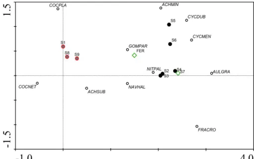
22 Figure 2: Results of DCA analysis of the relationship between the sampling sites and the corresponding species of diatoms. Red line circles are marine samples, black circles are estuaries samples and green rhombs are river samples and the empty circle is the diatom’s species
Based on the distribution of the diatom samples along the first axis of DCA (detrended correspondence analysis) depicted in figure 1, which can be interpreted as corresponding to a salinity gradient, it is possible to understand that there is a clear separation between marine diatoms to brackish and fresh diatoms, although the same does not happen between fresh and brackish points. The two samples taken from rivers could not be distinguished from those taken from estuaries on the basis of diatom composition. Increasing the sample size from rivers could help in such distinction. In figure 2, we have the results of DCA analysis of the relationship between the sites and the species of diatoms that characterize them, being possible to assert that: the sites nº 2 and nº 3 have a high affinity to the Nitzschia palea species; the Ferreira River has a high affinity to the Gomphonema parvulum species; the site nº 5 and nº 6 have affinity to Cyclostephanos dubius e Cyclotella meneghiniana respectively; and the marine sites have a relation with Cocconeis placentula and
Cocconeis neothumensis species.
Figure 1: Results of DCA analysis of all the sites according with the diatoms identification. Red line circles are marine samples, black circles are estuaries samples and green rhombs are river samples
23
Thirteen genera were represented by just one species of diatoms. Another twenty genera had a number less or equal to five species of diatoms. Six genera had more than ten species, such as the genera Navicula (46 species), Nitzschia (23),
Achnanthes (18) and Fragilaria (17). Fragilaria genus represent 16.73% of the total
diatoms observed, followed by Navicula (12.93%), Achnanthes (12.69%),
Cyclostephanos (11.18%), Cyclotella (11.10%), Cocconeis (9.08%) and Nitzschia (7.42%).
Genus Nº of taxa % RA? Achnanthes 18 12,69% Actinocyclus 2 0,24% Actinoptychus 1 0,67% Amphora 6 0,68% Anomoeoneis 1 0,013% Anorthoneis 1 0,013% Asterionella 1 1,66% Asteromphalus 1 0,051% Aulacoseira 3 4,51% Bacillaria 1 0,65& Caloneis 2 0,052% Chaetoceros 1 0,026% Cocconeis 7 9,08% Coscinodiscus 1 1,06% Cyclotella 9 11,10% Cyclostephanos 2 11,18% Cymbella 14 1,42% Diatoma 4 0,50% Dimeregramma 2 0,026% Diploneis 6 0,25% Epithemia 3 0,04% Eunotia 4 0,51% Genus Nº of taxa % RA% Fragilaria 17 16,73% Frustulia 2 0,065% Gomphoneis 2 0,104% Gomphonema 13 2,73% Gyrosigma 2 0,052% Hantzschia 1 0,065% Meridion 2 0,052% Navicula 46 12,93% Neidium 2 0,039% Nitzschia 23 7,42% Opephora 2 0,98% Paralia 1 0,013% Pinnularia 10 0,29% Pleurosigma 2 0,026% Rhaphoneis 1 0,013% Rhoicosphenia 2 0,19% Stauroneis 4 0,039% Stephanodiscus 2 0,14% Surirella 5 0,73% Tabellaria 2 0,84% Thalassionema 1 0,091% Toxonidea 1 0,026%
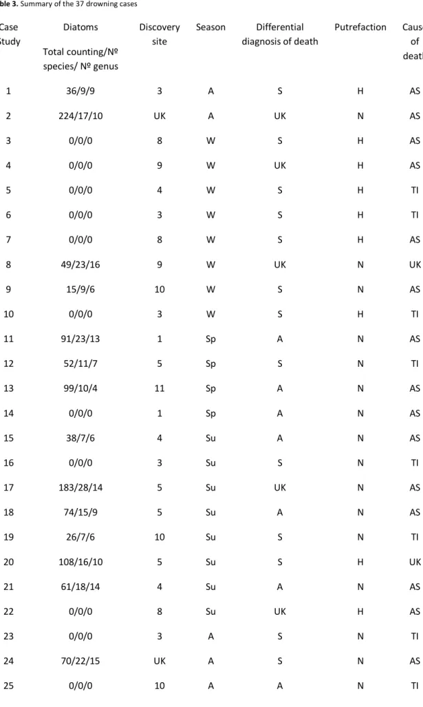
24 Table 3. Summary of the 37 drowning cases
Case Study Diatoms Total counting/Nº species/ Nº genus Discovery site Season Differential diagnosis of death Putrefaction Cause of death 1 36/9/9 3 A S H AS 2 224/17/10 UK A UK N AS 3 0/0/0 8 W S H AS 4 0/0/0 9 W UK H AS 5 0/0/0 4 W S H TI 6 0/0/0 3 W S H TI 7 0/0/0 8 W S H AS 8 49/23/16 9 W UK N UK 9 15/9/6 10 W S N AS 10 0/0/0 3 W S H TI 11 91/23/13 1 Sp A N AS 12 52/11/7 5 Sp S N TI 13 99/10/4 11 Sp A N AS 14 0/0/0 1 Sp A N AS 15 38/7/6 4 Su A N AS 16 0/0/0 3 Su S N TI 17 183/28/14 5 Su UK N AS 18 74/15/9 5 Su A N AS 19 26/7/6 10 Su S N TI 20 108/16/10 5 Su S H UK 21 61/18/14 4 Su A N AS 22 0/0/0 8 Su UK H AS 23 0/0/0 3 A S N TI 24 70/22/15 UK A S N AS 25 0/0/0 10 A A N TI
25 26 10/6/5 10 A UK H UK 27 0/0/0 4 A UK H AS 28 102/13/6 10 A S H AS 29 5/2/2 UK W S N AS 30 13/7/7 UK W S H AS 31 5/3/2 UK W UK N AS 32 30/13/12 UK W A N AS 33 40/17/14 8 W UK N AS 34 0/0/0 4 W S N TI 35 11/7/6 UK W A N AS 36 0/0/0 10 W A N AS 37 3/3/3 10 W UK N AS
Discovery site: 1- Luz beach, 2- Delta of the of estuary, 3- Downstream Arrábida bridge, 4- Downstream Luis I bridge, 5- Areinho beach, 6- Arnelas beach, 7- upstream the dam of Crestuma, 8- Póvoa de Varzim beach, 9- Titan beach in Matosinhos, 10- Ferreira River, 11- Private lake in Arcozelo and 12 - unknown; Season: W- Winter, Sp- Spring, Su - Summer, A - autumn; Differential diagnosis of death: S - Suicide, H - Homicide, A - Accident and UK- Unknown; Putrefaction: H- High, N- Normal, L- Low; Cause of death: TI- Traumatic injuries, AS - Asphyxia by drowning, UK - Unknown
26 Cases Total
Countin g of Diatoms
The species, number and organ where it was found the Diatoms.
Corresponding with the discovery
site* Right Lung Left Lung stomach contents Other organs
1 36 Achnanthes clevei (2) Aulacoseira distans (2) Cocconeis placentula (3) Cyclotella meneghiniana (1) Cyclostephanos dubius (5) Fragilaria fasciculata (15) Navicula duerrenbergiana (3) Nitzschia palea (4) Stauroneis phoenicenteron (1) None none No No Yes Yes Yes Yes No Yes No 2 224 Epithemia adnata (4) Fragilaria pinnata (1) Stauroneis kriegerii (1) Achnanthes conspicua (4) Achnanthes lanceolata (2) Achnanthes minutissima (7) Cyclotella meneghiniana (8) Epithemia adnata (11) Amphora pediculus (1) Cyclotella meneghiniana (4) Epithemia adnata (2) Epithemia sorex (3) Fragilaria crotonensis (6)
none It wasn't possible
27 Epithemia sorex (9) Fragilaria crotonensis (4) Frafilaria eliptica (25) Fragilaria pinnata (11) Navicula capitata (1) Navicula cuspidata (1) Navicula cryptotenella (7) Nitzschia inconspicua (1) Nitzschia palea (20) Pinnularia viridis (4) Stauroneis kriegerii (5) Stauroneis phoenicenteron (2) Frafilaria eliptica (42) Gomphonema accuminatum (2) Navicula cryptotenella (2) Navicula pygmaea (1) Nitzschia inconspicua (4) Nitzschia palea (23) Pinnularia viridis (2) Stauroneis phoenicenteron (3) 8 49 Achnanthes clevei (2) Achnanthes oblongella (1) Amphora coffeaeformis (2) Astrionella formosa (1) Bacilaria paradoxa (2) Bacilaria paradoxa (1) Cocconeis scutellum (2) Cyclotella meneghiniana (2) Cyclotella ocellata (1) Cymbella minuta (1)
None none Yes
Yes No No No No No Yes No No
28 Climaconeis fasciculata (1) Cocconeis neothumensis (2) Cocconeis placentula (1) Cocconeis scutellum (3) Cyclotella ocellata (2) Cymbella minuta (2) Epithemia adnata (1) Fragilaria crotonensis (2) Frafilaria eliptica (3) Fragilaria pinnata (1) Gomphonema rhombicum (1) Navicula lesmonensis (1) Nitzschia constricta (1) Nitzschia parvula (1) Tabellaria floculosa (3) Thalassionema nitzschioides (1) Frafilaria eliptica (4) Pinnularia appendiculata (1) Tabellaria floculosa (2) No Yes Yes Yes No No No Yes No No No No No Yes Yes No No Yes Yes No
29 Cocconeis scutellum (1) Fragilaria crotonensis (1) Fragilaria ulna (1) Frustulia vulgaris (2) Gomphonema truncatum (1) Nitzschia palea (2) Pinnularia viridis (1) No No No No Yes Yes Yes 11 91 Achnanthes oblongella (1) Amphora sp. (1) Bacilaria paradoxa (1) Cocconeis placentula (2) Cyclotella meneghiniana (1) Cyclotella ocellata (1) Navicula menisculos (1) Bacilaria paradoxa (3) Cocconeis scutellum (2) Cyclotella meneghiniana (1) Actinoptychus senarius (3) Aulacoseira distans (3) Cyclostephanos dubius (25) Cyclotella steligera (1) Cyclostella meneghiniana (23) Diploneis interrupta (1) Fragilaria capucina (1) Frafilaria eliptica (7) Frustulia vulgaris (1) Frustulia rhomboides (1) none No No No Yes Yes No No No Yes Yes No Yes Yes No Yes No Yes No Yes No
30 Gomphonema rhombicum (1) Navicula cuspidata (1) Navicula lesmonensis (1) Navicula pygmaea (3) Nitzschia constricta (1) Nitzschia parvula (3) Nitzschia sigmoidea (1) No No No No No No No 12 52 Cocconeis placentula (1) Cyclotella meneghiniana (5) Cyclotella steligera (1) Cyclostephanos dubius (15) Cyclotella meneghiniana (5) Cyclostephanos dubius (6) Achnanthes brevipes (1) Achnanthes lanceolata (1) Climaconeis fasciculata (1) Cocconeis placentula (10) Cocconeis scutellum (3) Fragilaria capucina (1) Fragilaria fasciculata (1) Nitzschia constricta (1) None Yes Yes No Yes Yes Yes No Yes No Yes No No No No 13 99 Frafilaria eliptica (20) Gomphonema parvulum (2) Fragilaria capucina (2) Frafilaria eliptica (35) Achnanthes lanceolata (1) Achnanthes minutissima (2) None Yes No Yes Yes Yes Yes
31 Achnanthes oblongella (1) Achnanthes subatomoides (2) Cyclotella meneghiniana (1) Fragilaria capucina (2) Frafilaria eliptica (23) Fragilaria pinnata (1) Gomphonema gracile (4) Gomphonema parvulum (3) Yes No No Yes Yes No No No 15 38 Cyclotella meneghiniana (1) Cyclostephanos dubius (4) Aulacoseira granulata (1) Cocconeis placentula (1) Cyclotella meneghiniana (1) Cyclostephanos dubius (14) Fragilaria capucina (1) Fragilaria crotonensis (5) Cyclotella meneghiniana (1) Cyclostephanos dubius (6) Fragilaria crotonensis (2) Gomphonema parvulum (1) None Yes Yes No Yes Yes Yes Yes Yes Yes Yes Yes Yes 17 183 Achnanthes delicatula (2) Achnanthes lanceolata (1) Achnanthes subatomoides (1) Achnanthes delicatula (2) Achnanthes lanceolata (6) Achnanthes minutissima (5) Cyclotella meneghiniana (2) Cyclostephanos dubius (2) Fragilaria crotonensis (4) None Yes Yes No Yes Yes Yes Yes Yes Yes
32 Cocconeis placentula (3) Cyclotella meneghiniana (4) Cyclostephanos dubius (18) Cymbella minuta (1) Eunotia bilunaris (1) Fragilaria capucina (1) Fragilaria crotonensis (11) Fragilaria pulchella (1) Navicula capitata (1) Navicula leptostriata (3) Navicula pygmaea (1) Nitzschia palea (1) Nitzschia sigmoidea (2) Achnanthes subatomoides (3) Aulacoseira granulata (1) Cocconeis placentula (4) Cyclotella meneghiniana (9) Cyclostephanos dubius (48) Cymbella minuta (3) Fragilaria arcus (1) Fragilaria capucina (3) Fragilaria crotonensis (13) Fragilaria pulchella (1) Fragilaria ulna (1) Frustulia rhomboides (1) Gomphonema gracile (1) Gomphonema parvulum (7) Gomphonema rhombicum (1) Navicula capitata (3) Navicula crytocephala (1) Yes Yes Yes No No Yes Yes No Yes No Yes Yes Yes No No Yes Yes Yes No No Yes Yes No Yes No Yes No Yes No Yes
33 Navicula lesmonensis (1) Navicula leptostriata (1) Navicula menisculos (1) Nitzschia palea (2) Pleurosigma angulatum (1) Tabellaria floculosa (2) No Yes Yes No No 18 74 Achnanthes brevipes (1) Amphora pediculus (1) Aulacoseira distans (1) Cyclotella meneghiniana (3) Cyclostephanos dubius (16) Fragilaria crotonensis (16) Navicula halophila (3) Navicula pygmaea (2) Nitzschia constricta (1) Nitzschia palea (1) Achnanthes hintzii (1) Cyclotella meneghiniana (1) Cyclostephanos dubius (9) Fragilaria crotonensis (5) Frafilaria eliptica (9) Fragilaria fasciculata (1) Fragilaria ulna (2) Gomphonema angustum (1) none None No Yes Yes Yes Yes Yes No Yes Yes Yes Yes Yes Yes Yes No No Yes Yes
34 19** 26 Cyclotella steligera (1) Navicula halophila (1) Amphora coffeaeformis (1) Cocconeis scutellum (4) Cyclotella steligera (7) Fragilaria capucina (8) Gomphonema herculeana (1) Navicula cryptotenella (1) Navicula halophila (2) None None No No No No No Yes No No No 20 108 Achnanthes hintzii (4) Achnanthes minutissima (1) Aulacoseira distans (2) Cocconeis neothumensis (1) Cyclotella meneghiniana (13) Cyclostephanos dubius (15) Cymbella gracilis (1) Cymbella hillardii (1) Fragilaria crotonensis (14) Fragilaria fasciculata (6) Achnanthes delicatula (1) Cyclotella meneghiniana (4) Cyclostephanos dubius (13) Eunotia minor (1) Fragilaria crotonensis (12) Navicula pygmaea (1) Nitzschia constricta (1) Achnanthes hintzii (1) Cyclotella meneghiniana (4) Cyclostephanos dubius (6) Fragilaria crotonensis (1) None Yes Yes Yes No Yes Yes No No Yes No Yes Yes Yes No Yes Yes Yes Yes Yes Yes
35 Navicula menisculos (4)
Nitzschia palea (1)
Yes Yes 21 61 Cyclostephanos dubius (7) Achnanthes clevei (1)
Actinoptychus senarius (1) Astrionella formosa (3) Aulacoseira distans (1) Bacilaria paradoxa (1) Cocconeis placentula (1) Cocconeis scutellum (3) Cyclotella species (3) Cyclostephanos dubius (18) Eunotia minor (1) Fragilaria fasciculata (1) Gomphonema olivaceum (1) Navicula capitata (1) Navicula cryptotenella (1) Navicula halophila (1) Cocconeis placentula (3) Cocconeis scutellum (1) Cyclostephanos dubius (8) None Yes No Yes Yes No Yes Yes No No Yes No Yes No Yes Yes Yes Yes No Yes
36 Navicula pygmaea (1) Nitzschia palea (2) Tabellaria floculosa (1) No Yes No 24*** 70 Achnanthes clevei (2) Achnanthes hintzii (2) Actinoptychus senarius (1) Aulacoseira distans (5) Cocconeis placentula (9) Cyclotella meneghiniana (2) Cyclostephanos dubius (4) Epithemia adnata (2) Fragilaria virescens (1) Navicula pygmaea (1) Nitzschia palea (1) Thalassionema nitzschioides (1) Achnanthes brevipes (1) Achnanthes clevei (1) Achnanthes hintzii (2) Achnanthes lanceolata (1) Achnanthes minutissima (1) Actinoptychus senarius (1) Amphora coffeaeformis (2) Asterophalus sp. (1) Astrionella formosa (1) Aulacoseira distans (6) Cocconeis placentula (12) Cyclotella meneghiniana (2) Cyclostephanos dubius (2) Epithemia adnata (1)
None None Yes
Yes Yes Yes Yes Yes Yes No No No Yes No Yes Yes Yes Yes No Yes No Yes Yes Yes Yes No No No
37 Eunotia minor (1) Fragilaria arcus (1) Fragilaria pinnata (1) Navicula arenaria (2) No No 26 10 Achnanthes clevei (1) Achnanthes minutissima (2) Cymbella amphicephala var hercynica (1)
Eunotia minor (3)
Gomphonema angustum (2) Stauroneis kriegerii (1)
None None None No
Yes No Yes No Yes 28** 102 Achnanthes oblongella (1) Achnanthes subatomoides (3) Frafgilaria construens (1) Gomphonema parvulum (1) Navicula clementis (1) Pinnularia gibba (2) Achnanthes lanceolata (1) Achnanthes minutissima (1) Achnanthes oblongella (1) Achnanthes subatomoides (3) Gomphonema parvulum (3) Gomphonema truncatum (1) Achnanthes lanceolata (4) Achnanthes subatomoides (1) Fragilaria capucina (1) Navicula cryptotenella (1) None Yes Yes No Yes Yes No Yes Yes Yes Yes Yes Yes Yes Yes Yes No
38 Navicula clementis (2) Navicula halophila (1) Nitzschia palea (1) Pinnularia gibba (1) Yes No Yes No 29*** 5 Achnanthes minutissima (3) Aulacoseira distans (2)
None None None Yes
Yes 30*** 13 Achnanthes lanceolata (1) Actinoptychus senarius (2) Cocconeis placentula (1) Cyclotella meneghiniana (1) Cyclostephanos dubius (3) Fragilaria crotonensis (1) Cocconeis placentula (1) Cyclostephanos dubius (1) Fragilaria crotonensis (1) Navicula pupula (1)
None None Yes
Yes Yes Yes Yes Yes Yes Yes Yes Yes
31*** 5 Fragilaria crotonensis (2) Fragilaria fasciculata (1) Nitzschia palea (1)
None None Yes No
Yes 32*** 30 Amphora pediculus (1) Aulacoseira distans (2) Cyclotella meneghiniana (1) Achnanthes clevei (1) Actinoptychus senarius (2) Aulacoseira distans (3) Aulacoseira distans (1) Cyclostephanos dubius (1) Opephora pacifica (1) None Yes Yes Yes Yes Yes Yes Yes Yes Yes
39 Cyclostephanos dubius (1) Diploneis interrupta (1) Fragilaria crotonensis (3) Fragilaria fasciculata (1) Opephora pacifica (1) Cyclostephanos dubius (2) Diatoma mesodon (1) Fragilaria crotonensis (3) Navicula minima (1) Opephora pacifica (1) Thalassionema nitzschioides (2) Yes No Yes No Yes Yes No No Yes No 33 11 Actinoptychus senarius (4) Amphora pediculus (1) Astrionella formosa (1) Aulacoseira distans (2) Cocconeis placentula (1) Cyclotella steligera (1) Fragilaria capucina (3) Frafilaria eliptica (8) Gomphonema parvulum 82) Nitzschia palea (2) Tabellaria floculosa (1) Achnanthes minutissima (1) Astrionella formosa (1) Aulacoseira distans (1) Cyclotella ocellata (1) Cyclotella steligera (2) Cyclostephanos dubius (2) Fragilaria bidens (1) Fragilaria capucina (3) Navicula rhynchocephala (1) Opephora pacifica (1) None None No No Yes No Yes No Yes No Yes Yes No Yes Yes No No No Yes No Yes No No
40 * The corresponding to the discovery site had made by the specific seasonal database.
** This case occurs in Summer or Autumn although the water sample collection was made only once in the Winter.
*** In this case we made the comparison with marine sites, because there is strong evidence that drowning had occurred at the sea. 35*** 3 Achnanthes minutissima (1) Achnanthes subatomoides (1) Navicula halophila (1) Nitzschia palea (2) Nitzschia sigmoidea (2) Achnanthes minutissima (1) Cymbella minuta (1) Navicula halophila (1) Pinnularia gibba (1)
None None Yes
Yes Yes Yes No Yes No Yes No 37 Achnanthes lanceolata (1) Navicula halophila (1) Stauroneis kriegerii (1)
None None Yes
No Yes
41
We performeda seasonal water sample collection and analyzed the distribution of the diatoms according to the sampling sites and the corresponding drowning cases that occurred in those periods.
In the Winter
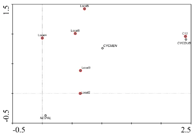
In Figure 3, the DCA analysis shows the relationship between the known cases of drowning, the water samples collected during the winter and the diatom species that caracterize each site’s sample. According to the distribution of the diatom samples along the second axis of DCA represented in Figure 3, this axis can be interpreted as corresponding to the salinity gradient, by the fact that local 2 is nearest to the sea and local 6 is nearest to the river. We can find an affinity of C31 with the site nº 3, for C30 we observed the definition for Cyclostephanos dubius species. In Figure 1, we defined that this species is associated to local 5 so it is possible that C30 occured near site 5.
In DCA analysis of cases and water samples that were collected in winter time, it was not possible to observe a difference between brackish and fresh water as we analyze the distribution by the salinity gradient in axis 2. It is possible to correlate the case C33 to the local 1 (marine site), C8 to the local 4 (brackish site) and C30 to the Local 7 (fresh site). We observed a high affinity between C31 and the species Fragilaria
crotonensis, C30 and Cyclostephanos dubius, C9 and Achnanthes minutissima, C37 and Navicula halophila/ Achnanthes lanceolata, Local 8 and Cocconeis placentula,
Figure 3: Results of DCA analysis of estuary samples and case samples at winter time. Red line circles are the case samples, black circles are the diatom species
42 Figure 4: Results of DCA analysis of case samples, water samples and diatoms species that occur in winter time. Red line circles are marine cases and water samples, black circles are estuaries’ cases and water samples, green rhombs are river cases and water samples, the yellow line are the cases with unknown occurrence place and the black empty circle lines are the diatoms’ species
43
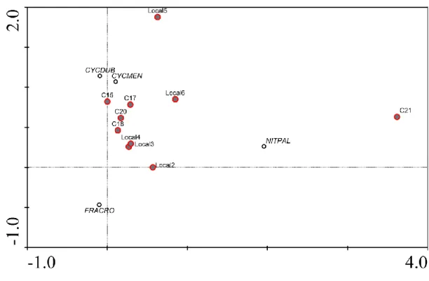
In the Spring
In Figure 5, the DCA analysis shows the relationship between the cases of drowning and the water samples collected, both in estuaries. The case C12 has a high affinity with Cyclostephanos dubius.
Figure 5: Results of DCA analysis of estuary’s samples and case samples that occur in spring time. Red line circles are the samples, black circle are the diatom species
44
In Figure 6, the results of DCA analysis show that the water samples are organized in the second axis by the salinity gradient, so we can observe that Cocconeis
placentula species have a higher affinity to the marine water samples. The case C11, in
spite of being a marine case, appeared to be closer to brackish samples than marine ones. Also, C11 had a high correlation with Cyclostephanos dubius. The sites 2 and 3 had a high affinity with the species Nitzschia palea and Cyclotella meneghiniana. The specific case C13 corresponded to Arcozelo site and, in the Figure 6, it is clear that they have a corresponding principal species who related that site with Fragilaria elliptica.
Figure 6: Results of DCA analysis of cases, water samples and diatoms species connection that occur with them in spring time. Red line circles are marine cases and water samples, black circles are estuaries cases and water samples, green rhombs are river cases and water samples and the yellow line is the case and the site related to Arcozelo place
45
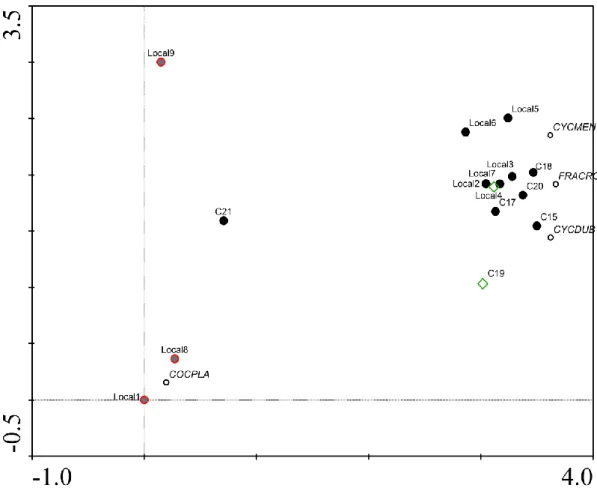
In the Summer
In Figure 7, the DCA analysis exposes the relationship between the cases of drowning in estuaries and water samples collected in the estuaries during the summer. The cases C15, C17, C18 and C20 are included around the sites 4 and 6, this cases have affinity with Cyclostephanos dubius and Cyclotella meneghiniana.
Figure 7: Results of DCA analysis of estuaries’ samples and cases’ samples that occur in summer time. Red line circles are the samples, black circles are the diatoms species
46
In DCA analysis of cases and water samples that were collected in summer time there are no differences between brackish and fresh water, and we can observe the distribution by the salinity gradient in axis 1. It is possible to correlate the site 5 with
Cyclotella meneghiniana, the site 3 is related with cases 18 and 20 and the diatom
species that characterize them is Fragilaria crotonensis. The site 4 is similar to C17, the case C15 is characterized by Cyclostephanos dubius and the marine sites 1 and 8 are characterized by Cocconeis placentula.
Figure 8: Results of DCA analysis of cases, water samples and diatoms species connection that occur with them in summer time. Red line circles are marine cases and water samples, black circles are estuaries cases and water samples, green rhombs are river cases and water samples
47
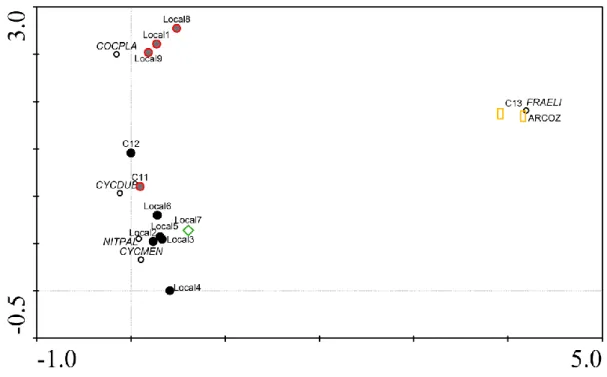
In the Autumn
In Figure 9, the DCA analysis shows the relation between the cases of drowning in estuaries with the water samples collected in estuaries during Autum. It was possible to observe that the case C24 had an affinity to Cocconeis placentula species, the case C1 had an affinity to Fragilaria fasciculata, the site 3 showned high affinity to
Cyclostephanos dubius and Cyclotella meneghiniana. The site 6 was defined by Fragilaria crotonensis and Aulacoseira granulta.
Figure 9: Results of DCA analysis of estuary samples and case samples that occur in autumn time. Red line circles are the water samples, black circles are the diatoms species
48
In DCA analysis of cases and water samples that occurred in autumn time there was no difference between brackish and fresh water, but we can see a difference between the fresh and marine samples. We can observe the distribution by the salinity gradient in axis 2. It was possible to correlate the site 9 with Achnanthes brevipes, the site 6 and 7 with Fragilaria crotonensis, the site 3 with Nitzschia palea, the site 1 with
Cocconeis placentula. The case C28 is correlated with Achnanthes subatomoides, which
is a characteristic species of fresh waters, the case C26 with Eunotia minor, also characteristic of fresh waters and the case C1 with Fragilaria fasciculata.
Figure 10: Results of DCA analysis of the relation between cases, water samples and diatoms species that occur in autumn time. Red line circles are marine cases and water samples, black circles are estuaries cases and water samples, green rhombs are river cases
49
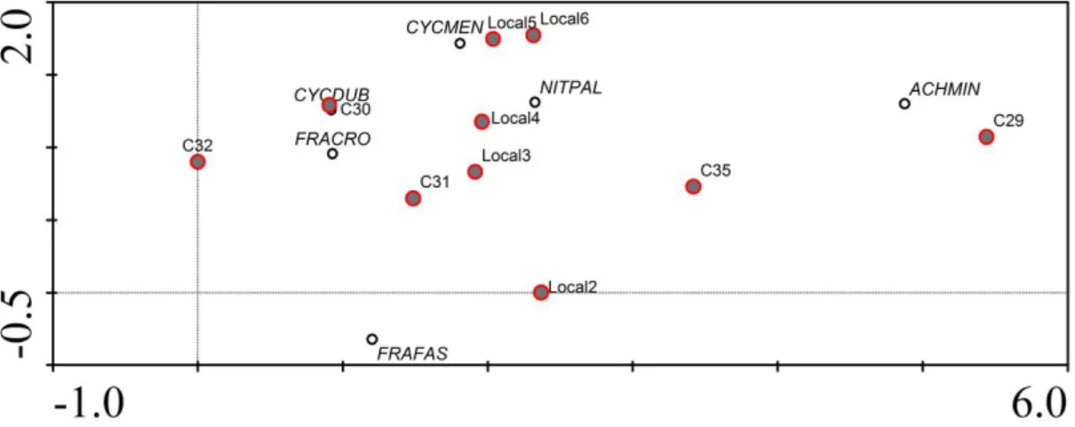
All seasons
In Figure 11 of DCA analysis on the cases and the correlation with diatoms species, it is possible to observe that the case C8 was very similar to C30. The first case occurred in a marine medium and the second in an unknown place. The case C11 had a high resemblance to Cyclotella meneghinia, which seems to be incoherent because the case C11 occurred in a marine site and this species is naturally from fresh water. The case C15 had an affinity with Fragilaria exigua and Cyclotella species, this case occurred in a brackish place and this species are characteristic of fresh water. The case C21 had an affinity with Fragilaria fasciculate; This case occurred in brackish waters and this species is characteristic of fresh waters. The case C24 had parity to Cocconeis
placentula and Aulacoseira distans. In this case, we had no information where it
occurred but these species are defined as marine. The case C29 had an affinity to
Achnanthes minutissima species, which are defined as belonging to fresh waters so
C29 probably occurred in a fresh water medium.
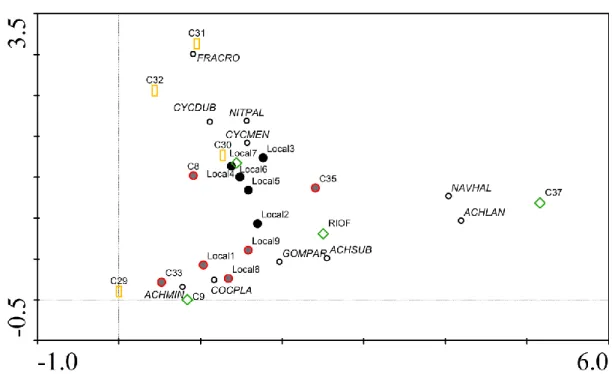
In Figure 12 of DCA analysis on the cases, the sites and the diatoms’ species, it was possible to observe that cases C24, C31, C32 were near to the sites 1 and 8, (marine sites), the cases C8 and C30 were near to the site S9 (a marine site) and the cases C26 and C28 were similar to the composition of diatoms of Ferreira river.
50 Figure 11: Results of DCA analysis of cases and diatoms species correlation that occurs during a year of water samples collection. Red line circles are marine cases, black circles are estuaries cases, green rhombs are river cases, the yellow rectangles are unknown cases and the black empty lines are diatoms species
51 Figure 12: Results of DCA analysis of cases and water samples and diatoms species correlation that occurred during a year. Red line circles are marine cases, black circles are estuaries cases, green rhombs are river cases, the yellow rectangles are unknown cases and the red circle is the Arcozelo site
Figure 13: Results of DCA analysis of cases, water samples and diatoms species correlation that occurred during a year. Red line circles are marine cases, black circles are estuaries cases, green rhombs are river cases, the yellow rectangles are unknown cases and the red circle is the Arcozelo site
53
e) Discussion
In our study, we had 37 drowning cases and in 63% of those it was possible to find diatoms (Table 4).
- Drowning in estuary sites
In the analysis of the cases in which the drowning was known to have occurred in estuaries sites, we found C1, C15, C17, C18 and C21.
Case C1 occurred in autumn. Considering the family and police information we knew that the drowning occurred in site 3 and, according to the analysis of Figures 9 and 10, the species Fragilaria fasciculata characterized this case. These species normally exist in fresh waters. By analyzing table 4, there are four species that don't have correspondence to this site, although we found this species in the sites around site 2 and 1. In Figure 12, it is clear that this case was similar to the composition of the estuary samples.
Cases C15, C17 and C18 were close to each other and all cases occurred during summer. According to the family and police information, the drowning of C15 occurred in site 4 and the other two occurred in site 5. The species that characterized these cases are Cyclostephanos dubius, Fragilaria crotonensis, Cyclotella species and
Achnanthes lanceolata. All of them had affinity to the estuaries samples. Through the
analysis of table 4, we found C15 and C18 to have a total correspondence of species with the ones found in site 4 and 5 respectively, for the case C17 the species that didn't had corresponding to the site 5, were included in site 6.
Case C21 occurred in summer and, according to the family and police information, the drowning occurred near site 4. In Figure 8, this case was not associated with any location and, in the analysis of all seasons, Figures 11 and 13, this case had high association to the species Fragilaria fasciculata, characteristic from fresh waters. According to table 4 the species Cyclostephanos dubius is the one with a higher expression. The species that were not associated on this site are present in marine sites.
- Drowning in river sites
In the analysis of the cases in which the drowning was known to have occurred in river sites, we identified C9 and C28.
The case C9 occurred during winter. In Figure 4, this case was very similar to marine samples and with a high affinity to Achnanthes minutissima. Although we saw in Figure 12 that this case is very similar to case C21 (estuary case), in Figure 13 the species most similar is Achnanhtes lanceolata. Both are typical of fresh waters.
54
Case C28 occurred in summer. According to Figure 8, Achnanthes subatomoides is characteristic. In Figure 12 this case is very similar to the samples taken from Ferreira river, and in Figure 13 the species most distinguishable are Cyclotephanos dubius and
Achnanthes subatomoides, both characteristic of fresh waters. This confirms that, in
this case, the body was recovered from a river and, in the samples analyzed, we found species from the river. In table 4 there are some species that don't have correspondence, although this can be explained by the absence of samples taken during summer, given that, in Ferreira river, we just took a sample in the winter.
- Drowning in an unknown specific site
In this study we had 6 cases (C24, C29, C30, C31, C32 and C35) with an unknown specific location of death, but we tried to define a possible location, by the analysis of diatoms species.
The case C24 occurred in autumn, the body was recovered near the mouth of the estuary and, by the examination of the Figure 10, it is possible to see that local 1 is very similar to the species found in this case. So, it is possible to say that the drowning happened, in the sea. To support this, the species that characterized this case are
Aulacoseira distans and Cocconeis placentula (Figure 11), both species specific of sea
waters. With the analysis of table 4, we compared to local 1. In a total of 22 species identified, 10 had no corresponding to site 1, although 4 of them were found in marine sites.
The case C29 occurred in the winter. The body was recovered in the Cabedelo beach, in sea water, and it is possible to observe, in Figure 4, that Achananthes
minutissima is the species that characterized this case. This specie is found in fresh
waters. In Figure 12, it is possible to see that this case appeared near the other cases related to fresh and brackish waters.
In case C30, the body was recovered in Sindicato beach in Valadares. In Figure 3, there’s an affinity between this case and the species Cyclotephanos dubius and
Cyclotella meneghiniana, both characteristic from fresh waters. In Figure 4 we
observed that this case was very similar to site 7 (river sample). However, when we looked at the analysis of all seasons, we saw that C30 was almost equal to C8 (Figure 11), which was a marine case. Nevertheless, in Figure 12, case C30 was near C8 and estuaries sites. The exact location were the death occurred can not be defined precisely by the analysis of the graphics. If we observe table 4, we find complete matches comparing the species found in this case and the one found in site 1, mostly close to Sindicato beach.
Cases C31 and C32 were both corpses recovered from the beach (sea water) and they were very close. In Figure 4, we observed that they are represented by the