Mestrado Integrado em Medicina Veterinária
Relatório de Estágio
Évora, 2019
Este relatório inclui as críticas e as sugestões feitas pelo júri
Orientação | Prof. Doutora Catarina Lavrador
Dr. Steve Carter
ESCOLA DE CIÊNCIAS E TECNOLOGIA
DEPARTAMENTO DE MEDICINA VETERINÁRIA
New insights on the age for neutering
dogs
Mestrado Integrado em Medicina Veterinária
Relatório de Estágio
Évora, 2019
Este relatório inclui as críticas e as sugestões feitas pelo júri
Ana Barradas Saraiva
Orientação | Prof. Doutora Catarina Lavrador
Dr. Steve Carter
ESCOLA DE CIÊNCIAS E TECNOLOGIA
DEPARTAMENTO DE MEDICINA VETERINÁRIA
I Acknowledgements
I would like to show my appreciation and gratitude to all of those who have accompanied me throughout this accomplishment. An enormous thank you
To my parents, for their unconditional love and support, for your investment in my academic knowledge and for all the amazing experiences you were able to provide me. I hope you feel proud of the person you brought up and it is to you that I dedicate this thesis to.
To my brother, who is the greatest of friends and companions – my pride and joy. To my grandparents, who support and love me unconditionally.
To my supervisor Prof. Dr. Catarina Lavrador for her availability, encouragement and kindness, not only throughout the duration of this traineeship and thesis, but also during my academic path. To my supervisor, Dr. Steve Carter for the lifetime opportunity, for all the professional and personal knowledge and for your kindness. A great thank you to his family, especially Helen and Alfred, who made me feel at home since the very first day in the United Kingdom.
To all the team of Priory Vet Surgeons who taught me new work methods and made me feel very welcome, with all the laughter and biscuits! Besides being an essential support, you were a family away from home. A very big thank you to Kathryn who believed in my surgical skills, to Rebecca who was a constant inspiration, Beth who is the sweetest and neatest surgeon I have ever known, Dermot for letting me get my hands dirty, Adrian for all his book recommendations and Brendan for sharing the beginning of this journey. To April, Chris, Emma, Georgia, Eve, Laura A., Laura H., Lauren and Sam for being the best nurses, thank you for all your patience and joyful mood. To Dr. Maria João Carvalho, for being a role model and for teaching me since day one.
To my dearest friends, Aninhas, Joana, José, Gonçalo, Maria and Teresa for always being there. To all my academic family, with whom I shared the last six years of this veterinary course in laughter. A special thank you goes to Tita, Daniela, Carolina, Bárbara, Carina, Andreia, Vânia, Beatriz, Rodrigo, André, Fábio, Julieta, Ana and Margarida.
To Bel, for all his kindness, comfort and love.
II Abstract
The current report was carried out for the completion of the Master’s degree in veterinary Medicine and is divided into two parts. The first part is formed by a description of the cases followed during the four-month traineeship completed at Priory Veterinary Surgeons. The most prevalent medical field was dermatology, with a relative frequency of 26% (n= 474), followed by gastroenterology representing 18% of total cases. The second part is composed of a review of the current literature on the proposed benefits and detriments of gonadectomy in dogs and whether performing it at a specific age causes or prevents specific health issues.
Keywords: canine, gonadectomy, pre-pubertal neutering, post-pubertal neutering, benefits, detriments
Resumo - Novas perspetivas quanto à idade de esterilização em cães
O presente relatório foi realizado no âmbito do Mestrado Integrado em Medicina Veterinária e está dividido em duas partes. A primeira parte é constituída por uma descrição da casuística observada durante o estágio curricular de quatro meses realizado no Priory Veterinary
Surgeons. A área mais prevalente foi dermatologia, com uma frequência relativa de 27% (n=480),
seguida da gastroenterologia, que representou 18% dos casos totais. Na segunda parte é realizada uma revisão da literatura atual sobre os benefícios e malefícios inerentes à gonadectomia em cães e se a sua realização em determinada idade específica causa ou impede problemas de saúde específicos.
Palavras-chave: cão, gonadectomia, esterilização pré-pubertal, esterilização pós-pubertal, benefícios, malefícios
III
Table of contents
Table of contents ... III
List of tables ... V
List of graphics ... VII
List of figures ... VII
List of acronyms and abbreviations ... VIII
Part I: Report on activities performed during the traineeship ... 1
1. Introduction ... 1
2. Case-by-case description ... 1
2.1. Case distribution regarding animal species ... 1
2.2. Case distribution regarding clinical area ... 2
2.2.1. Preventive medicine ... 2
2.2.2. Internal Medicine ... 6
2.2.2.1. Cardiology and Pneumology ... 7
2.2.2.2. Dermatology ... 11
2.2.2.3. Endocrinology ... 13
2.2.2.4. Gastroenterology and annexes ... 15
2.2.2.5. Immunology ... 17
2.2.2.6. Infectology and parasitology... 20
2.2.2.7. Musculoskeletal system ... 21
2.2.2.8. Neurology ... 24
2.2.2.9. Nephrology and Urology ... 25
2.2.2.10. Oncology ... 28
2.2.2.11. Ophthalmology ... 30
2.2.2.12. Theriogenology & Neonatology ... 32
2.2.2.13. Toxicology ... 34
2.2.3. Surgery ... 35
2.2.3.1. Soft tissue surgery ... 35
2.2.3.2. Minimally invasive surgery ... 38
2.2.3.3. Orthopaedic surgery ... 38
2.2.4. Other procedures ... 39
IV
1. Introduction ... 41
2. Defining pre-pubertal neutering ... 41
3. Surgical Techniques ... 42
3.1. Closed versus open orchiectomy ... 42
3.2. Laparoscopic ovariectomy (LOV) versus open ovariohysterectomy (OVH) ... 43
4. Elective gonadectomy indications ... 46
4.1. Societal benefits ... 46
4.2. Medical indications in males ... 46
4.2.1. Benign prostatic hyperplasia ... 47
4.2.2. Prostatitis ... 47
4.2.3. Prostatic cavitary lesions - abscesses and cysts ... 49
4.2.4. Cryptorchidism ... 50
4.2.5. Testicular tumours ... 50
4.2.6. Perianal adenomas ... 50
4.3. Medical indications in females ... 51
4.3.1. Pregnancy and parturition-related disorders ... 51
4.3.2. Mammary gland neoplasms ... 52
4.3.3. Other reproductive neoplasia ... 53
4.3.4. Pyometra ... 54
4.3.5. Pseudo-pregnancy ... 56
4.4. Behavioural Benefits ... 56
5. Risks according to the age of neutering ... 57
5.1. Early-age Gonadectomy Surgical and Anaesthetic Risk ... 57
5.1.1. Intraoperative considerations ... 57
5.1.2. Postoperative complications ... 58
5.2. Effects on metabolism/Obesity ... 58
5.3. Diabetes mellitus ... 59
5.4. Acquired urethral sphincter mechanism incompetence in neutered bitches ... 60
5.5. Musculoskeletal disorders ... 61
5.5.1. Canine hip dysplasia ... 62
5.5.2. Elbow dysplasia ... 63
V
5.6. Oncologic considerations ... 64
5.6.1. Prostatic neoplasia in neutered males ... 64
5.6.2. Transitional cell carcinoma non-prostatic ... 65
5.6.3. Osteosarcoma ... 66
5.6.4. Haemangiossarcoma ... 66
5.6.5. Lymphoma ... 67
5.6.6. Mast cell tumours ... 68
5.7. Auto-immune disorders ... 69
6. Validity of study results & Potential future research ... 70
7. Discussion ... 71
8. Conclusion ... 75
VI List of tables
Table 1: Case distribution regarding animal species and clinical area ... 2
Table 2: Case distribution regarding Preventive Medicine ... 3
Table 3: Core and non-core vaccinations and timings for canines ... 5
Table 4: Core and non-core vaccinations and timings for felines ... 6
Table 5: Case distribution regarding Internal Medicine Clinical Areas ... 7
Table 6: Case distribution regarding Cardiology ... 7
Table 7: Case distribution regarding Pneumology ... 10
Table 8: Case distribution regarding Dermatology ... 11
Table 9: Case distribution regarding Endocrinology ... 13
Table 10: FHT diagnostic groups and management options ... 14
Table 11: Case distribution regarding Gastroenterology and annexes ... 15
Table 12: Case distribution regarding Immunology... 18
Table 13: Subtypes of idiopathic immune mediated polyarthritis ... 19
Table 14: Case distribution regarding Infectology and Parasitology ... 20
Table 15: Case distribution regarding the Musculoskeletal system ... 21
Table 16: Case distribution regarding Neurology ... 24
Table 17: Case distribution regarding Nephrology ... 26
Table 18: CKD Staging criteria, adapted from IRIS, 2019 ... 27
Table 19: CKD Substaging criteria, adapted from IRIS, 2019 ... 27
Table 20: Case distribution regarding Urology ... 28
Table 21: Case distribution regarding Oncology ... 28
Table 22: Case distribution regarding Ophthalmology ... 30
Table 23: Case distribution regarding Theriogenology and Neonatology ... 32
Table 24: Case distribution regarding Toxicology ... 34
Table 25: Case distribution regarding surgical practice ... 35
Table 26: Case distribution regarding Soft Tissue Surgery ... 35
Table 27: Case distribution regarding minimally invasive surgery ... 38
Table 28: Case distribution regarding Orthopaedic Surgery ... 38
Table 29: Case distribution regarding imaging procedures ... 39
Table 30: Case distribution regarding other medical procedures ... 40
Table 31: Benefits of gonadectomy in females ... 72
Table 32: Benefits of gonadectomy in males ... 73
VII List of graphics
Graphic 1: Case distribution regarding Animal species ... 2
List of figures
Figure 1: Cranial drawer test, adapted from Fossum, 2013 ... 23 Figure 2: Tibial compression test, adapted from Fossum, 2013 ... 23 Figure 3: Ovaries' surgical approach in an open ovariohysterectomy, adapted from Tobias and Johnston, 2012 ... 44 Figure 4: Uterus’ surgical approach in an open ovariohysterectomy, adapted from Tobias and Johnston, 2012 ... 44
VIII List of acronyms and abbreviations
ACVIM: American College of Veterinary Internal Medicine
CDMVD: Chronic degenerative mitral valve disease
CEH: Cystic endometrial hyperplasia CKD: Chronic kidney disease CO2: Carbon dioxide
CVD: Central vestibular disease CVHD: Chronic valvular heart disease CRI: Constant rate infusion
CT: Computed tomography scan ECG: Electrocardiogram
FeLV: Feline Leukaemia Virus FHT: Feline hyperthyroidism Fi: Absolute frequency
FISS: Feline injection site sarcoma FIV: Feline Immunodeficiency Virus FSH: Follicle-stimulating hormone FR(%): Relative frequency in percentage IRIS: International Renal Interest Society kg: Kilograms
LH: Luteinizing hormone LOV. Laparoscopic ovariectomy
NSAID’s: Nonsteroidal anti-inflammatory drugs
MCT: Mast cell tumour
mg/dL: Milligrams per decilitre mmHg: Millimetres of mercury n: Absolute frequency
OV: Ovariectomy
OVH: Open ovariohysterectomy
PLI: Serum pancreatic lipase
immunoreactivity
PVD: Peripheral vestibular disease SDMA: Symmetric dimethylarginine T3: Triiodothyronine
T4: Thyroxine
TTA: Tibial tuberosity Advancement TB: Tuberculosis
UP/C: Urine protein:creatinine ratio
WSAVA: World Small Animal Veterinary Association
1 Part I: Report on activities performed during the traineeship
1. Introduction
The present master thesis was written following a four-month traineeship, from the beginning of September 2018 until the end of December 2018, at Priory Veterinary Surgeons, a mixed small animal and equine practice with four different branches located in Reigate, Tadworth, Redhill and Banstead, United Kingdom.
This first part includes a description of the activities developed during the traineeship, with a casuistry description accompanied by a brief bibliographical review of one of the most clinically relevant pathologies in each area.
2. Case-by-case description
In this section, the clinical cases data are processed statistically regarding preventive medicine, internal medicine, surgery and other followed diagnostic procedures.
In each of the previously mentioned areas, statistical data are presented in tables by their absolute (Fi) and relative (FR) frequencies and were studied regarding each taxonomic family (for canines and felines) and group of animals (for exotic animals). The exotic animals taken into account included guinea pigs (Cavia porcellus) and rabbits (Oryctolagus cuniculus).
Regarding these data, it is important to acknowledge that the number of cases does not correspond to the total of cases of any of the practices. The total number of animals is lower than the number of cases observed, since some patients had several co-pathologies and the same animal may have been accounted in more than one clinical area.
2.1. Case distribution regarding animal species
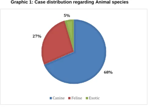
By observing the data in table 1 and respective graphic 1, a total of 1027 different animals were observed. Canines (Canis lupus familiaris) were the most frequent observed species, with 703 cases, representing 68% (n= 1027), whilst feline (Felis catus) represented 27% (n=1027), with 277 cases witnessed. When it came to exotic animals, 47 patients were observed, representing 5% (n=1027) of cases.
2 Graphic 1: Case distribution regarding Animal species
Internal medicine was the most prevalent group, with 474 cases, representing 46% (n=1027) of total cases observed. It is followed by preventive medicine, with 401 cases which is 39% (n=1027) and by surgery with 152 surgeries performed, which represents 15% (n=1027) of casuistry.
Table 1: Case distribution regarding animal species and clinical area
2.2. Case distribution regarding clinical area
2.2.1. Preventive medicine
Preventive medicine acts such as vaccination, deworming, microchipping and issuing of passports are of utmost importance as they prevent diseases (including zoonosis) and allow an epidemiological control, contributing to public health and animal health. Besides these reasons, it constitutes an important opportunity to establish effective communication between the vet and the owner.
Canine Feline Exotic
Total FR (%) Fi FR (%) Fi FR (%) Fi FR (%) Preventative Medicine 236 34% 149 54% 16 34% 401 39% Internal Medicine 369 52% 90 32% 15 32% 474 46% Surgical Medicine 98 14% 38 14% 16 34% 152 15% Total 703 100% 277 100% 47 100% 1027 100% 68% 27% 5%
3 In the United Kingdom, all dogs are legally required to have a microchip and to wear a collar with the name and the address of the owner inscribed on the collar or on a plate or badge attached to it (“The Control of Dogs Order” 1992; “The Microchipping of Dogs” 2015).
Table 2: Case distribution regarding Preventive Medicine Preventive
Medicine
Canine Feline Exotic
Total FR (%) Fi FR (%) Fi FR (%) Fi FR (%) Vaccination 157 67% 95 64% 11 69% 263 66% Worming 12 5% 10 7% 0 0% 22 5% Electronic identification 10 4% 13 9% 0 0% 23 6% 6 month health-check up 52 22% 31 21% 5 31% 88 22% Passport Issuance 5 2% 0 0% 0 0% 5 1% Total 236 100% 149 100% 16 100% 401 100%
In table 2 are enumerated the preventive acts carried out – the most frequent act was vaccination, representing 66% (n=401). Prevention and treatment against internal and external parasites is included in deworming. At Priory Veterinary Surgeons it is highly emphasized the importance of six-monthly health checks ups, and such are enumerated as well in table 1.
According to the WSAVA (World Small Animal Veterinary Association) guidelines for the vaccination of dogs and cats, there are different types of vaccines - infectious and non-infectious vaccines, core vaccines and non-core vaccines (Day et al. 2016).
Infectious vaccines are made of attenuated organisms or recombinant vectored vaccines, in order to reduce virulence but still be capable of inducing immunity by infection and replication in the animal, without any significant pathology or clinical signs. These vaccines are very effective when inducing mucosal immunity, hence they boost cell-mediated and humoral immunity and will usually protect with a single dose. Non-infectious vaccines incorporate an antigenically intact but inactivated pathogen, which is incapable of infecting or replicating in the animal, neither inducing pathology. These vaccines depend upon an adjuvant and multiple doses to increase efficiency (Day et al. 2016; Day 2017).
Core vaccines are defined as those which all dogs and cats must receive at recommended intervals, regardless of circumstances or geographical location, hence they provide protection from life-threatening diseases with global significance (Day et al. 2016).
For dogs, vaccines against canine adenovirus, canine distemper virus and the variants of canine parvovirus type 2 are the ones considered core. For cats, core vaccines protect from feline herpesvirus-1, feline calicivirus (two types of virus that may cause cat flu) and feline parvovirus (which causes feline panleukopenia). However, it needs to be stressed that in areas of the world where rabies is still endemic, vaccination against it must be defined as a core vaccine
4 for both dogs and cats and is also usually required for international pet travel (Day et al. 2016). In Portugal, immunization against rabies is legally required (“Decreto-Lei No 313/2003”, 2003), whereas in the United Kingdom it is not.
Non-core vaccination is defined as the vaccines required only by animals whose environment, lifestyle or geographical location puts them at risk for specific infections (Day 2017). The non-core vaccination for dogs protects the species from Leptospira spp, Bordetella
bronchiseptica, canine parainfluenza virus, Borrelia and Leishmania canis. The main non-core
feline vaccines are against the feline leukaemia virus (FeLV), Chlamydia felis and Bordetella bronchiseptica (Day et al. 2016).
As the vast majority of puppies and kittens are protected by maternal antibodies in the first weeks of life, only by 8-12 weeks of age will passive immunity have waned enough to a level that enables active immunization by vaccines (Day et al. 2016; Day 2017).
In regard to the beginning of both puppy and kitten vaccination, guidelines state that initial core and non-core vaccination should start between eight and nine weeks of age, then giving them a booster three to four weeks later. Another core booster vaccine should be given at 16 weeks or older and a fourth booster at 26 weeks in order to protect those who failed to respond due to blocking maternal antibodies. Twelve months after the last primary series of core vaccination, (if not given at 26 weeks, as these may go to an adult plan), non-core vaccines can be administered (Day et al. 2016; Day 2017).
The canine revaccination should be done annually, however the components differ each year - infectious core vaccines can be given at intervals of 3 years in any adult dog, but non-infectious non-core vaccines (except rabies) or vaccines that contain bacterial antigens, should be given every year in adult dogs (Day et al. 2016; Day 2017).
At Priory Veterinary Surgeons, vaccinating against kennel cough (caused by Bordetella and Parainfluenza) is recommended. The intra-nasal kennel cough vaccination is administered every year. Leishmaniasis is not a frequent condition observed in the United Kingdom, however vaccines are available, starting with three injections, three weeks apart, followed by annual boosters (Day 2017). The global core and non-core vaccinations and timings for canines are demonstrated in Table 3.
5 Table 3: Core and non-core vaccinations and timings for canines
Feline revaccination is assessed by the risk of exposure of each individual - a high-risk cat is an indoor/outdoor cat, a cat that lives in a multicat household or visits a boarding cattery regularly, whereas low risk cats are defined as lonely, indoor-only cat that never visits a boarding cattery (Day 2017).
A low risk cat is given core vaccines every three years but non-core vaccination is not required after the first year. A high-risk cat should have annual boosters for feline calicivirus, feline herpesvirus type 1, Chlamydia felis and Bordetella bronchiseptica before any regular annual boarding at a cattery (as most robust immunity occurs within a 3-month period) and triennial booster for feline parvovirus, FeLV and rabies. The feline vaccinations and their respective timings are demonstrated in Table 4 (Day 2017).
All types of vaccinations in cats have been considered to be linked to the pathogenesis of the feline injection site sarcoma (FISS), especially the ones containing adjuvant, as FeLV and rabies vaccines. FISS is a chronic inflammation that develops into infiltrative malignant mesenchymal cells tumour. As for these types of tumours radical surgical resection is advised, guidelines state that the riskier vaccines should be administered into anatomical sites that would facilitate the future removal. Some recommend that FeLV vaccine should be administered as distal as possible into the left hindlimb and rabies into the right hindlimb (Axiak 2012; Day et al. 2016).
Vaccination Weeks of age Years of age
3 8 12 16 26 52 Core vaccination Canine adenovirus x x x x X (if not given at 26) Triennial vaccination ( at 4, 7, 10, 13 years of age) Canine distemper virus x x x x X (if not given at 26) Canine parvovirus type 2 x x x x X (if not given at 26) Rabies virus (in endemic countries) x x Non-core vaccination Leptospira x x x Annual vaccination Bordetella bronchiseptica x x Canine parainfluenza virus x x Borrelia x Leishmania x
6 Table 4: Core and non-core vaccinations and timings for felines
In regard to rabbit vaccination, the recommendation is to vaccinate domestic rabbits against Myxomatosis and the two strains of Rabbit Viral Haemorrhagic Disease (RHD) caused by RHDV-1 and RHDV-2. Vaccinations start at 6 weeks old with a single combined vaccine, against Myxomatosis and one strain of RHD (RHD virus type 1), two weeks after they should be given a vaccine against RHD-1 and RHD-2. An annual booster is recommended for both diseases (BSAVA 2018; NOAH 2014).
2.2.2. Internal Medicine
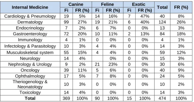
Internal Medicine cases are divided into 13 different areas, which are represented in table 5. The most prevalent medical field was dermatology, with a relative frequency of 26% (n= 474), followed by gastroenterology representing 18% of the total cases. In all clinical areas, the most representative group was the canine group, except for cardiology, pneumology, nephrology and urology areas, in which feline group was more represented. Exotic medicine was mostly represented in the cardiology, pneumology, dermatology and gastroenterology areas.
A brief bibliographical review of one relevant disease of each area will be developed, whether by it being the most prevalent disease in the particular clinical field or by it being author’s preference.
Weeks of age Years of age
low-risk cat Years of age high-risk cat 8 12 16 26 52 Core vaccination Feline herpesvirus type 1 x x x x X (if not given at 26) Triennial vaccination ( at 4, 7, 10, 13 years of age) Annual vaccination Feline calicivirus x x x x X (if not given at 26) Feline parvovirus x x x x X (if not given at 26) Triennial vaccination ( at 4, 7, 10, 13 years of age) Rabies virus (in endemic countries) x x Non-core vaccination Feline leukaemia virus x x x Not recommended for low-risk cats Annual vaccination Chlamydia felis x x x Bordetella x
7 Table 5: Case distribution regarding Internal Medicine Clinical Areas
2.2.2.1. Cardiology and Pneumology
Cardiology and pneumology completed 8% (n=474) of the medical practice, with 12 cases (Table 5). The most common conditions were dilated cardiopathy and chronic degenerative mitral valve disease, both with 33% (n=12), as described in table 6.
Table 6: Case distribution regarding Cardiology
Cardiology Canine Feline Total FR (%)
Fi FR (%) Fi FR (%)
Dilated cardiopathy 2 25% 2 50% 4 33%
Hypertrophic cardiomiophaty 0 0% 2 50% 2 17%
Heart failure 1 13% 0 0% 1 8%
Pericardic effusion 1 13% 0 0% 1 8%
Chronic degenerative mitral valve disease
stage B 3 38% 0 0% 3 25%
stage D 1 13% 0 0% 1 8%
Total 8 100% 4 100% 12 100%
Chronic valvular heart disease (CVHD), also known as degenerative valvular disease or endocardiosis (Ljungvall and Häggström 2015) is more likely to affect the left atrioventricular/mitral valve, with or without involving the right atrioventricular/tricuspid valve (Atkins et al. 2009).
Myxomatous degeneration is a pathological change in the organization of the connective tissue of the heart valves (Ljungvall and Häggström 2015), which includes collagen changes in
Internal Medicine Canine Feline Exotic Total FR (%)
Fi FR (%) Fi FR (%) Fi FR (%)
Cardiology & Pneumology 19 5% 14 16% 7 47% 40 8%
Dermatology 99 27% 19 21% 6 40% 124 26%
Endocrinology 7 2% 6 7% 0 0% 13 3%
Gastroenterology 72 20% 10 11% 2 13% 84 18%
Immunology 4 1% 0 0% 0 0% 4 1%
Infectology & Parasitology 10 3% 4 4% 0 0% 14 3%
Musculoskeletal system 55 15% 4 4% 0 0% 59 12%
Neurology 14 4% 0% 0 0% 15 3%
Nephrology & Urology 9 2% 21 23% 0 0% 30 6%
Oncology 39 11% 5 6% 0 0% 40 9% Ophthalmology 17 5% 7 8% 0 0% 24 5% Theriogenology & Neonatology 10 3% 0 0% 0 0% 10 2% Toxicology 14 4% 0 0% 0 0% 14 3% Total 369 100% 90 100% 15 100% 474 100%
8 cellular and intercellular components as well as endothelial thickening. These deformations limit the valves coaptation, which progresses to valvular regurgitation into the atrium and leads to an increase on cardiac work and consequently to a ventricular remodelling and dysfunction (Atkins et al. 2009).
Slight to moderate regurgitation is often compensated for years by cardiac dilation, eccentric hypertrophy, increased force of contraction, increased heart rate, fluid retention and neurohormonal modulation of cardiovascular function. When valvular regurgitation can no longer be compensated, the increased venous pressure and the reduced cardiac output lead to congestive heart failure (Ljungvall and Häggström 2015).
There is still no known cause for this disease, however it looks like it has a genetic component in some breeds, being Cavalier King Charles Spaniels predisposed to develop it at a young age. CVHD is also more prevalent in males and in smaller breeds. However, whereas the disease is slow and relatively predictably progressive in dogs under 20 kilograms, with most of them being diagnosed with mitral valve regurgitation years before the clinical onset of heart failure, when it occurs in larger breeds, the progression appears to be faster (Atkins et al. 2009). The most important clinical finding of chronic degenerative mitral valve disease (CDMVD) is the characteristic left apical systolic heart murmur. Thoracic radiographs are the first indicated diagnostic tests (as they can rule out non cardiac causes, assess the cardiac size by vertebral heart score and also indicate decompensated cardiac heart failure if there is pulmonary congestion and oedema present), however echocardiography is the test of choice for demonstrating the valve lesion, as it detects the thickened atrioventricular valve and identifies the regurgitant jet as well as changes in structure and contractile function of the heart chambers. The differential diagnosis must exclude anaemia, bacterial endocarditis, secondary mitral regurgitation due to dilated cardiomyopathy and primary respiratory disease (Ljungvall and Häggström 2015). After the diagnosis, CDMVD should be staged according to an ACVIM system that classifies the four basic stages of heart disease and failure and helps guiding the clinical approach for each(Atkins et al. 2009; Ljungvall and Häggström 2015).
Stage A patients are at high risk for developing heart disease but presently hold no identifiable structural disorder of the heart (e.g., Cavalier King Charles Spaniel without a heart murmur). No treatment is indicated in stages A or B1, however these animals should be subjected to yearly cardiac evaluations (Atkins et al. 2009; Ljungvall and Häggström 2015).
In Stage B patients clinical signs are not present, but they do present with a systolic click (early stage) or murmur, revealing structural heart disease. Stage B1 are asymptomatic patients that have hemodynamically insignificant valve regurgitation, with no radiographic or echocardiographic evidence of cardiac remodelling in response to CVHD. Stage B2 are asymptomatic patients that have hemodynamically significant valve regurgitation, with radiographic or echocardiographic evidence of cardiac remodelling in response to CVHD (Atkins et al. 2009; Ljungvall and Häggström 2015).
9 Stage C patients have past or current clinical signs of heart failure associated with structural heart disease.
Stage D patients have end-stage disease with clinical signs of heart failure caused by CVHD that are refractory to standard therapy and require specialized treatment so that they remain clinically comfortable (Atkins et al. 2009; Ljungvall and Häggström 2015).
In stage B2, according to a recent study published by the Journal of Veterinary Internal Medicine in 2016, the administration of pimobendan to dogs without any other cardiovascular therapy results in the prolongation of the preclinical period, prolonging the onset of congestive heart failure by 15 months and reducing its risk by one-third. Angiotensin converting enzyme inhibitors (ACEI) and ß-blockers are other therapeutic possibilities for patients with clinically relevant left atrial enlargement on initial examination or on successive monitoring examinations. Changing to a diet with mild dietary sodium restriction and adequate protein and calories for maintaining optimal body condition, might also be an option at this stage (Atkins et al. 2009; Ljungvall and Häggström 2015).
In acute stage C, furosemide (doses related to the severity of clinical signs and response to therapy), pimobendan, oxygen supplementation, paracentesis or thoracocentesis (if effusions are causing respiratory distress), sedation (if anxiety reflects in dyspnoea) are recommended. Combining the administration of enalapril with furosemide improves pulmonary capillary wedge pressure, so it is also a possibility in these cases, as well as nitroglycerin ointment. In chronic stage C, medical therapy with furosemide, pimobendan and an ACEI (e.g. enalapril) is a indicated. Other possibilities include spironolactone (as an aldosterone antagonist), digoxin, ß-blockers (e.g. atenolol, as a protection on myocardial function and remodeling), antiarrhythmic medication, (to maintain sinus rhythm, e.g. diltiazem), cough suppressants and maintaining adequate calorie intake (Atkins et al. 2009).
In acute stage D, it is recommended to increase the dose and frequency of furosemide, spironolactone, oxygen supplementation or even mechanical ventilatory assistance, paracentesis or thoracocentesis as needed. Sodium nitroprusside, dobutamine, hydralazine, amlodipine, direct vasodilators, sildenafil, bronchodilators are also possibilities to add on. In chronic stage D, increasing the dose and frequency of furosemide, and administering spironolactone is indicated. Other possibilities include hydrochlorthiazide, pimobendan, digoxin, sildenafil, cough suppressants and bronchodilators (Atkins et al. 2009).
Although open-heart surgical repair of myxomatous mitral valve disease in dogs has been described with success, there is no treatment that reverses the myxomatous disease. Prognosis and progression of the disease depends on the age, left atrial size, heart rate, presence of other diseases and the development of complications (such as tendinous chord rupture, intracardiac thrombus) (Atkins et al. 2009; Ljungvall and Häggström 2015).
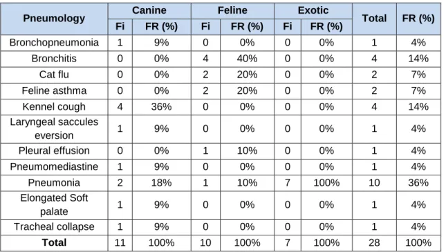
10 Table 7: Case distribution regarding Pneumology
Pneumology Canine Feline Exotic Total FR (%)
Fi FR (%) Fi FR (%) Fi FR (%) Bronchopneumonia 1 9% 0 0% 0 0% 1 4% Bronchitis 0 0% 4 40% 0 0% 4 14% Cat flu 0 0% 2 20% 0 0% 2 7% Feline asthma 0 0% 2 20% 0 0% 2 7% Kennel cough 4 36% 0 0% 0 0% 4 14% Laryngeal saccules eversion 1 9% 0 0% 0 0% 1 4% Pleural effusion 0 0% 1 10% 0 0% 1 4% Pneumomediastine 1 9% 0 0% 0 0% 1 4% Pneumonia 2 18% 1 10% 7 100% 10 36% Elongated Soft palate 1 9% 0 0% 0 0% 1 4% Tracheal collapse 1 9% 0 0% 0 0% 1 4% Total 11 100% 10 100% 7 100% 28 100%
Table 7 shows the absolute frequencies and respective percentages of the cases observed in the area of pneumology, according to the species. Pneumonia was the most common condition, with 36% of cases, followed by bronchitis in cats and kennel cough in dogs, both with 14%.
Infectious tracheobronchitis, commonly denominated kennel cough or canine respiratory disease complex (CRDC), is an acute highly contagious respiratory infection/complex that affects canines. Paroxysmal cough is often the only clinical sign, although it could be accompanied by fever, anorexia and lethargy. In complicated infections, nasal and ocular discharge, respiratory distress and pneumonia (which might become life threatening) may also occur (Ford 2004).
This condition has a multifactorial aetiology, caused by one or a combination of bacterial and/or viral agents - Bordetella bronchiseptica and canine parainfluenza virus are the two most common, however canine adenovirus-2, canine distemper virus and canine herpesvirus, as well as environmental conditions can be involved. All are capable of colonizing the epithelium of the upper respiratory tract and causing a high-pitched cough often described as goose or seal honk. These pathogens especially threaten puppies (who are more severely affected and are at significant risk of dying if not treated) and dogs in shelters, kennels and veterinary hospitals (Ford 2004).
Diagnosis is often facilitated by the acknowledgement of recent contact with other dogs (whether or not having cough) within the previous 10 days. In addition, a positive and rapid response to cough suppressants (as an anti-inflammatory or antitussive medication) supports the diagnosis of an uncomplicated infectious tracheobronchitis with an excellent prognosis. If there is any respiratory distress or pneumonia, hospitalization and aerosol therapy is recommended.
11 Thoracic radiographies may be helpful to rule out pneumonia in a dog with fever or anorexia (Ford 2004).
Although vaccination stimulates immune response, it does not eliminate the risk of infection nor the development of subclinical to mild infection. Once exposed, intra-nasally vaccinated canines have significantly lower rates of coughing, nasal discharge and retching, compared to unvaccinated ones. Vaccination is recommended at least 5 days prior to a known or potential exposure (Ellis et al. 2016).
2.2.2.2. Dermatology
Table 8: Case distribution regarding Dermatology
Dermatology Canine Feline Exotic Total FR (%)
Fi FR (%) Fi FR (%) Fi FR (%)
Acral lick dermatitis 2 2% 0 0% 0 0% 2 2%
Anal glands abscess 4 4% 0 0% 0 0% 4 3%
Anal glands furunculosis 1 1% 0 0% 0 0% 1 1% Atopy 10 10% 0 0% 0 0% 10 8% Burn 0 0% 1 5% 0 0% 1 1% Contact dermatitis 1 1% 0 0% 0 0% 1 1% Chronic Allergy 10 10% 0 0% 0 0% 10 8% Dermatitis 7 7% 3 16% 0 0% 10 8%
Ear foreign body 1 1% 0 0% 0 0% 1 1%
Eosinophilic complex 0 0% 1 5% 0 0% 1 1% Environmental allergy 1 1% 0 0% 0 0% 1 1% External otitis 33 33% 2 11% 0 0% 35 28% Facial dermatitis 1 1% 0 0% 0 0% 1 1% Flea Allergy dermatitis 1 1% 2 11% 0 0% 3 2% Food allergy 1 1% 0 0% 0 0% 1 1% Harvest mites 2 2% 0 0% 0 0% 2 2% Hotspot 1 1% 0 0% 0 0% 1 1%
Nail bed infection 1 1% 0 0% 0 0% 1 1%
Otohematoma 2 2% 0 0% 0 0% 2 2% Pyoderma 1 1% 0 0% 0 0% 1 1% Skin Abscess 2 2% 1 5% 4 67% 7 6% Subcorneal pustular dermatosis 1 1% 0 0% 0 0% 1 1% Tick granuloma 1 1% 0 0% 0 0% 1 1% Wound 15 15% 9 47% 2 33% 26 21% Total 99 100% 19 100% 6 100% 124 100%
12 Dermatology comprised 26% (n=474) of the medical practice, with 124 cases (Table 5), having been the most prevalent area. Absolute frequencies and respective percentages of the cases observed in the area of dermatology, are shown in table 8. The most common condition observed was otitis externa with 35 cases, making 28% of total dermatologic maladies.
Otitis externa is one of the most common pathologies presented by dogs regarding the dermatology area. It is characterized as an acute or chronic inflammation of the external ear canal, in which aetiology can be divided into primary (direct cause of inflammation) and secondary (perpetuating or predisposing factors that contribute to ear disease, prevent resolution and/or lead to recurrence) causes (Scott 2015; Paterson 2016).
Primary causes include allergy (food, contact, atopy), endocrine (hypothyroidism, hyperadrenocorticism), immune-mediated (pemphigus foliaceus, lupus erythema multiforme, vasculitis, drug eruption), keratisation disorders (sebaceous adenitis, primary idiopathic seborrhoea), ectoparasites (Otodectes cynotis, Demodex species), foreign body and idiopathic (juvenile cellulitis particularly in young puppies, idiopathic glandular hyperplasia in spaniels). (Paterson 2016). Frequent swimming, pendulous ear pinnae, presence of hair within the canal, stenotic ear canals and excessive moisture, systemic disease (renal, pancreatic or hepatic), obstructive ear disease (neoplasia, polyps, cysts) are predisposing factors in the establishment of an otitis externa (Scott 2015; Paterson 2016).
Secondary infection with yeast (Malassezia and Candida species) or bacteria (Staphylococcus spp., Streptococus spp., Corynebacterium spp., Enterococcus spp.,
Pseudomonas spp., Proteus spp., Escherichia coli) develops after inflammation trigged by
primary causes, usually combined with perpetuating and predisposing factors. Although treatment against these is essential, if it does not treat the primary cause, it will not resolve the ear disease (Scott 2015; Paterson 2016).
Owners report head rubbing, ear scratching and/or head shaking, and animals show erythema and oedema of the ear canal, auricular discharge (brown-black “coffee grounds” most of the times indicate infestation with Otodectes cynotis, also known as ear mites; brown or gray discharge suggest Malassezia and yellow-greenish discharge points towards bacterial infection), erosions and ulcers (which are associated with Pseudomonas infection), pain when opening the mouth, facial paralysis, head tilt and/or signs of generalized skin disease (Scott 2015; Paterson 2016).
Cytology, bacterial culture and sensitivity testing may be of help in establishing the diagnosis and the correct therapy. The majority of acute otitis can be treated with manual cleaning and a polyvalent topical ear product, which usually includes a glucocorticoid, an antimicrobial, and/or an antifungal. In regard to chronic cases, the underlying causes should be recognised and treated whilst managing the secondary infection (Nuttall 2016; Paterson 2016)
13 Pathological changes in the canal wall, glandular tissue and in the tympanum, as well as an acute or chronic otitis media, are perpetuating factors of otitis externa and should be addressed too (Nuttall 2016; Paterson 2016).
Vertical ear canal ablation, which was performed during the traineeship, is recommended in irreversible hyperplastic otitis (characterized by palpable mineralization and complete occlusion of the external ear canal by hyperplastic tissue) when the horizontal canal is healthy, which allows complete excision of vertical canal tissue with less postoperative pain and exudate than a total ear canal ablation(Bacon 2012; Scott 2015)
2.2.2.3. Endocrinology
Endocrinology comprised 3% (n = 474) of the medical practice, with 13 cases (Table 5). Table 9 displays the absolute frequencies and respective percentages of the cases observed in the area of endocrinology. Diabetes mellitus was the most common condition observed with 31% (n=13), followed by hyperadrenocorticism and hyperthyroidism observed both with 23% (n=13) of total cases witnessed in this medical field. A brief bibliographical review on hyperthyroidism will be made due to the author’s preference on this topic.
Table 9: Case distribution regarding Endocrinology
Endocrinology Canine Feline Total FR (%)
Fi FR (%) Fi FR (%) Diabetes mellitus 2 29% 2 33% 4 31% Exocrine pancreatic insufficiency 1 14% 1 17% 2 15% Hyperadrenocorticism 3 43% 0 0% 3 23% Hyperthyroidism 0 0% 3 50% 3 23% Hypothyroidism 1 14% 0 0% 1 8% Total 7 100% 6 100% 13 100%
Hyperthyroidism, also known as thyrotoxicosis, results from an excess of active thyroid hormones (T3/triiodothyronine and T4/thyroxine) circulating in the blood stream. It usually affects cats from eight years old and beyond, with no sex predilection (Mooney and Peterson 2004; Daminet 2015)
The most common cause of this disease is a benign thyroid neoplasia (adenoma) or an adenomatous hyperplasia in one or more commonly both thyroid lobes, although a thyroid carcinoma is also a rare but possible cause. When it comes to dogs, hyperthyroidism is due to a functional thyroid carcinoma(Mooney and Peterson 2004; Daminet 2015)
Feline hyperthyroidism (FHT) is suspected when the animal presents with weight loss, polyphagia, polyuria, polydipsia, tachypnoea, tachycardia, increased vocalization and activity,
14 gastrointestinal disorders, palpably enlarged thyroid glands and/or unkempt hair coat. If a high total T4 serum concentration is concurrent with one or more of the previous signs, the diagnosis is confirmed. Differential diagnosis must take into account Diabetes mellitus, inflammatory bowel disease, gastrointestinal lymphoma, gastrointestinal malabsorption, chronic kidney disease and parasitism. As older cats are the most affected, age related comorbidities such as heart disease, hypertension, retinopathy, chronic kidney disease, gastrointestinal dysfunction and Diabetes
mellitus may be present. A simple approach to diagnosis and management by categorizing the
animal into one of six diagnostic groups, is demonstrated in table 10 (Daminet 2015; Carney et al. 2016).
Table 10: FHT diagnostic groups and management options
Group Clinical Presentation Options
Classical clinical disease Clinical signs Elevated T4 No identifiable concurrent disease Treat for FHT
Clincal FHT with one or more concurrent disease Clinical signs Elevated T4 identifiable concurrent disease
Treat for FHT and manage concurrent disease Possible FHT with probable non-thyroid disease Clinical signs Normal T4
Test for T4 and fT4ed Test for differential
diagnosis
Consider T3 suppression or thyroid scintigraphy Enlarged thyroid without
clinical signs
No clinical signs
Normal T4 Monitor clinical signs and T4 serum concentration Subclinical FHT
No clinical signs but
suggestive physical findings
Elevated T4 Confirm T4 - if elevated,
treat for FHT Clinically normal
No clinical signs and no suggestive physical findings Elevated T4
There are four treatment options, which should be applied according to each patient’s characteristics. Treatment with radioactive iodine or surgical thyroidectomy are the two definitive cures recommended for fairly young and healthy cats. However, for geriatric cats, cats with concurrent non-thyroidal disease and those whose owners decline definitive therapy, the long-term medical management with anti-thyroid drugs (carbimazole or methimazole) or an iodine-restricted diet are the possible therapies. Nevertheless, monitoring the patient is essential to assess efficacy and detect iatrogenic hypothyroidism (Daminet 2015; Carney et al. 2016).
Cats without concurrent chronic kidney disease have a median survival of up to 5 years, but cats with a comorbid disease have shorter survival times(Carney et al. 2016).
15
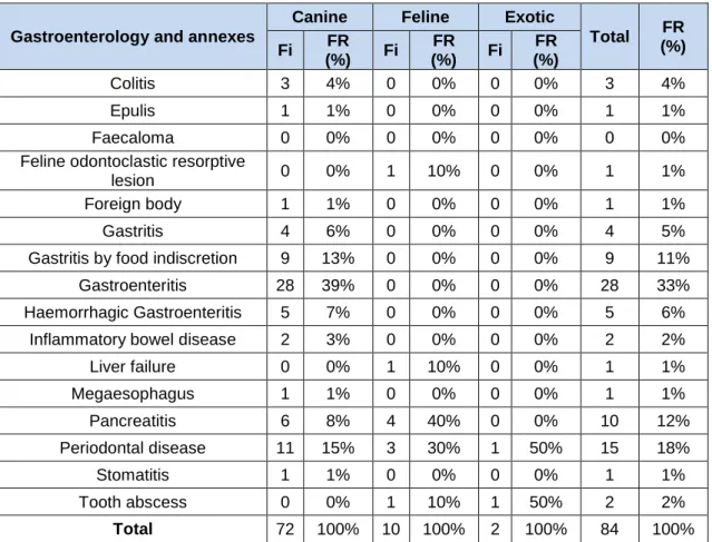
2.2.2.4. Gastroenterology and annexes
With a relative frequency of 27% (n=474), 84 cases of the gastroenterology medical field were seen (Table 5). Absolute frequencies and respective percentages of the cases observed this area are shown in table 11. The most common condition observed was gastroenteritis with 28 cases (33%), followed by periodontal disease with 18% (n= 84) and pancreatitis with 12% (n= 84) of gastroenterology conditions in total. A brief bibliographical review on pancreatitis will be made due to the author’s preference on this topic.
Table 11: Case distribution regarding Gastroenterology and annexes
Gastroenterology and annexes
Canine Feline Exotic
Total FR (%) Fi FR (%) Fi FR (%) Fi FR (%) Colitis 3 4% 0 0% 0 0% 3 4% Epulis 1 1% 0 0% 0 0% 1 1% Faecaloma 0 0% 0 0% 0 0% 0 0%
Feline odontoclastic resorptive
lesion 0 0% 1 10% 0 0% 1 1%
Foreign body 1 1% 0 0% 0 0% 1 1%
Gastritis 4 6% 0 0% 0 0% 4 5%
Gastritis by food indiscretion 9 13% 0 0% 0 0% 9 11%
Gastroenteritis 28 39% 0 0% 0 0% 28 33%
Haemorrhagic Gastroenteritis 5 7% 0 0% 0 0% 5 6%
Inflammatory bowel disease 2 3% 0 0% 0 0% 2 2%
Liver failure 0 0% 1 10% 0 0% 1 1% Megaesophagus 1 1% 0 0% 0 0% 1 1% Pancreatitis 6 8% 4 40% 0 0% 10 12% Periodontal disease 11 15% 3 30% 1 50% 15 18% Stomatitis 1 1% 0 0% 0 0% 1 1% Tooth abscess 0 0% 1 10% 1 50% 2 2% Total 72 100% 10 100% 2 100% 84 100%
Common in both dogs and cats, pancreatitis is usually classified as acute or chronic and mild or severe. The acute inflammation of the pancreas is associated with high mortality, however it is possible to witness a complete recovery of pancreas structure and function. In the other hand, chronic pancreatitis might lead to progressive loss of the pancreas normal function, causing pain and therefore reducing quality of life (Watson 2015). However, recurrent reversible acute pancreatitis may result in progressive and irreversible chronic disease (Xenoulis 2015).
Mild pancreatitis refers to a disease where there is no multi-organ failure, whereas severe pancreatitis includes a multisystem failure and/or pancreatic or peri-pancreatic necrosis (Mansfield and Beths 2015).
16 The gold standard for definitive diagnosis of pancreatitis and its definition as acute or chronic disease is histological – whereas in an acute form neutrophilic inflammation, oedema and necrosis are present, in a chronic form, there are permanent, irreversible and progressive histopathological changes, such as fibrosis and acinar loss (Watson 2015; Xenoulis 2015). Although a definitive diagnostic and classification in acute or chronic pancreatitis is important for therapy management and prognosis, histology is not often performed as it is an invasive procedure and it has associated morbidity. Therefore, a presumptive diagnosis may be done on the basis of functional changes and clinical findings together with abnormal serum pancreatic lipase immunoreactivity (PLI) concentration and abdominal ultrasonography findings (enlargement of the pancreas, fluid accumulation around the pancreas, necrosis if hypoechoic, fibrosis if hyperechoic, dilated pancreatic duct). Complete blood count, serum biochemistry profile and urinalysis should always be performed in dogs and cats suspected of having pancreatitis and abdominal radiography might be performed to exclude other diseases (Steiner 2015; Xenoulis 2015).
Although many studies show environmental and genetic influences in pancreatitis pathophysiology, aetiology is often seen as idiopathic. Generally, the inflammation starts with premature and excessive activation of pancreatic digestive enzymes which may be a result of oxidative stress or hypotension. The pancreatic proteases then stimulate the migration of neutrophils to the pancreas with subsequent worsening inflammation in the pancreatic and peri-pancreatic area which leads to progression and complications(Steiner 2015)
Domestic shorthaired and Siamese cats, Miniature schnauzers, Border Collies, Cavalier King Charles and English cocker spaniels are more commonly affected. However being overweight, having hypertriglyceridaemia, being a male, having been submitted to previous surgery, having concurrent endocrine diseases, drug administration (azathioprine or potassium bromide) and dietary indiscretions increase the risks of acute fatal pancreatitis. On the other hand, in cats, risk factors include high fat diets, hypercalcaemia, blunt abdominal trauma and infections (such as Toxoplasma gondii and Amphimerus pseudofelineus). Feline pancreatitis can occur simultaneously with inflammatory bowel disease and/or cholangitis; so-called triaditis (Steiner 2015; Watson 2015).
Clinical signs vary greatly – most dogs with chronic pancreatitis present with subclinical disease or intermittent mild non-specific signs, whereas animals with acute pancreatitis may display anorexia, weakness, vomiting, diarrhoea, icterus, hypothermia or fever, bleeding diathesis and/or abdominal pain in numerous combinations (Xenoulis, 2015) and in severe cases cardiovascular shock, disseminated intravascular coagulation, multi-organ failure and even death may be observed. (Watson 2014, Xenoulis 2015). It is important to consider that additional clinical signs might also be present, in consequence of other concurrent or complicating disease, such as exocrine pancreatic insufficiency or Diabetes mellitus. Clinical presentation of feline pancreatitis is mostly anorexia, lethargy, dehydration, icterus, hypothermia or fever, signs of
17 abdominal pain and frequently a palpable abdominal mass, with less predominance of gastrointestinal signs (Steiner 2015; Xenoulis 2015; Watson 2015).
A therapeutic approach to mild acute pancreatitis, starts with intravenous fluid replacement with Ringer’s solution (which by increasing pH prevents further trypsin activation) and an anti-emetic therapy with maropitant. If there is any suspicion of bacterial translocation or infected pancreatic necrosis, a course of antibiotics should be started, preferably with amoxicillin-clavulanate or ticarcillin. Reduction of gastric acidity should also be addressed, omeprazole being the first drug of choice. In unresponsive cases, hydrocortisone in a low dose can be considered. (Steiner 2015; Watson 2015).
Patients with mild disease, should be fasted until they regain appetite and eat voluntarily. However, if they reach five days of anorexia, enteral feeding is the next step. In contrast, in dogs with severe disease, a naso-oesophageal or oesophageal feeding tube should be placed as soon as possible. High fat diets should be avoided as many of the animals are hyperlipidaemic (Xenoulis et al 2008; Mansfield and Beths 2015).
In mild pancreatitis, non-steroidal anti-inflammatory agents, buprenorphine or methadone are recommended for pain management and a lidocaine and ketamine constant rate infusion (CRI) should be added in a moderate disease case. In case of severe to excruciating pain, an epidural morphine or fentanyl infusion in combination with a lidocaine/ketamine CRI is the recommended analgesia. If there is any unexpected exacerbation of the pain, assessment for pancreatic fluid collection should be made. Gabapentin may be given with any combination for any level of pain. Currently, there is no recommendation for surgical intervention in the management of pancreatitis (Xenoulis 2015)
The long-term management of chronic pancreatitis involve a fat restricted diet, resolving concurrent diseases that may trigger an acute episode and monitoring PLI concentration. Drugs associated with pancreatitis should also be avoided and there is still no evidence about pancreatic enzymes oral supplements for chronic patients (Xenoulis et al 2008).
Prognosis is directly related to the severity of the disease - animals with mild, uncomplicated disease, the prognosis is usually good. However, in severe disease outcome should be guarded, not to mention that exocrine pancreatic insufficiency and Diabetes mellitus may be long-term sequelae of chronic disease (Xenoulis et al 2008).
2.2.2.5. Immunology
Immunology comprised 1% (n = 474) of the medical practice, with three cases (Table 5). Table 12 displays the absolute frequencies and respective percentages of the four cases observed in the area of immunology.
18 Table 12: Case distribution regarding Immunology
Immunology Canine
Fi FR (%)
Chronic polyarthritis 1 25%
Immune-mediated hemolytic anemia 1 25%
Immune-mediated polyarthritis 1 25%
Symetric lymphoid onchodistrophy 1 25%
Total 4 100%
Polyarthritis is characterized by an inflammation of more than two joints and is classified as infectious, reactive and immune-mediated, which is in turn subcategorized into erosive (or deforming) and non-erosive(or non-deforming) form (Johnson and Mackin 2012b; Mahony 2015). Although it may affect any dog or cat of any age or sex, classical lameness, reluctance to walk, stiffness and occasionally nonspecific signs of systemic disease (fever, inappetence, lethargy) may be more often be presented by young adults (Mahony 2015; Stone 2017).
Infectious etiologies for polyarthritis include Lyme disease, Erlichiosis, Anaplasma
phagocytum, Corynebacterium, Mycoplasma, Leishmania, Bartonella, Calicivirus infection in cats,
amongst others (Mahony 2015).
Reactive polyarthritis can develop due to vaccine or drug induced responses. Vaccine-induced polyarthritis is a possible reaction whether after a first vaccination or a booster. Recent studies suggest that canine distemper virus may be implicated, though Akitas seem to be predisposed to this adverse reaction. Treatment with doxycycline and NSAID’s usually resolves the clinical signs. Drug-induced polyarthritis is widely described after sulfonamide treatment, especially in Doberman pinschers. Additionally, polyarthritis secondary to erythropoietin, penicillin, cephalosporins and phenobarbital administration has also been described (Johnson and Mackin 2012b).
A type III hypersensitivity reaction may possibly be the cause behind immune-mediated non-deforming polyarthritis - immune complexes accumulated in the joint space activate the complement which leads to neutrophilic inflammation and consequent tissue damage. Non erosive immune mediated polyarthropathy is most commonly idiopathic. However it can also be specific to the Shar-Pei and Akita breeds, or a result of polymyositis syndrome, a meningitis-arthritis or may be associated with systemic lupus erythematosus (Johnson and Mackin 2012b; Mahony 2015).
Young large breed and sporting females, such as Retrievers, Cocker Spaniels and German Shepherds, seem most frequently affected by idiopathic polyarthritis, which is classified into four subtypes, as shown in Table 13 (Johnson and Mackin 2012b).
19 Table 13: Subtypes of idiopathic immune mediated polyarthritis
Subtype of idiopathic immune mediated polyarthritis
Type I No underlying disease or aetiology diagnosed
Type II Reaction to an infectious or inflammatory disease distant from the joint (pancreas, heart, urinary tract, spine, uterus, etc.)
Type
III/Enteropathic Presence of gastrointestinal or hepatic disease
Type IV Caused by neoplasia outside the joints
Classical clinical signs that reveal polyarthritis/polymyositis syndrome are a muscle atrophy and fibrosis following a polyarthritis accompanied by focal or generalized muscle pain and swelling. Therapy with cyclophosphamide combined with prednisone has had some success in resolving the symptomatology. Juvenile-onset polyarthritis in Akitas is characterized by cycles of fever combined with painful and swollen joints that spontaneously resolve after 24 to 48 hours. It usually onsets between nine weeks and eight months of age. Similarly, familial Shar-Pei fever is characterized by intermittent recurring attacks of inflammation and fever due to a genetic mutation, however, in contrast to polyarthritis in Akitas, the onset of that disease can strike whether youngsters or adults (Johnson and Mackin 2012b).
Systemic lupus erythematosus is uncommon, typically starting between two and four years of age due to various and simultaneous immunologic reactions. Clinical signs consist of non-erosive polyarthritis with shifting lameness, haemolytic anaemia, glomerulonephritis, fever, skin lesions and lymphadenopathies (Johnson and Mackin 2012b).
On the other hand, erosive joint polyarthritis seems to have a type IV hypersensitivity component, combined with chronic synovitis and the presence of antibodies against distemper virus, type II collagen, immunoglobulins M, G and A. The presence of anti-IgA antibodies in the joint fluid is the primary factor to differentiate rheumatoid arthritis from osteoarthritis, thus its quantity can be used as a prognosis indicator (Johnson and Mackin 2012b) . This deforming form is very rare - rheumatoid arthritis is the most frequent, nevertheless erosive polyarthritis of greyhounds, Felty’s syndrome (triad composed by rheumatoid arthritis, splenomegaly and neutropenia) and feline periosteal proliferative polyarthritis (periosteal new bone formation and erosions) have also been described. Radiographic findings that indicate subchondral bone destruction can take up to six months to become visible. Dogs primarily diagnosed with the non-deforming form in which clinical signs persist, should be periodically reassessed (Johnson and Mackin 2012b; Mahony 2015).
The diagnosis for an immune-mediated polyarthritis is only definitive after exclusion of other causes of joint disease, such as the conditions described above, and a favourable response
20 to immunosuppressive treatment. Further confirmatory testing by arthrocentesis and joint fluid analysis may be performed(Johnson and Mackin 2012a; Stone 2017).
Treatment must be decided according to the underlying aetiology, though NSAIDs can always be administered to treat inflammation and remove pain. In cases of idiopathic immune-mediated polyarthritis, a course of prednisolone in immunosuppressive doses is recommended until the resolution of clinical signs. Afterwards, the dosage should be slowly reduced, and the finish of the course might be between four to six months, nevertheless there are dogs who need life-long treatment to remain in remission. It is imperative to exclude bacterial infection before any glucocorticoid administration. If there is any glucocorticoid adverse reaction, azathioprine or cyclophosphamide may be options to consider. In addition, antioxidant supplementation may be a way of preventing hyaluronic acid degradation (Mahony 2015).
Long-term prognosis for erosive arthritis is poor, because it affects quality of life due to intense pain. On the other hand, non-deforming polyarthritis is associated with a fair to good prognosis. Although drug and vaccine induced polyarthritis have an excellent recovery, polyarthritis/polymyositis syndrome, meningitis-arthritis and Akitas with juvenile-onset polyarthritis have frequent relapses (Johnson and Mackin 2012b; Mahony 2015).
2.2.2.6. Infectology and parasitology
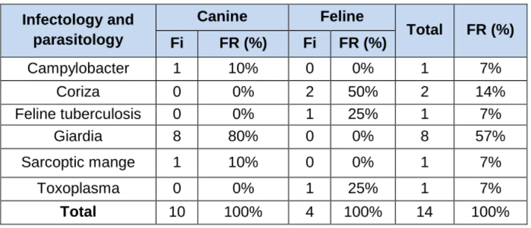
With a relative frequency of 3% (n=474), 14 cases corresponding to the infectology and parasitology medical field were seen (Table 5). Absolute frequencies and respective percentages of the cases observed this area are shown in table 14. The most common condition observed was infection by Giardia with 8 cases (57%, n= 14), followed by Coriza infection, which represented 14% (n= 14) of total cases. A brief bibliographical review on feline tuberculosis will be made due to the author’s preference on this topic.
Table 14: Case distribution regarding Infectology and Parasitology Infectology and parasitology Canine Feline Total FR (%) Fi FR (%) Fi FR (%) Campylobacter 1 10% 0 0% 1 7% Coriza 0 0% 2 50% 2 14% Feline tuberculosis 0 0% 1 25% 1 7% Giardia 8 80% 0 0% 8 57% Sarcoptic mange 1 10% 0 0% 1 7% Toxoplasma 0 0% 1 25% 1 7% Total 10 100% 4 100% 14 100%
21 Feline tuberculosis (TB) is a new fatal threat as five clinical cases due to Mycobacterium
bovis were confirmed in 2018 in the United Kingdom. They occurred in young, mostly pedigree
cats with no history of outdoor access. (O’Halloran, et al. 2018, Veterinary Times 2018)
Furthermore, all these cases were atypical, as they involve a rare gastrointestinal form of
M. bovis, instead of the cutaneous lesions at bite and fight sites where the bacteria typically grow.
Clinical signs are gastrointestinal, that might be suggestive of abdominal lymphadenopathy or feline infectious peritonitis as so is important to do a differential diagnosis. Although unclear, the only common denominator that makes sense for experts is that all five cats were fed the same commercial raw frozen food diet.(O’Halloran et al. 2018; “Investigations under Way in Wake of Feline TB Outbreak” 2018). The TB case witnessed during the traineeship correlates to all this information, and although tests were ordered to confirm it, the samples taken were not sufficient. The kitten did not survive for further sampling.
Although TB is potentially treatable with months of antibiotics, it might be fatal in humans and animals, as it is a zoonosis.
2.2.2.7. Musculoskeletal system
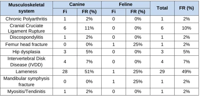
With a relative frequency of 12% (n=474), 59 cases involving conditions of the musculoskeletal system were witnessed during the traineeship (Table 5). Absolute frequencies and respective percentages of the cases observed in this medical field are shown in table 15. The most common condition was idiopathic lameness with 49% (n=59), followed by osteoarthritis with 17% (n=59) and cranial cruciate ligament rupture representing 10% (n=59) of total cases. A brief bibliographical review on the cranial cruciate ligament rupture will be made due to the author’s preference on this topic.
Table 15: Case distribution regarding the Musculoskeletal system Musculoskeletal system Canine Feline Total FR (%) Fi FR (%) Fi FR (%) Chronic Polyarthritis 1 2% 0 0% 1 2% Cranial Cruciate Ligament Rupture 6 11% 0 0% 6 10% Discospondylitis 1 2% 0 0% 1 2%
Femur head fracture 0 0% 1 25% 1 2%
Hip dysplasia 3 5% 0 0% 3 5% Intervertebral Disk Disease (IVDD) 4 7% 0 0% 4 7% Lameness 28 51% 1 25% 29 49% Mandibular symphysis fracture 0 0% 1 25% 1 2% Myositis/Tendinitis 1 2% 0 0% 1 2%
22
Osteoarthritis 9 16% 1 25% 10 17%
Patellar luxation 2 4% 0 0% 2 3%
Total 55 100% 4 100% 59 100%
Complete or partial rupture of the cranial cruciate ligament is widely common in dogs of all ages, mostly due to progressive degeneration or trauma. Nevertheless, female large breed dogs, Rottweilers, Newfoundlands, Staffordshire Terriers and Boxers are more prone to develop this condition. When found in small breed dogs, this condition is more likely to happen later in life, whereas large breed dogs are affected earlier. In addition, obesity, neutering and systemic diseases such as hyperadrenocorticism, cutaneous asthenia or any autoimmune disease are demonstrated to be risk factors for the onset of this ligament rupture (Kowaleski and Boudrieau 2012; Linn 2015).
Progressive degeneration that leads to a rupture of the cranial cruciate ligament is due to a weakness in the ligament properties which make it impossible to withstand the forces applied. This lack of collagen fibre maintenance and loss of fibroblasts is seen earlier and more pronounced in dogs weighing more than 15kg. Although pathophysiology is yet to be fully understood, risk factors seem to include obesity and abnormal conformation, such as a non-standard gait and an increased tibial plateau. Acute traumatic rupture of the cranial cruciate ligament may be caused by a traumatic hyperextension, excessive limb loading and/or excessive internal rotation of the tibia (Kowaleski and Boudrieau 2012)
The general signs depend on the severity - non-weight bearing lameness with dramatic pain is compatible with a complete rupture, whereas a subtle lameness only following exercise is more likely to be a partial tear (Kowaleski and Boudrieau 2012; Linn 2015).
Physical examination findings include stifle pain with flexion and extension, joint effusion adjacent to the patellar tendon, crepitus whilst manipulating, stifle rotated and more flexed when walking, asymmetrical sitting position with one stifle abducted and clicking if a meniscal tear is present. In addition, chronicity might have led to a quadriceps muscle atrophy and periarticular fibrosis on the medial side of the stifle (Kowaleski and Boudrieau 2012; Linn 2015).
There are two tests to evaluate the stifle joint stability, the cranial drawer and the tibial compression tests. Though a positive test is diagnostic of a rupture, a negative test does not rule it out.
Cranial drawer test is performed with the patient in lateral recumbency, the examiner positions the thumb and forefinger of one hand on the femur to hold it stable – thumb placed behind the lateral febella and the index finger over the patella, with the remaining fingers wrapped around the thigh. The second hand is placed on the tibia, with the thumb behind the fibula and the index finger over the tibial crest. This second hand applies a force to the tibia, moving it forward and backward with the remaining fingers wrapped around the tibial shaft, as exemplified in figure 1– any significant motion in the sagittal plane is considered abnormal. Tibia must not be allowed to rotate internally as motion of soft tissues would be misinterpreted, however in puppies,
23 a small degree of physiologic stifle instability may exist (Kowaleski and Boudrieau 2012; Schulz 2013).
Figure 1: Cranial drawer test, adapted from Fossum, 2013
For the tibial compression test, the patient is standing or in lateral recumbency and the examiner, located caudal or caudal and lateral, places the index finger on the tibial tuberosity, the palm with the remaining fingers grasp the distal quadriceps and maintain stifle joint extension. The second hand grasps the foot at the metatarsals from the plantar surface and flexes the hock, as shown in figure 2.The index finger of the first hand will feel pressure from the patella in a healthy knee, whereas with a cruciate rupture, the tibial crest will advance forward (Kowaleski and Boudrieau 2012; Schulz 2013)
Figure 2: Tibial compression test, adapted from Fossum, 2013
When diagnosing, it is important to include patellar luxation, degenerative joint disease, meniscal injury, fractures, lumbosacral disease, hip dysplasia, neoplasia and inflammatory arthritis in the differentials. Therefore, radiographic examination is helpful when ruling out these disorders. When radiographs are performed, a typical destruction or compression of the infrapatellar fat pad and a soft tissue opacity in the lateral view, consistent with joint effusion are observed. There may also be detectable osteophyte formation. In some cases, stifle joint