João Pedro Leitão Guerra
Mestrado em Bioquímica
Departamento de Química e Bioquímica 2017
Orientador:
João Morais Cabral, Investigador Principal, IBMC - i3s
Biochemical
Characterization
of KhtTU, a
K
+
/H
+
Antiporter
from Bacillus
subtilis
Todas as correções determinadas pelo júri, e só essas, foram efetuadas. O Presidente do Júri,
FCUP/ICBAS Biochemical Characterization of KhtTU, a K+/H+ Antiporter from Bacillus subtilis v
Agradecimentos
Gostaria de expressar o mais profundo agradecimento:A João Morais Cabral, pela oportunidade de realizar este projecto e por toda a confiança, orientação e influência no meu desenvolvimento pessoal e profissional;
A Carol Harley, por todo o apoio, paciência e acima de tudo por todos os ensinamentos e conselhos ao longo deste ano;
Ao Celso Duarte, pelo companheirismo, assistência, motivação e partilha de conhecimentos e amizade;
Aos restantes membros do grupo: Andreia, Rita, Artur, João Jorge e Patrícia, pelo fantástico ambiente de equipa, amizade e apoio;
A todos os meus amigos, em especial ao Vasco Fontes, Ana Spencer, Daniel Botto, Pedro Rodrigues, Ricardo Martins e Pedro Baptista, Padrinhos e Afilhados, responsáveis pelos momentos mais inesquecíveis, por estarem presentes todos os dias, perto ou longe;
Ao Diogo Gomes, pela maior das amizades, desde sempre;
À minha Mãe, Pai, Irmão e Avós, por todo o amor e carinho, por tudo aquilo que sou e alcancei.
FCUP/ICBAS Biochemical Characterization of KhtTU, a K+/H+ Antiporter from Bacillus subtilis vi
Resumo
O potássio (K+) é o catião intracelular mais abundante em todos os domínios da Vida. De modo a garantir a concentração apropriada deste elemento, todos os organismos tiveram que desenvolver sistemas proteicos responsáveis pela homeostasia de K+. No organismo modelo de bactérias Gram-positivas Bacillus subtilis estão presentes diversos transportadores de potássio. Um destes é o antiportador de K+/H+ KhtTU, um transportador envolvido na regulação do pH e da pressão osmótica deste organismo. Este antiportador é composto pela proteína transmembranar KhtU, responsável pelo fluxo de iões e pela proteína citosólica reguladora KhtT. A principal intenção deste projecto foi a de caracterizar as propriedades moleculares deste antiportador.
Durante o curso deste trabalho foram estabelecidos os protocolos de expressão e purificação em larga-escala da proteína KhtT e do complexo KhtTU. O mensageiro secundário di-AMP cíclico (c-di-AMP), essencial para alguns processos celulares deste organismo incluindo a homeostasia de potássio, foi identificado como ligando da KhtT. A caracterização das propriedades moleculares desta interação demonstrou que a mesma acontece com alta afinidade e especificidade e estabiliza a estrutura da proteína. Ensaios de calorimetria de titulação isotérmica (ITC) definiram o valor de 138 nM como a constante de dissociação desta ligação. Ensaios funcionais usando vesículas invertidas contendo o complexo KhtTU revelaram que o c-di-AMP actua como activador do antiporte, aumentando a sua constante de velocidade de fluxo quase 10 vezes. Com este ensaio foi estimado o valor de 1.97 μM para o EC50 de
activação. Foram ainda feitos ensaios cristalográficos na tentativa de obter a estrutura da KhtT com c-di-AMP associado, que resultaram na determinação e optimização de uma condição. Vários conjuntos de dados com resolução até 2.3 Å foram recolhidos mas a determinação da estrutura da proteína não foi concluída.
Palavras-chave: Homeostasia de potássio, antiportador, proteína de membrana,
FCUP/ICBAS Biochemical Characterization of KhtTU, a K+/H+ Antiporter from Bacillus subtilis vii
Abstract
Potassium (K+) is the most abundant intracellular cation in all cells. In order to ensure proper intracellular levels of this element all organisms evolved a potassium homeostasis system. Bacillus subtilis, a Gram-positive bacterium, possesses several potassium transporters. The KhtTU protein complex is a B. subtilis K+/H+ antiporter, a still largely uncharacterized protein with established roles in alkali resistance and osmoregulation. This antiporter is composed by the membrane protein KhtU, responsible for ion flux, and the cytosolic regulatory protein KhtT. The major aim of this project was to characterize the molecular properties of this antiporter.
This work established large-scale expression and purification protocols for KhtT and KhtU (as a KhtTU complex). Cyclic di-AMP (c-di-AMP), an important second messenger in Gram-positive bacteria and potassium homeostasis, was identified as a ligand of KhtT. Biochemical characterization of this interaction showed that binding occurs with high affinity (dissociation constant of 138 nM determined by ITC), specificity (cyclic di-GMP does not bind) and induces a structural stabilization of the protein. Functional assays of KhtTU in everted vesicles revealed that c-di-AMP acts as an activator of antiporter activity, enhancing transport rate almost 10-fold. Using this assay it was determined that the EC50 of activation for c-di-AMP is 1.97 μM.
Crystallization trials for KhtT in the presence of c-di-AMP were carried out and one workable condition was optimized. Several datasets with resolution as high as 2.3 Å were collected. However, the determination of the protein structure was not concluded.
Keywords: Potassium homeostasis, antiporter, membrane protein, KhtTU, KhtT, KhtU,
FCUP/ICBAS Biochemical Characterization of KhtTU, a K+/H+ Antiporter from Bacillus subtilis viii
Table of Contents
Agradecimentos v
Resumo vi
Abstract vii
Table of Contents viii
List of Tables x
List of Figures xi
List of Abbreviations xiii
1. Introduction 1
1.1. Potassium in Biology 1
1.2. Potassium Homeostasis in Bacillus subtilis 1
1.3. The khtSTU Operon 4
1.4. Current Knowledge of the KhtTU Transporter 5
1.5. Cyclic di-AMP and K+ homeostasis 6
1.6. Importance of Studying Potassium Transporters 7
2. Objectives 8
3. Materials and Methods 9
3.1. Reagents, Growth Media and Competent Cells 9
3.2. Molecular Biology 9
3.3. Small-scale Protein Expression Tests 11
3.4. Pull-down Assay of KhtT and KhtS 12
3.5. KhtT Expression and Purification Protocol 12
3.5.1. Native KhtT 12
FCUP/ICBAS Biochemical Characterization of KhtTU, a K+/H+ Antiporter from Bacillus subtilis ix
3.6. KhtTU Expression and Purification Protocol 14
3.7. Ligand Testing by Thermal Shift Assay 15
3.8. Isothermal Titration Calorimetry (ITC) 15
3.9. Controlled Proteolysis Assay 16
3.10. KhtTU Everted Vesicles and Antiporter Activity Assay 16
3.11. Crystallography 17
3.12. Cyclic-di-AMP Binding Site Comparison 18
4. Results & Discussion 19
4.1. Protein Expression and Purification 19
4.1.1. KhtT Expression and Purification 19
4.1.2. KhtS Expression Testing 21
4.1.3. KhtU Expression Vector Testing 24
4.1.4. KhtTU Expression and Purification 25
4.1.5. Expression of the Selenomethionine Variant of KhtT 28
4.2. Ligand Testing by Thermal Shift Assay 29
4.3. Isothermal Titration Calorimetry Assays 31
4.4. Controlled Proteolysis Assay 34
4.5. KhtTU Antiporter Activity Assay 35
4.6. Crystallography Studies on KhtT 41
4.7. Cyclic di-AMP Binding Site Comparison 45
5. Conclusions 47
6. Future Perspectives 49
7. References 50
FCUP/ICBAS Biochemical Characterization of KhtTU, a K+/H+ Antiporter from Bacillus subtilis x
List of Tables
Table 3.1 - Composition of growth media used in cell cultures used throughout this work. 9
Table 3.2 - Genotype information of the E. coli strains used throughout this work. 9
Table 3.3 - Primers and templates used to prepare the vectors used in biochemical studies on
KhtT, KhtU, KhtS and KhtTU. 9
Table 4.1 - Melting temperature [(Tm (ºC)] values obtained for KhtT in the presence
of different ligands. 30
Table 4.2 - Thermodynamic parameters determined for the binding interaction between KhtT
(monomer) and c-di-AMP calculated by a global analysis of 5 different runs. 32 Table 4.3 – Kinetic parameters determined by 1- or 2-phase exponential fitting of the KhtTU
antiporter assay curves. 37
Table 4.4 - Summary of the statistical analysis of two of the datasets collected using
KhtT+c-di-AMP crystals. 43
Appendix:
Table 8.1 – Standards used for the calibration of a Superdex 200 10/300 GL gel filtration
column (GE Healthcare). 54
Table 8.2 - Heavy atom stock solutions used in heavy atom derivatization of native
KhtT+c-di-AMP crystals. 55
Table 8.3 – Composition of handmade screens around condition D10 (D10.1 to D10.3). 56
FCUP/ICBAS Biochemical Characterization of KhtTU, a K+/H+ Antiporter from Bacillus subtilis xi
List of Figures
Figure 1.1 - Schematic representation of the general assembly of a potassium channel. 2 Figure 1.2 - Representation of the RCK homodimer unit of a prokaryotic potassium channel. 2 Figure 1.3 - Organization and schematic diagram of the khtSTU operon locus on the Bacillus
subtilis chromosome. 4
Figure 1.4 - Structural formula of a cyclic di-AMP molecule. 6 Figure 4.1 - SDS-PAGE gels of KhtT expression tests in BL21(DE3) and Rosetta E.coli strains. 19 Figure 4.2 - SDS-PAGE gel of the overall extraction and purification protocol for KhtT. 20 Figure 4.3 - Chromatogram of KhtT size exclusion chromatography and SDS-PAGE gel of peak
fractions. 20
Figure 4.4 - SDS-PAGE gels of KhtS expression and KhtT co-expression tests in BL21(DE3) and
Rosetta E.coli strains. 21
Figure 4.5 - SDS-PAGE gel of His6-tagged KhtT and KhtS co-expression pull-down assay. 22
Figure 4.6 - SDS-PAGE gels of His-tagged cytS expression tests in BL21(DE3) and Rosetta E. coli
strains. 23
Figure 4.7 - Western Blot visualization of His-tagged cytS expression tests in E. coli BL21(DE3)
samples. 23
Figure 4.8 - SDS-PAGE gel and Western Blot of the small-scale purification test results for KhtU
expression vectors (KhtU; KhtU+KhtT and KhtTU). 24
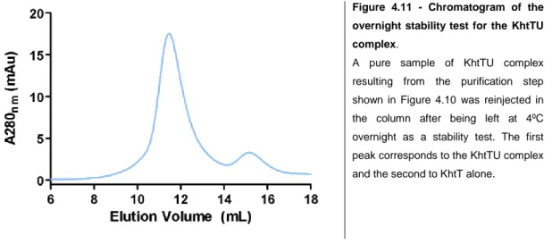
Figure 4.9 - SDS-PAGE gel of KhtTU complex extraction and IMAC purification steps. 26 Figure 4.10 - Chromatogram and corresponding SDS-PAGE gel of KhtTU complex gel filtration. 26 Figure 4.11 - Chromatogram of the overnight stability test for the KhtTU complex. 27 Figure 4.12 - SDS-PAGE of the extraction and IMAC purification steps of the selenomethionine
variant of KhtT (SeMet KhtT). 28
Figure 4.13 - Thermal shift assay ligand testing results for KhtT supplemented with nucleotide
ligands. 30
Figure 4.14 - Thermal shift assay ligand testing results for KhtT supplemented with bacillithiol. 30 Figure 4.15 - Representative ITC curves of interaction studies of c-di-AMP and KhtT. 32 Figure 4.16 - Binding signature (Gibbs Free Energy ΔG, binding enthalpy ΔH and entropy
FCUP/ICBAS Biochemical Characterization of KhtTU, a K+/H+ Antiporter from Bacillus subtilis xii
Figure 4.17 - Representative ITC curves of interaction studies for KhtT and c-di-GMP. 33 Figure 4.18 - SDS-PAGE gels of KhtT controlled proteolysis assays in the presence and absence of
c-di-AMP. 34
Figure 4.19 – Cartoon schematics and representative trace of the KhtTU antiporter assay steps. 35 Figure 4.20 - Comparison of K+/H+ antiporter activity of KhtTU in the presence of di-AMP and
c-di-GMP against basal activity in everted vesicles. 36
Figure 4.21 – Fitting process of the antiporter assay curves. 37 Figure 4.22 - Kinetic curves for the titration of KhtTU vesicles with c-di-AMP. 38 Figure 4.23 - Dose-response curve of the titration of KhtTU vesicles with c-di-AMP. 38 Figure 4.24 - Cation/Proton antiport activity of everted vesicles containing KhtTU in different
monovalent cation gradients with and without 10 μM c-di-AMP. 39 Figure 4.25 - Comparison of K+/H+ antiporter activity of everted vesicles containing KhtTU in the
presence of reduced bacillithiol (BSH) and its oxidized form ethylmaleimide-bacillithiol
(E-BSH) against basal activity. 40
Figure 4.26 - Crystals obtained in the initial crystallization screening for KhtT incubated
with c-di-AMP. 41
Figure 4.27 - Examples of KhtT + c-di-AMP crystals obtained with the handmade reproduction
screens around conditions D10 and F11 from Molecular Dimensions’ Wizard I&II screening
kit. 42
Figure 4.28 - Diffraction pattern of one of the KhtT + c-di-AMP crystals collected using
synchrotron radiation at the Soleil Synchrotron in Paris, France. 43 Figure 4.29 - Examples of SeMet-KhtT + c-di-AMP crystals obtained using handmade screens
around condition F10 of Molecular Dimensions’ Wizard I&II kit. 44 Figure 4.30 - Comparison of the c-di-AMP binding sites between the crystallographic structure of
KtrA of S.Aureus and a 3-D model of KhtT. 45
Figure 4.31 - Sequence alignment of the RCK_C segments of S. aureus KtrA and B. subtilis KhtT. 46
Appendix:
Figure 8.1 - Calibration curve for Superdex 200 10/300 GL gel filtration column (GE Healthcare). 54 Figure 8.2 - Representative curve of KhtT ITC Buffer being titrated with 300 μM c-di-AMP. 54 Figure 8.3 - Ponceau staining of the Western Blot membrane shown in Figure 4.8. 55
FCUP/ICBAS Biochemical Characterization of KhtTU, a K+/H+ Antiporter from Bacillus subtilis xiii
List of Abbreviations
Å Ångström
A280nm Absorbance at a wavelength of 280 nanometers
aa Aminoacids
ACMA 9-Amino-6-Chloro-2-Methoxyacridine ADP Adenosine diphosphate
ATP Adenosine triphosphate BSA Bovine Serum Albumin BSH Bacillithiol (reduced)
Ca2+ Calcium ion
c-di-AMP Cyclic di-adenosine monophosphate c-di-GMP Cyclic di-guanosine monophosphate
cm Centimeter
CPA Cation-Proton Antiporter family CPA2 Cation-Proton Antiporter 2 subfamily
CVs Column Volumes
cytS 84-aminoacids long segment between amino acids 29 and 112 of KhtS
Da Dalton
DDM n-Dodecyl-β-D-maltopyranoside DNA Deoxyribonucleic acid
DSF Differential Scanning Fluorimetry DTT Dithiothreitol
E-BSH Ethylmaleimide-Bacillithiol (oxidized form of BSH) ECL Enhanced chemoluminescence
EC50 Half maximal effective concentration
H+ Hydrogen ion / Proton HCl Hydrochloric acid
HEPES 4-(2-hydroxyethyl)-1-piperazineethanesulfonicacid His/His6 Hexahistidine tag
IMAC Immobilized Metal Affinity Chromatography IPTG Isopropyl β-D-1-thiogalactopyranoside ITC Isothermal Titration Calorimetry k / kfast / kslow Rate constants
K+ Potassium ion
K2HPO4 Potassium phosphate dibasic
FCUP/ICBAS Biochemical Characterization of KhtTU, a K+/H+ Antiporter from Bacillus subtilis xiv
kcal Kilocalories
KCl Potassium Chloride KD Dissociation constant
KH2PO4 Potassium phosphate monobasic
LB Lysogeny broth
LBK Lysogeny broth with potassium
Li+ Lithium ion
M Molar
MCS Multiple Cloning Site
mg Miligram
MgCl2 Magnesium Chloride
mL Mililiter
mM Milimolar
Na+ Sodium ion
NaCl Sodium Chloride
NAD+ Nicotinamide adenine dinucleotide, oxidized NADH Nicotinamide adenine dinucleotide, reduced NaOH Sodium hydroxide
NH4+ Ammonium ion
Ni2+ Nickel ion
NiNTA Nickel-nitrilotriacetic acid
nm Nanometer
OD Optical density
ON Overnight
ORF Open Reading Frame
PCR Polymerase Chain Reaction PDB Protein Data Bank
PEG Polyethylene Glycol
PMSF Phenylmethane sulfonyl fluoride psi Pounds per square inch
Rb+ Rubidium ion
RbCl Rubidium Chloride
RCK Regulator of conductance of K+
RCK_C C-terminal subdomain of an RCK domain RCK_N N-terminal subdomain of an RCK domain RFU Relative fluorescence units
FCUP/ICBAS Biochemical Characterization of KhtTU, a K+/H+ Antiporter from Bacillus subtilis xv
RNA Ribonucleic acid rpm Revolutions per minute SD Standard Deviation SDS Sodium dodecyl sulfate
SDS-PAGE Sodium dodecyl sulfate polyacrylamide gel electrophoresis SEC Size-exclusion chromatography
SeMet Selenomethionine variant SL-BSH S-lactoyl-bacillithiol
T Temperature
TB Terrific Broth TBS Tris-buffered saline
TCEP Tris(2-carboxyethyl)phosphine TFA Trifluoroacetic acid
Thr Thrombin
Tm (ºC) Melting Temperature
Tris Tris(hydroxymethyl)aminomethane
tRNA Transfer RNA
TSA Thermal Shift Assay
α Anti
Δ Variation
ε Molar extinction coefficient
λ Wavelength μcal Microcalories μg Microgram μL Microliter μM Micromolar μM Micromolar
FCUP/ICBAS Biochemical Characterization of KhtTU, a K+/H+ Antiporter from Bacillus subtilis xvi
FCUP/ICBAS Biochemical Characterization of KhtTU, a K+/H+ Antiporter from Bacillus subtilis 1
1. Introduction
1.1. Potassium in Biology
The ionic content of the cytosol is usually much different from that of the extracellular fluid. In essentially all cells cytosolic pH is maintained close to 7.2 and the concentration of potassium (K+) is much higher than sodium (Na+) 1. In fact, K+ is the most abundant intracellular cation in both bacteria and eukaryotes 2. All living cells accumulate K+ to achieve internal concentrations of this cation higher than and relatively independent of those outside 3. In Escherichia coli, for example, K+ is accumulated up to 400 mM 4. In Bacillus subtilis, a potassium pool of about 350-600 mM has been reported 5,6. In human cells, its levels are kept around 140 mM, while other cations such as sodium (Na+) and calcium (Ca2+) have much lower intracellular concentrations (12 mM and <0.2 μM, respectively)1
.
Potassium serves multiple purposes: besides acting as a compatible solute to counteract osmotic stress7–9 and being required for maintaining intracellular pH 10,11, it also affects gene expression12, biofilm formation13, activation of intracellular enzymes14 and is necessary for ribosome function 15,16. In more advanced organisms, such as mammals, potassium is also used to generate a membrane potential, critical for highly important processes such as neurotransmission17,18, muscle contraction 19,20 and heart function 21,22.
One of the factors that determine K+ levels in bacteria is cell turgor pressure. A significant increase in medium osmolarity stimulates K+ influx. This increase in the potassium pool serves as the first line of defense against loss of water and leads to a regain of turgor 9. Conversely, when cells adapt to media of lower osmolarity, the increase in turgor stimulates K+ efflux 3. Changes in cellular K+ concentration alter turgor while turgor is implicated as a signal-controlling activity of K+ transport systems 2. In order to ensure the appropriate intracellular levels of potassium all organisms had to evolve potassium homeostasis systems, regulating both its influx and efflux.
FCUP/ICBAS Biochemical Characterization of KhtTU, a K+/H+ Antiporter from Bacillus subtilis 2
1.2. Potassium Homeostasis in Bacillus subtilis
Since the external concentration of potassium is usually low (only 100 μM to 10 mM in typical bacterial habitats), efficient uptake and intracellular enrichment of the ion is necessary. Most bacteria possess multiple potassium uptake systems to allow efficient transport under different environmental conditions 4.
Many of the prokaryotic ligand-gated K+ channels and some eukaryotic channels are composed of two components: a functional domain consisting of a transmembrane tetrameric pore through which ions flow and a regulatory component consisting of cytosolic ligand-binding domains called “regulator of K+ conductance” (RCK) domains 23, in an assembly similar to the examples shown in Figure 1.1.
Figure 1.1 - Schematic representation of the general assembly of a potassium channel. It is usually composed of two parts: a transmembrane component containing the pore through which potassium ions pass and an oligomeric peripheral regulator component usually containing an RCK domain (regulator of conductance of K+). Legend: Side views of MthK (PDB code: 1LNQ), Slo2 (PDB code: 5A6E), GsuK (PDB code: 4GX1) and ADP-bound KtrAB. Membrane domains in grey; flexible linker or extended helices in yellow; octameric RCK domains in blue. The grey bar represents the membrane. Adapted from 24.
The basic assembly unit of these regulatory domains is a homodimer of RCK subunits. Each one of these domains can be subdivided into two subdomains: the N-terminal subdomain (termed RCK_N) and the C-N-terminal subdomain (RCK_C), usually connected by a linker region 25, as shown in Figure 1.2. Four of these homodimer units then assemble as an octamer, acquiring the architecture shown above.
Figure 1.2 - Representation of the RCK homodimer unit of a prokaryotic potassium channel.
Depiction of the homodimer unit of the RCK domain of MthK, a K+ channel from
Methanothermobacter thermoautotrophicus (PDB
code: 1LNQ). Four of these homodimers bind to each other, assembling the regulatory component of the channel, in the form of an octamer. Each monomer is colored in either cyan or green.
FCUP/ICBAS Biochemical Characterization of KhtTU, a K+/H+ Antiporter from Bacillus subtilis 3
Different ligands have been identified for RCK domains present in the potassium transport machinery of different organisms, such as ATP/ADP26, NADH/NAD+ 27 or divalent cations such as Ca2+ 28. A general mechanism has been proposed for the way RCK domains regulate the activity of the channels 24. In brief, the binding of a ligand to these domains leads to a conformational change that is propagated to its transmembrane counterpart, leading to a more open state of the channel, increasing the flow of K+ 23,24.
In the Gram-positive model organism Bacillus subtilis three different potassium importers have been found: KtrAB and KtrCD 8, which consist of a transmembrane channel-like subunit (KtrB or KtrD) and a peripheral regulatory subunit (KtrA or KtrC); and KimA 29. The KtrA and KtrC regulatory subunits of the KtrAB and KtrCD transporters are RCK domains and exhibit the ability to control the activity of the transporter subunit by binding nucleotides 25. Crystal structures of KtrAB have been obtained (PDB code: 4J7C) 26,30, revealing a complex architecture of a dimer of KtrB bound to a cytosolic octameric ring formed by homodimeric KtrA units (see Figure 1.1, to the right).
Thus, Bacillus subtilis is equipped to acquire potassium from the extracellular media. However, prolonged high levels of potassium ions are detrimental to cellular physiology, mostly when turgor is dangerously high. At this point, potassium efflux is required 9. The only well-characterized K+ efflux systems in bacteria are the E. coli glutathione-gated KefB and KefC systems.31,32 These are potassium-proton (K+/H+) antiporters composed of transmembrane subunits (KefC or KefB) bound to cytoplasmic ancillary subunits (KefF or KefG) 33. In contrast, potassium extrusion in B. subtilis is not completely understood, with two putative K+ export pathways proposed: the YugO channel13,34 and the cation-proton antiporter KhtTU 35.
FCUP/ICBAS Biochemical Characterization of KhtTU, a K+/H+ Antiporter from Bacillus subtilis 4
1.3. The khtSTU Operon
The cation/proton antiporters (CPAs) superfamily of proteins is generally divided into 4 subfamilies. One of which is the CPA2 subfamily. This family of antiporters includes proteins present in different bacteria, archaea and eukaryotes such as the aforementioned E. coli K+/H+ antiporters KefFC and KefGB, the Enterococcus
hirae Na+/H+ antiporter NapA or the Bacillus pseudofirmus OF4 NH4+(K+)/H+ antiporter
AmhMT. 35 These antiporters have roles in resistance to diverse cytotoxic cations, alkali resistance, electrophile resistance and osmoregulation.
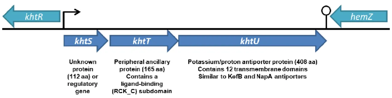
The khtSTU operon of Bacillus subtilis encodes potassium-transport-related proteins that exhibit sequence similarity to CPA2 family members. 36 According to two reference genome databases for Bacillus subtilis, SubtiList 37 and SubtiWiki 38, this operon consists of 3 different ORFs: khtU, khtT and khtS (Figure 1.3). The khtU gene encodes a transmembrane protein with high sequence similarity to the cation/proton antiporter family, with a theoretical size of 44 kDa and 408 amino acids per subunit. Located upstream of khtU, two other genes appear in this operon. First, the operon exhibits the khtT gene, which codes for an ancillary hydrophilic regulator protein (18.5 kDa, 165 amino acids per subunit). Interestingly, khtT only exhibits a partial RCK domain: only the RCK_C subdomain is present, at its C-terminal. Finally, the operon also contains khtS; which is still unclear whether this region codes for a 13 kDa, 112 amino acids protein or is a regulatory element of the operon.
Figure 1.3 - Organization and schematic diagram of the khtSTU operon locus on the Bacillus subtilis
FCUP/ICBAS Biochemical Characterization of KhtTU, a K+/H+ Antiporter from Bacillus subtilis 5
1.4. Current Knowledge of the KhtTU Transporter
The khtSTU operon was referenced in some studies on B. subtilis’ transcriptomic which mention increased transcription in response to heat shock 39, alkaline stress 40, and cell wall stress 41. Fujisawa et al. published a study 36 stating that the operon was upregulated during salt shock and alkali pH, which supports the idea that this protein is part of an osmotic and alkali stress response. Furthermore, it was suggested that both KhtT and KhtS are peripheral ancillary proteins and form a complex with KhtU regulating its activity. More recently, another study by the same group 35 proposed that the protein complex is composed solely by KhtT and KhtU and that the protein complex functions as a K+/H+ antiporter mechanism. The reasons behind the non-inclusion of KhtS were not specified. Using an antiport activity assay, they concluded that KhtU does not have antiporter activity when expressed alone, and that only the KhtTU complex is active. They also showed that activity of the KhtTU antiporter is dependent on pH, showing that activity is null at pH 7.5 and increases with alkaline pH, being highest at pH 9.0. Regarding the selectivity of KhtTU, their results reveal that Rb+/H+ antiport is also possible, while Na+/H+ or Li+/H+ antiport are not. Altogether, these results indicate that the KhtTU antiporter acts as a K+/H+ antiporter that is involved in adaptation to osmotic pressure (by exporting K+) and alkaline stress (by importing H+).
It has also been suggested that KhtTU is involved in methylglyoxal resistance in
Bacillus subtilis 42. Methylglyoxal is a toxic, endogenous by-product of glycolysis that
damages DNA and proteins ultimately leading to cell death. In E.coli the Kef proteins (KefC and KefB) are involved in adaptation to exposure to toxic electrophiles, including methylglyoxal 43,44. Increased K+/H+ antiport activity of Kef proteins by glutathione adducts, such as S-lactoyl-glutathione, causes cytoplasmic acidification, enhancing electrophile resistance and methylglyoxal detoxification 45,46. As such, and due to the sequence similarity between KhtU and KefC it was suggested that KhtTU had a similar role in B. subtilis. Glutathione is absent in Gram-positive bacteria, instead bacillithiol (BSH) is the major reducing agent in B. subtilis. In parallel to the Kef systems of
E. coli, it was proposed that KhtTU is activated by S-lactoyl-bacillithiol (SL-BSH), an
intermediate formed during detoxification pathways 42. However, this hypothesis lacks experimental evidence and there is still no proof that bacillithiol is capable of binding to KhtTU, let alone of activating it.
FCUP/ICBAS Biochemical Characterization of KhtTU, a K+/H+ Antiporter from Bacillus subtilis 6
1.5. Cyclic di-AMP and K
+homeostasis
Cyclic nucleotides that act as second messenger molecules play key roles in signaling pathways that sense environmental changes. The recently discovered cyclic di-AMP (c-di-AMP) (Figure 1.4) is an essential second messenger present in (but not limited to) Gram-positive bacteria such as Bacillus
subtilis 47,48. Intracellular concentrations
between 2 and 5 μM have been measured in cytoplasmic extracts of this bacterium 49.
Figure 1.4 - Structural formula of a
cyclic di-AMP molecule. Adapted from 50.
C-di-AMP is implicated in a variety of functions in the cell, including cell wall metabolism, DNA repair, sporulation, control of gene expression and potassium homeostasis 51. Interestingly, c-di-AMP modulates potassium homeostasis by binding both proteins and RNA molecules. The ktrAB and kimA genes (responsible for expression of the high-affinity potassium uptake proteins mentioned in previous subchapters) are under the control of a riboswitch that responds to c-di-AMP, inhibiting transcription 52. In fact, transcription of these genes is only possible at low intracellular c-di-AMP 52. In addition, c-di-AMP acts as a ligand of the regulatory ancillary protein of the KtrAB system KtrA 53. This protein assembles as a tetramer of dimeric RCK domains and c-di-AMP binds at the dimer interface 54. This interaction between c-di-AMP and the RCK_C domain of KtrA in not limited to B. subtilis, being also found in S. aureus, S. pneumoniae or C. glutamicum 48. Overall, this suggests that c-di-AMP has a role in controlling K+ homeostasis, mostly through inhibiting its uptake beyond normal levels. Conversely, high levels of K+ also increase c-di-AMP levels in the cell, suggesting that the system is bidirectionally regulated 52.
FCUP/ICBAS Biochemical Characterization of KhtTU, a K+/H+ Antiporter from Bacillus subtilis 7
1.6. Importance of Studying Potassium Transporters
As disclosed in the previous sections, potassium transporters play an essential role throughout all domains of Life. In nerve and muscle cells of higher organisms, their crucial role in K+ homeostasis implicates these proteins in the control of action potential frequency and duration, and ultimately in the regulation of neurotransmitter release and hormone secretion, epithelial electrolyte transport, cell proliferation, apoptosis, and tumor progression 55–58.
A relevant application of the knowledge acquired from potassium transporters is related to antibiotic resistance. Antibiotic resistance is a major healthcare problem and there is a pressing need to identify new antibacterial strategies 59. The physiological importance of potassium homeostasis for the survival of bacteria together with the fact that the bacterial machinery that regulates potassium transport is biochemically different from the machinery in mammalians makes bacterial potassium transporters potential new targets for antibacterial strategies. Detailed characterization of the machinery is a crucial first step for the development of these strategies. In fact, investigation of regulatory proteins present in the Ktr or Trk systems has already shown potential in the study of antibiotic susceptibility in known pathogens such as
S.aureus 60, S. enterica 61 or M. smegmatis 62, related to M. tuberculosis.
In conclusion, the biochemical and biophysical study of potassium transporters is of indisputable importance. Visualization of regulatory subunits and the analysis of their binding sites through crystallography would prove a valuable approach in exploring the modulation of transporter activity.
FCUP/ICBAS Biochemical Characterization of KhtTU, a K+/H+ Antiporter from Bacillus subtilis 8
2. Objectives
The main goal for this work was to characterize the molecular properties of the KhtTU antiporter of Bacillus subtilis with particular emphasis on elucidating the role of regulatory protein KhtT. We were interested in answering such questions as: what is the molecular basis of the transporter activity and how is it regulated? If it is regulated by a ligand, what residues comprise its binding site? What is the architecture of the regulatory domain and what are the differences and similarities between KhtT and other RCK domains? What is the function of this antiporter in the potassium homeostasis machinery? Also, what explains its selectivity towards K+? And is it involved in methylglyoxal detoxification through bacillithiol signaling?
Accordingly, the first sub-goal was to design and build an expression vector system for heterologous expression of all the proteins of the khtSTU operon and to determine the optimal expression and purification conditions for each protein. Also, since KhtT and KhtU are expected to act together, the optimization of a protocol for the expression of a KhtTU complex was also an objective.
Subsequently, two different approaches were taken in order to investigate the biochemical and biophysical properties of the KhtU antiporter and its regulatory component KhtT: the study of its functional properties (which consisted on ligand screening by differential scanning fluorimetry, followed by ligand affinity determination by calorimetry and characterization by antiporter activity assays) was complemented with the study of its structural properties (which consisted on attempting to obtain its three-dimensional structure through crystallography and x-ray diffraction).
FCUP/ICBAS Biochemical Characterization of KhtTU, a K+/H+ Antiporter from Bacillus subtilis 9
3. Materials and Methods
3.1. Reagents, Growth Media and Competent Cells
Bacterial growth media was prepared according to Table 3.1; sterilization was achieved by autoclaving for 20 minutes at 121ºC. Information about the E. coli competent cell strains used in molecular biology or small and large-scale protein expression protocols in this work is displayed in Table 3.2. Bacillithiol (BSH) was purchased as (BSH)2 2xTFA salt from Vanderbilt Institute of Chemical Biology,
Vanderbilt University, USA.
Table 3.1 - Composition of growth media used in cell cultures used throughout this work. Lysogeny Broth (LB) 10 g/L Tryptone, 5 g/L Yeast Extract, 10 g/L NaCl
LB with K+ (LBK) 10 g/L Tryptone, 5 g/L Yeast Extract, 10 g/L KCl
Terrific Broth (TB) 20 g/L Tryptone, 24 g/L Yeast Extract, 0.4% Glycerol, 17 mM KH2PO4, 72 mM K2HPO4
2xYT 16 g/L Tryptone, 10 g/L Yeast Extract, 5 g/L NaCl, pH 7.0
Table 3.2 - Genotype information of the E. coli strains used throughout this work. DH5α F– endA1 glnV44 thi-1 recA1 relA1 gyrA96 deoR nupG purB20 φ80dlacZΔM15 Δ(lacZYA-argF)U169, hsdR17(rK–mK+), λ– XL1-Blue recA1 endA1 gyrA96 thi-1 hsdR17 supE44 relA1 lac [F´ proAB lacIq Z∆M15 Tn10 (Tetr )]
BL21 (DE3) F- ompT lon hsdSB (rB-mB-) gal dcm (DE3)
Rosetta™ F- ompT hsdSB(rB- mB-) gal dcm (DE3) pRARE (CamR)
B834 (DE3) F- ompT hsdSB(rB- mB-) gal dcm met (DE3)
KNabc Derivative of TG1 cells (ΔnhaA, ΔnhaB, ΔchaA)
3.2. Molecular Biology
All DNA constructs (see Table 3.3) were created using traditional molecular biology techniques. Insert amplification was performed by using 25-30 cycles of polymerase chain reaction (PCR) with Bacillus subtilis genomic DNA, the oligos shown in the table, and either Pfu or Exact Run DNA polymerases (Thermo Scientific). PCR products were then digested for 2 hours at 37ºC using the appropriate restriction enzymes, as were their respective vectors. Digestion was followed by gel purification and a ligation reaction incubating T4 DNA Ligase and both digests at 4ºC overnight. Ligation reactions were then transformed into DH5α or XL1-Blue cells and plated onto LB-agar plates with the appropriate antibiotic. 3-4 colonies of each plate were selected, inoculated in 5-10 mL of liquid LB with antibiotic and grown at 37ºC, 225 rpm overnight. A second PCR reaction was performed on each culture to determine whether cloning had been successful. DNA from 2 positive clone cultures was extracted using NZYTech’s MiniPrep Kit and sent to sequencing (GATC Biotech, Germany).
FCUP/ICBAS Biochemical Characterization of KhtTU, a K+/H+ Antiporter from Bacillus subtilis 10
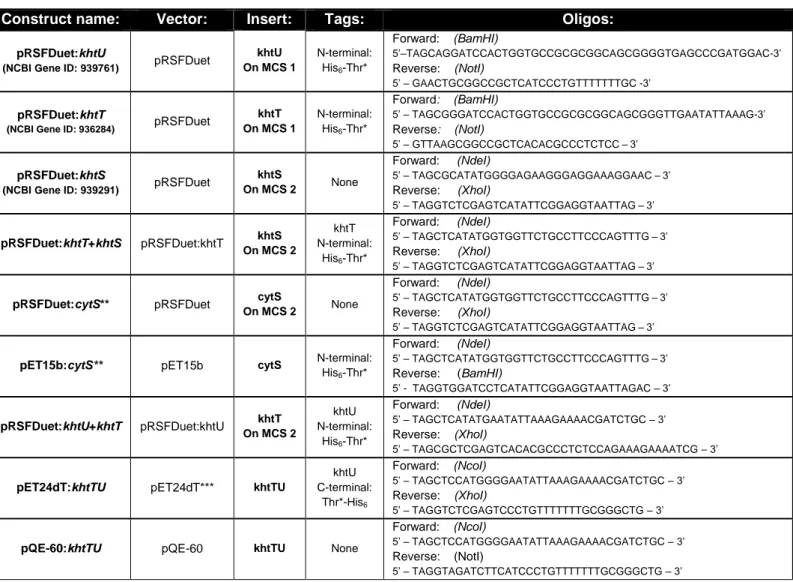
Table 3.3 - Primers and templates used to prepare the vectors used in biochemical studies on KhtT, KhtU, KhtS and KhtTU.
Construct name: Vector: Insert: Tags: Oligos:
pRSFDuet:khtU
(NCBI Gene ID: 939761) pRSFDuet
khtU On MCS 1 N-terminal: His6-Thr* Forward: (BamHI) 5’–TAGCAGGATCCACTGGTGCCGCGCGGCAGCGGGGTGAGCCCGATGGAC-3’ Reverse: (NotI) 5’ – GAACTGCGGCCGCTCATCCCTGTTTTTTTGC -3’ pRSFDuet:khtT
(NCBI Gene ID: 936284) pRSFDuet
khtT On MCS 1 N-terminal: His6-Thr* Forward: (BamHI) 5’ – TAGCGGGATCCACTGGTGCCGCGCGGCAGCGGGTTGAATATTAAAG-3’ Reverse: (NotI) 5’ – GTTAAGCGGCCGCTCACACGCCCTCTCC–3’ pRSFDuet:khtS
(NCBI Gene ID: 939291) pRSFDuet
khtS On MCS 2 None Forward: (NdeI) 5’ – TAGCGCATATGGGGAGAAGGGAGGAAAGGAAC–3’ Reverse: (XhoI) 5’ – TAGGTCTCGAGTCATATTCGGAGGTAATTAG–3’ pRSFDuet:khtT+khtS pRSFDuet:khtT khtS On MCS 2 khtT N-terminal: His6-Thr* Forward: (NdeI) 5’ – TAGCTCATATGGTGGTTCTGCCTTCCCAGTTTG–3’ Reverse: (XhoI) 5’ – TAGGTCTCGAGTCATATTCGGAGGTAATTAG–3’
pRSFDuet:cytS** pRSFDuet cytS
On MCS 2 None
Forward: (NdeI)
5’ – TAGCTCATATGGTGGTTCTGCCTTCCCAGTTTG–3’ Reverse: (XhoI)
5’ – TAGGTCTCGAGTCATATTCGGAGGTAATTAG–3’
pET15b:cytS** pET15b cytS N-terminal:
His6-Thr* Forward: (NdeI) 5’ – TAGCTCATATGGTGGTTCTGCCTTCCCAGTTTG–3’ Reverse: (BamHI) 5’ - TAGGTGGATCCTCATATTCGGAGGTAATTAGAC – 3’ pRSFDuet:khtU+khtT pRSFDuet:khtU khtT On MCS 2 khtU N-terminal: His6-Thr* Forward: (NdeI) 5’ – TAGCTCATATGAATATTAAAGAAAACGATCTGC – 3’ Reverse: (XhoI) 5’ – TAGCGCTCGAGTCACACGCCCTCTCCAGAAAGAAAATCG – 3’
pET24dT:khtTU pET24dT*** khtTU
khtU C-terminal: Thr*-His6 Forward: (NcoI) 5’ – TAGCTCCATGGGGAATATTAAAGAAAACGATCTGC – 3’ Reverse: (XhoI) 5’ – TAGGTCTCGAGTCCCTGTTTTTTTGCGGGCTG – 3’
pQE-60:khtTU pQE-60 khtTU None
Forward: (NcoI)
5’ – TAGCTCCATGGGGAATATTAAAGAAAACGATCTGC – 3’ Reverse: (NotI)
5’ – TAGGTAGATCTTCATCCCTGTTTTTTTGCGGGCTG – 3’
* “Thr” symbolizes a thrombin cleavage site (nucleotide sequence CTGGTGCCGCGCGGCAGC, coding for the amino acid sequence LVPR↓GS, recognized by this protease) embedded in the oligo, to be placed between the polyhistidine tag and the protein sequence.
** “cytS” is the name chosen to designate the 84-aminoacids long segment between amino acids 29 and 112 of khtS, believed to be a hydrophilic, cytosolic part of KhtS, according to structure prediction tools and by previous studies 36.
*** This vector derives from pET-24d but contains a thrombin cleavage site between the stop codon and the polyhistidine tag.
FCUP/ICBAS Biochemical Characterization of KhtTU, a K+/H+ Antiporter from Bacillus subtilis 11
3.3. Small-scale Protein Expression Tests
Protein expression levels for each construct were tested by changing a number of variables: Use of different E. coli strains [BL21(DE3) or Rosetta (strain that supplies extra tRNAs)]; Different growth media [Lysogeny Broth (LB), 2xYT, Terrific Broth (TB), see media composition in section 3.1.]; Different times and temperatures of induction [3 hours at 37ºC or overnight at 20ºC (the latter with induction of heat and cold-shock chaperones by 1% ethanol and ice-bath treatment, respectively]. Induction started by addition of 0.5 mM of isopropyl-β-D-1-thiogalactopyranoside (IPTG).
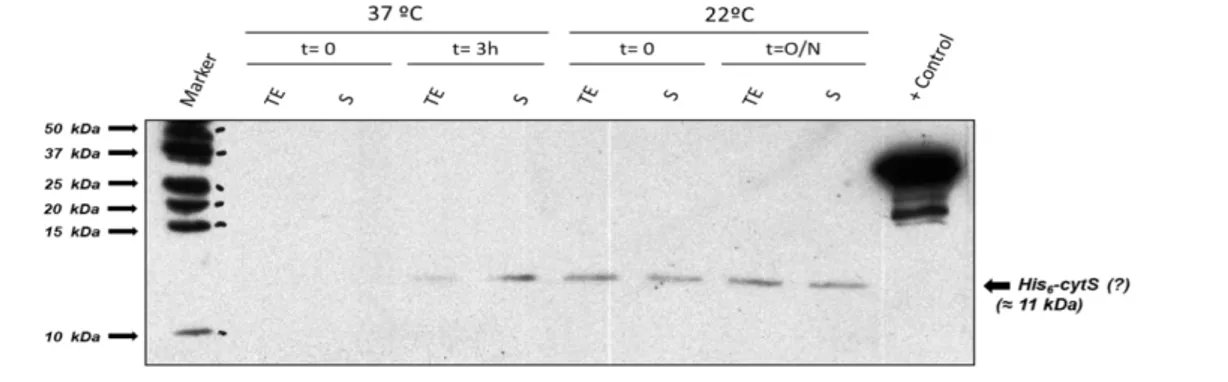
In the case of cytosolic proteins/domains, small-scale expression tests were performed by inoculating 50 mL of liquid media (containing antibiotic) with a resuspension of transformed colonies followed by whatever set of conditions being tested. In the end, cells were pelleted, resuspended in 1 mL of Expression Test Lysis Buffer (50 mM Tris-HCl pH 8.0; 150 mM KCl; 2.5 mM DTT in the presence of protease inhibitors, DNAse and lysozyme) and lysed by sonication cycles (Branson sonifier 250; 3-4 cycles of 15 seconds each, power level 3, duty cycle of 30%). After collecting samples of total extract (TE) (just after sonication) and supernatant (S) (after a 30 min, 12000 g centrifugation of the lysates) fractions, soluble protein levels of expression were assessed by SDS-PAGE. In the case of construct pET15b:cytS, protein levels were also visualized by Western Blotting, using a Mouse-α-His as primary antibody, a horseradish peroxidase conjugated Goat-α-Mouse as secondary antibody and the enhanced chemiluminescence (ECL) reagent method.
In the case of membrane proteins, small-scale expression tests were performed by inoculating 1 L of liquid media (containing antibiotic) with a resuspension of transformed colonies followed by growth until the OD600nm of the culture reached
1.0-1.1. At that point, protein expression was induced and after each induction period the cells were pelleted, resuspended in Membrane Protein Expression Test Lysis Buffer (50 mM HEPES pH 8.0; 210 mM NaCl; 90 mM KCl; 5 mM Imidazole pH 8.0 in the presence of protease inhibitors) and lysed using a prechilled cell cracker (Emulsiflex-C5 – Avestin). Extraction and solubilization of membrane proteins was done by adding 20 mM of n-Dodecyl-β-D-maltoside (DDM) and 5% glycerol to the lysates, followed by overnight rocking at 4ºC. The next day, lysates were centrifuged for 45 mins (25000 g at 4ºC) and the soluble fraction was loaded into an EconoPak column containing 1 mL of preequilibrated Ni2+ affinity bead resin. After washing the beads with 5 CVs of Wash Buffer (50 mM HEPES pH 8.0; 210 mM NaCl; 90 mM KCl; 5% Glycerol; 1mM DDM and 20 mM Imidazole pH 8.0;), the membrane protein of
FCUP/ICBAS Biochemical Characterization of KhtTU, a K+/H+ Antiporter from Bacillus subtilis 12
interest was eluted using increasing amounts of Imidazole in Elution Buffer (25 mM HEPES pH 8.0; 140 mM NaCl; 60 mM KCl, 5% Glycerol; 1mM DDM and 50-500 mM Imidazole pH 8.0). Expression levels for each construct were then assessed by SDS-PAGE and Western Blotting using Mouse-α-His as primary antibody, a horseradish peroxidase conjugated Goat-α-Mouse as secondary antibody and visualization by the enhanced chemiluminescence (ECL) reagent method.
3.4. Pull-down Assay of KhtT and KhtS
His-tagged KhtT was used as bait protein in a pull-down assay performed while testing for KhtS expression using the pRSFDuet:khtT+khtS construct. The pull-down assay was done using Millipore PureProteome™ Nickel Magnetic Beads. The protocol consisted on incubating approximately 1 mL of the lysates obtained in co-expression tests of KhtT and KhtS (section 3.3.) with 25 μL of nickel magnetic bead slurry pre-equilibrated with Tris-buffered saline (TBS) for 1 hour on a rotating mixer, at room temperature. Beads and supernatant were separated using the magnetic properties of the beads and a PureProteome™ Magnetic Stand. After removing the unbound fraction and washing the beads 5 times with 500 μL of TBS supplemented with 20 mM Imidazole, the beads were resuspended with 50 μL SDS-PAGE sample buffer, boiled for 5 minutes and loaded on an SDS-PAGE gel for analysis.
3.5. KhtT Expression and Purification Protocol
3.5.1. Native KhtT
The standard protocol used for large-scale native KhtT expression and purification used throughout this work consisted on the steps that follow: E. coli BL21(DE3) competent cells were transformed with pRSFDuet:khtT expression vector and plated onto LB plates containing 50 μg/mL kanamycin. The next day, all colonies of the plate were resuspended, inoculated into 1 L of liquid LB (with 50 μg/ml kanamycin) grown at 37ºC, 160 rpm in an orbital shaker until OD600nm reached 0.8, at
which point cells were induced with 0.5 mM IPTG and incubated 3 h further. Induced cultures were then pelleted at 3000 g for 20 minutes at 4ºC and stored at -20 ºC.
FCUP/ICBAS Biochemical Characterization of KhtTU, a K+/H+ Antiporter from Bacillus subtilis 13
Cell lysis was performed by resuspending the cells in KhtT Lysis Buffer (50 mM Tris-HCl pH 8.0; 150 mM KCl) followed by cell cracking using a prechilled Emulsifex-C5 (Avestin). Unwanted proteolytic digestion was inhibited by adding 1 mM phenylmethylsulfonyl fluoride (PMSF), 1 μg/mL Leupeptin and 1 μg/mL Pepstatin A to the cell homogenate. Crude cell extract was then clarified by centrifugation (45 minutes at 25000 g, 4ºC). Supernatant fraction was collected, incubated with preequilibrated NiNTA beads rocking for 90-120 minutes at 4ºC and loaded on a column. Beads were then washed with 10xCV of Wash Buffer (50 mM Tris-HCl pH 8.0; 300 mM KCl; 10 mM Imidazole), His-tagged KhtT was eluted by addition of Elution Buffer (50 mM Tris-HCl pH 8.0; 150 mM KCl; 50 mM Imidazole) and the amount of protein obtained was estimated by reading the absorbance of the eluate at 280nm, using a molar extinction coefficient [ε(KhtT monomer) = 9970 M-1 cm-1] predicted by the ProtParam software, a
tool from ExPASy’s resource portal. His-tagged KhtT protein fraction was then dialyzed overnight against KhtT Dialysis Buffer (20 mM Tris-HCl pH 8.0; 150 mM KCl in a volume ratio of 1:100 to 1:200) in the presence of 1-2 μg of Bovine Alpha Thrombin per mg of KhtT protein in order to cleave off the polyhistidine tag. The following day, dialyzed fraction was loaded onto a second preequilibrated NiNTA bead column to further select only untagged KhtT. Protein fraction was then concentrated using PALL or VivaSpin concentrators and injected in a preequilibrated size-exclusion chromatography Superdex 200 10/300 GL column (GE Healthcare) after passing through a 0.22 μm filter. Fractions containing pure KhtT were then pooled together and a final protein fraction was obtained in KhtT Buffer (20 mM Tris-HCl pH 8.0; 150 mM KCl). Small fractions were kept throughout the whole process for SDS-PAGE analysis.
3.5.2. Selenomethionine Variant of KhtT
The expression of a selenomethionine (SeMet) variant of KhtT was done using
E. coli B834(DE3) (a methionine auxotroph strain) and Molecular Dimension’s
SelenoMet™ commercial kit, which consisted in growing the cells in a synthetic M9 minimal media supplemented with glucose, vitamins and all the amino acids with the exception of L-methionine. To this media 4 mL of a 10 mg/mL L-Selenomethionine stock (from the kit) were added per liter of culture. From this point forward the expression and purification protocol was exactly the same as with the native KhtT, with the addition of 1 mM TCEP to every buffer as a reducing agent being the only difference.
FCUP/ICBAS Biochemical Characterization of KhtTU, a K+/H+ Antiporter from Bacillus subtilis 14
3.6. KhtTU Expression and Purification Protocol
The standard large-scale protocol for KhtTU complex expression and purification developed in this work consists on the following steps: E. coli BL21(DE3) competent cells were transformed with pET24dT:khtTU expression vector and plated onto LB plates containing 50 μg/mL kanamycin. The next day, all colonies of the plate were resuspended, inoculated into 1 L culture flasks with liquid 2xYT (with 50 μg/ml kanamycin) and grown in an orbital shaker at 37ºC, 160 rpm until OD600nm reached
1~1.1, at which point cells were induced with 0.5 mM IPTG and incubated 3 h further. Induced cultures were then pelleted for 20 mins at 3000 g, 4ºC and stored at -20 ºC.
Pellets were resuspended in KhtTU Lysis Buffer (50 mM HEPES pH 8.0; 100 mM NaCl; 50 mM KCl; 10 mM Imidazole pH 8.0) followed by cell cracking using a prechilled Emulsifex-C5 (Avestin). Unwanted proteolytic digestion was inhibited by adding 1 mM PMSF, 1 μg/mL Leupeptin and 1μg/mL Pepstatin A to the cell homogenate. At this point, 20 mM DDM and 5% glycerol were added to the lysate, which was then left rocking overnight at 4ºC for extraction of cellular membranes. The following day, the lysate was centrifuged (45 minutes, 25000 g, 4ºC) and the supernatant was loaded into a preequilibrated nickel affinity purification column. The flow-through was reloaded into the column to increase the probability of binding. Beads were then washed with 10xCV of Wash Buffer (50 mM HEPES pH 8.0; 210 mM NaCl; 90 mM KCl; 5% Glycerol; 1 mM DDM; 20 mM Imidazole pH 8.0) and bound KhtTU complex was eluted using Elution Buffer (25 mM HEPES pH 8.0; 140 mM NaCl; 60 mM KCl; 5% Glycerol; 1mM DDM; 150 mM Imidazole pH 8.0). Protein fraction was immediately diluted 1:5 in Elution Buffer without imidazole to reduce protein aggregation and dialyzed against KhtTU Dialysis Buffer (25 mM HEPES pH 8.0; 140 mM NaCl; 60 mM KCl; 5% Glycerol; 1mM DDM) in the presence of 1 μg of Bovine Alpha-Thrombin / mg protein. As with KhtT, protein amount was estimated by reading the absorbance of the eluate at 280 nm and using a predicted molar extinction coefficient [ε(KhtTU monomers) = 31400 M-1 cm-1]. The following day, dialyzed protein
was concentrated and injected in a preequilibrated size-exclusion chromatography Superdex 200 10/300 GL column (GE Healthcare). Fractions containing KhtTU protein complex in final KhtTU buffer (25 mM HEPES pH 8.0; 140 mM NaCl; 60 mM KCl; 1mM DDM) were pooled together, concentrated and re-dialyzed to reduce detergent levels to a minimum.
FCUP/ICBAS Biochemical Characterization of KhtTU, a K+/H+ Antiporter from Bacillus subtilis 15
3.7. Ligand Testing by Thermal Shift Assay
The thermal denaturation temperature of purified native KhtT was studied both with the absence and presence of potential ligands, in an attempt to identify ligands of this protein. Each sample consisted of a final concentration of 3 μM pure KhtT (in KhtT Buffer: 50 mM Tris pH 8.0; 150 mM KCl), a 2.5 X final concentration of Sypro Orange Protein Gel Stain and the final concentration of each ligand being tested varied between 0.1 mM and 10 mM. The total volume of each sample was either 40 or 50 μL. The experiment was then setup in a 96-well PCR White Plate (Bio-Rad) and performed in a iQ5 real-time PCR system (Bio-Rad) using the Cy3 filter specific for Sypro Orange detection (λex = 490 nm / λem = 575 nm), with the temperature varying from 25 ºC to 95 ºC in 0.5 ºC increments of 30 seconds each. Data was then analyzed using the CFX Manager Software (Bio-Rad).
C-di-AMP and c-di-GMP were obtained from BioLog in powder form and solutions were prepared by resuspension in 100 mM Tris-HCl pH 8.0. Reduced BSH was obtained by resuspending the salt in 20 mM HEPES-NaOH pH 7.5 and 2 mM TCEP. Oxidized BSH was obtained by overnight incubation of the reduced form with 10 mM N-ethylmaleimide, an alkylating reagent reactive towards thiols, rendering ethylmaleimide-bacillithiol (E-BSH). The resulting products were not evaluated.
3.8. Isothermal Titration Calorimetry (ITC)
Preparation of KhtT prior to ITC was done as described in section 3.5, with the addition of a second dialysis step the day before the assay was performed, against ITC Dialysis Buffer (50 mM HEPES pH 8.0; 150 mM KCl). Dialyzed protein was then centrifuged (30 minutes at 12000 g, 4ºC) to remove any possible aggregates and the ligands being tested (either c-di-AMP or c-di-GMP) were prepared by diluting stock aliquotes in ITC Dialysis Buffer. 3 subsets of experiments were performed: The first consisted on 20 or 30 μM KhtT samples in the cell titrated with 150 or 300 μM ligand samples in the syringe, respectively. The second subset consisted on a reverse titration: samples of 6 μM ligand in the cell were titrated with 175 μM KhtT set in the syringe. The last set of experiments consisted on control samples: ITC Dialysis Buffer in the cell and 300 μM ligand samples in the syringe, for reference.
Each experiment was executed in a MicroCal VP-ITC calorimeter (GE Healthcare), using 2 mL of cell sample volume and 500 μL of syringe sample volume, at 25 ºC (unless otherwise noted). Data were visualized and analyzed with Origin 7 Software (by OriginLab) and with AFFINImeter ITC Data Analysis Software [by Software for Science Developments (S4Sd)] using a single binding site model.
FCUP/ICBAS Biochemical Characterization of KhtTU, a K+/H+ Antiporter from Bacillus subtilis 16
3.9. Controlled Proteolysis Assay
The effect of c-di-AMP on KhtT’s proteolytic pattern was assessed by a controlled proteolysis assay. 15 μL samples containing 15 μg (50 μM) of pure protein were digested with commercial proteases (Trypsin, Chymotrypsin) at different protease-to-protein ratios (1:50; 1:100; 1:500; 1:1000) for 1 hour at room temperature in the absence or presence of 50 μM c-di-AMP. The reaction was stopped with the addition of SDS sample buffer followed by boiling and each sample was loaded onto a 17% SDS-PAGE gel for analysis of the digestion patterns.
3.10. KhtTU Everted Vesicles and Antiporter Activity Assay
Everted vesicles containing KhtTU antiporter were prepared by growing KNabc cells (a K+/H+, Na+/H+ and Ca+/H+ antiporters deficient E. coli strain) previously transformed with pQE60:khtTU in liquid LBK media (supplemented with 100 μg/mL ampicillin) until OD600nm reached 0.9-1.0, at which point cultures were induced with
0.5 mM IPTG, incubated at 37ºC for 3 hours and pelleted by a 20 minutes, 3000 g centrifugation. The cell pellet was then resuspended in minimum volume of Vesicle Lysis Buffer (10 mM Tris-HCl pH 8.0; 140 mM Choline Chloride, 0.5 mM DTT and 250 mM Sucrose), pelleted again and weighed. Cells were then resuspended in Vesicle Lysis Buffer and lysed by passage through a French Press at a pressure of 4000 psi in the presence of DNAse and 5 mM MgCl2. Unbroken cells were pelleted by
two sequential centrifugations of 15 min at 10000 g. Everted vesicles were extracted from the spin-cleared lysate by 1 hour centrifugation at 100.000 g, with the resulting pellet being resuspended once, washed and pelleted again. Vesicle pellet was resuspended in 1 mL Vesicle Lysis Buffer per gram of original wet cell weigh, total protein concentration was estimated using Bradford reagent method using bovine serum albumin (BSA) as standard and vesicles were stored in small aliquotes at -80ºC. The same protocol was applied using an empty pQE-60 vector in order to obtain vesicles without the antiporter as a negative control.
To perform the assay, vesicles were diluted in Vesicle Assay Buffer (15 mM Tris-HCl pH 8.5; 140 mM Choline Chloride; 5 mM MgCl2). Each sample
contained approximately 200 μg of total protein in a volume of 2 mL. 9-Amino-6-Chloro-2-Methoxyacridine (ACMA) was used as a fluorescence probe to quantify changes in transmembrane proton gradient; ACMA was added at a final concentration of 2 μM
FCUP/ICBAS Biochemical Characterization of KhtTU, a K+/H+ Antiporter from Bacillus subtilis 17
seconds before running the assay. Fluorescence was measured using a HORIBA Fluoromax-4 cuvette spectrofluorometer with constant stirring settings and excitation and emission wavelengths of 410 nm and 480 nm respectively. An integration rate of 0.2 seconds and a 2-nm slit were selected. A respiration-generated pH gradient was generated by addition of 4 mM D-Lactate, resulting in a quenching in ACMA fluorescence. KhtTU antiporter activity was studied by addition of a salt (KCl, RbCl or NaCl) and generation of a monovalent cation gradient. The effect of c-di-AMP and c-di-GMP on the function of KhtTU was studied by addition of the ligand to the sample cuvette seconds before the salt was added, initiating the antiporter activity. The effect of reduced bacillithiol (BSH) and its oxidized form (E-BSH) was also tested in this experiment. All assays were performed in triplicates and at room temperature. All curves were fitted with single or double exponentials using GraphPad Prism software.
3.11. Crystallography
Initial crystallization screens for native KhtT were performed using purified protein at a concentration of 8-10 mg/mL in KhtT buffer (20 mM Tris-HCl pH 8.0; 150 mM KCl), centrifuged for 30 minutes at 12000 g at 4ºC in a table-top centrifuge to clear any aggregates, and the following commercial screens: JBScreen Basic, JBScreen JCSG++ HTS L (Jena Biosciences); Morpheus, Wizard I&II, PACT Premier, MIDAS (Molecular Dimensions), Nextal Classics L Suite and Nextal Classics II Suite (QIAGEN). When testing or using the effect of a ligand, the same was added to the protein fraction and left incubating on ice for 30 minutes before setting up the plates.
Each initial screening drop consisted on 0.6 μL drops (0.3 μL protein + 0.3 μL reservoir solution) and 40 μL of reservoir solution and incubated at 4ºC and 20ºC. Any crystal-generating condition was optimized by fine tuning around each component of said condition (handmade screens varying precipitant concentrations, pH, drop size, protein-to-reservoir ratio; see Appendix, Table 8.3 and Table 8.4) and handmade screens were set up in 48-well plates with a drop size of 2 μL (1 μL protein + 1 μL reservoir solution) with 150 μL reservoir solution volume. Drops were set up using the sitting drop method with a protein crystallization robot (Oryx 4 by Douglas Instruments).
FCUP/ICBAS Biochemical Characterization of KhtTU, a K+/H+ Antiporter from Bacillus subtilis 18
The selenomethionine variant of KhtT was crystallized using the same handmade screens around hit conditions successfully used for native KhtT, in the exact same conditions. Heavy atom soaking of native KhtT crystals was performed by transferring them to a new replica drop with addition of 1 mM Heavy Atom Solution to the drop (description of each stock in Appendix, Table 8.2) followed by overnight incubation.
Native KhtT, SeMet-KhtT and heavy atom-soaked KhtT crystals were prepared for data acquisition by transferring them to cryoprotectant solutions with increasing percentage of cryoprotecting agent (up to 25% glycerol, ethylene glycol or PEG400) and ultimately flash-freezing them in liquid nitrogen. Data acquisition was done by lab colleagues at the Soleil Synchrotron in Paris, France at either the Proxima I or Proxima II beamlines.
3.12. Cyclic-di-AMP Binding Site Comparison
A three-dimensional model for the RCK_C domain of KhtT was obtained using Swiss-Model 63, a protein structure homology-modelling server and the structure of the RCK domain of MthK, a K+ channel from M. thermoautotrophicus 28 (PDB code: 4L74), as a template. This model was then compared with the structure of the RCK_C domain of KtrA of S. aureus bound to c-di-AMP 54 (PDB code: 4XTT), in order to try to estimate which residues may constitute the binding site in KhtT. The mapping of the residues that formed hydrogen bonds between c-di-AMP and the residues of each protein was done and visualized with PyMOL. A pairwise sequence alignment between KhtT and KtrA of S. aureus was also done via the Clustal Omega (EMBL-EBI) sequence alignment tool.
FCUP/ICBAS Biochemical Characterization of KhtTU, a K+/H+ Antiporter from Bacillus subtilis 19
4. Results & Discussion
4.1. Protein Expression and Purification
The first step in characterizing the biochemical and biophysical properties of the proteins involved in the khtSTU operon was to determine and optimize the conditions for large-scale expression and purification.
4.1.1. KhtT Expression and Purification
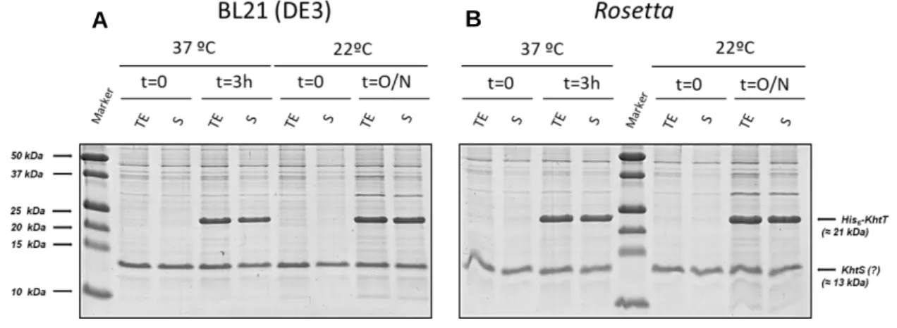
The best conditions for expression of soluble KhtT were tested as described in section 3.3. In short, the general strategy was growing different transformed E. coli strains in different media, temperature and/or induction periods. After growing the cells in different conditions, the level of soluble protein expression was assessed by SDS-PAGE analysis (Figure 4.1).
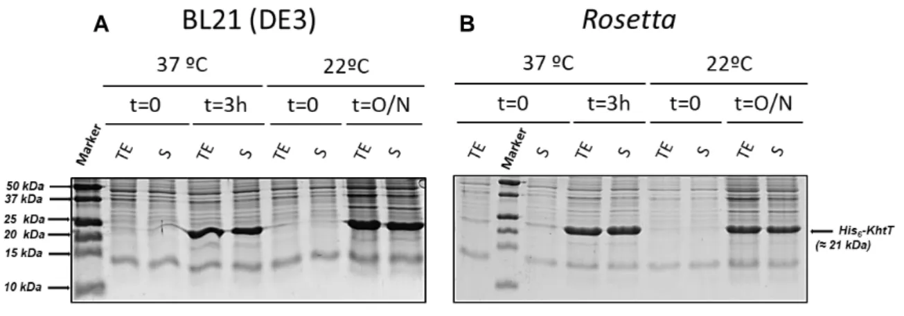
Figure 4.1 - SDS-PAGE gels of KhtT expression tests in (A) BL21(DE3) and (B) Rosetta E.coli strains. Cells were lysed by sonication and centrifuged. Pre-centrifugation Total Extract (TE) and post-centrifugation supernatant (S) samples were then loaded on a 17% SDS-PAGE gel and stained with NZYTech’s BlueSafe gel stain. Molecular weight protein markers are indicated on the left (Bio-Rad’s Precision Plus Protein™ All Blue Prestained Protein Standards); the expected position of His-tagged KhtT is marked on the right. Legend for each lane is displayed above each gel.
The gels show that KhtT is produced in both E. coli strains as a soluble protein in apparently large quantities regardless of time of induction. As such, the simplest conditions were selected to work with: 3 hours of induction at 37ºC. Large-scale expression and purification was then optimized, ultimately being standardized as described in section 3.5. The outcome of the first steps of KhtT purification protocol, consisting on cell lysis, lysate centrifugation, immobilized metal affinity chromatography (IMAC) using nickel beads and thrombin digestion are shown in Figure 4.2. Digested KhtT was then further purified using size exclusion chromatography; the chromatogram obtained is shown in Figure 4.3, together with a gel analysis of the peak fractions.
FCUP/ICBAS Biochemical Characterization of KhtTU, a K+/H+ Antiporter from Bacillus subtilis 20
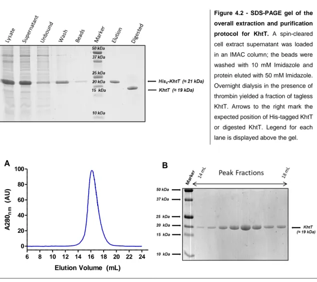
Figure 4.2 - SDS-PAGE gel of the overall extraction and purification protocol for KhtT. A spin-cleared cell extract supernatant was loaded in an IMAC column; the beads were washed with 10 mM Imidazole and protein eluted with 50 mM Imidazole. Overnight dialysis in the presence of thrombin yielded a fraction of tagless KhtT. Arrows to the right mark the expected position of His-tagged KhtT or digested KhtT. Legend for each lane is displayed above the gel.
Figure 4.3 - (A) Chromatogram of KhtT size exclusion chromatography and (B) SDS-PAGE gel of peak fractions. Protein peak elutes at approximately 16 mL of elution volume in the Superdex 200 10/300 GL column. The chromatogram also shows that there are no aggregates in the void volume (8 mL). Samples from each peak fraction (approximately 0.5 mL each) were loaded on a 17% SDS-PAGE gel and stained with NZYTech’s BlueSafe gel stain. Arrows to the left indicate protein markers (Bio-Rad’s Precision Plus Protein™ All Blue Prestained Protein Standards) and arrow to the right marks the expected position of KhtT in each lane.
KhtT’s size exclusion chromatography profile shows a single sharp peak suggesting that the protein is stable and monodisperse in the conditions used. There is no aggregate in the void volume (8 mL) and no other peak is observed (no contaminants). The comparison of the peak elution volume (16 mL) with a previously determined calibration curve for this column (see Appendix, Figure 8.1) allows an estimation of the oligomeric state of the protein. The apparent molecular weight is 38 kDa, corresponding to a dimer.
The protocol for expression and purification of KhtT yields approximately 10 mg of pure and well behaved protein per liter of culture.
6 8 10 12 14 16 18 20 22 24 0 20 40 60 80 100 Elution Volume (mL) A 2 8 0nm (A U ) A B
FCUP/ICBAS Biochemical Characterization of KhtTU, a K+/H+ Antiporter from Bacillus subtilis 21
4.1.2. KhtS Expression Testing
Expression tests for KhtS followed a slightly different approach. The double gene expression vector pRSFDuet:khtT+khtS was constructed for two reasons: first, since KhtS is coded in a separate promoter, its levels of expression could be assessed as in an individual construct. The second reason is that since this vector simultaneously codes for His6-tagged KhtT on one promoter and tagless KhtS on the
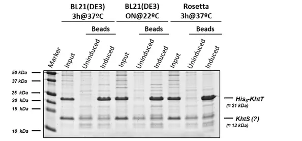
other, the hypothesis of an eventual complex formation by both proteins could also be tested by a pull-down assay (by binding KhtT to a bead resin and visualizing the proteins existing on the eluted fraction). Conditions for expression of KhtS using this construct were tested as shown above for KhtT. Soluble protein levels for each condition tested were assessed by SDS-PAGE (Figure 4.4).
Figure 4.4 - SDS-PAGE gels of KhtS expression and KhtT co-expression tests in (A) BL21(DE3) and (B) Rosetta E. coli strains. Cells were lysed by sonication and centrifuged. Pre-centrifugation total extract (TE) and post-centrifugation supernatant (S) samples were then loaded on a 17% SDS-PAGE gel and stained with NZYTech’s BlueSafe gel stain. Molecular weight protein markers are indicated on the left (Bio-Rad’s Precision Plus Protein™ All Blue Prestained Protein Standards); the expected positions of His-tagged KhtT and KhtS are marked on the right. Legend for each lane is displayed above each gel.
As expected, KhtT was expressed in high soluble quantities. On the other hand, it was not possible to observe an induced 13 kDa band corresponding to KhtS. There is a 13 kDa protein that appears in all conditions, prior and after induction. It is likely that this is a contaminant that could conceal a modest level of expression of KhtS. As such, a pull-down assay (Figure 4.5) using the lysates from the expression tests was performed as a way to clarify this uncertainty and to simultaneously test if KhtS can form a complex with KhtT.