UNIVERSIDADE NOVA DE LISBOA
INSTITUTO DE HIGIENE E MEDICINA TROPICAL
Activity of Compounds Isolated from Carpobrotus edulis on Efflux
Pumps of Bacteria and Cancer Cells
Ana Sofia Fernandes Martins
Thesis research submitted to the Instituto de Higiene e Medicina Tropical, Universidade Nova de Lisboa in partial fulfilment of the requirements for the granting of the degree of Doctor of Philosophy with specialization in the Biomedical Science of Microbiology
The Thesis research to be described was conducted at the Unit of Mycobacteriology, UPMM at the Instituto de Higiene e Medicina Tropical, Universidade Nova de Lisboa (IHMT, UNL) and supported by grant SFRH/BD/19445/2004 provided by Fundação para a Ciência e a Tecnologia (FCT) of Portugal.
SUPERVISOR:
Professor Doutor Leonard Amaral
Professor Catedrático Convidado e Director da Unidade de Micobactérias Unidade de Micobactérias, UPMM
Instituto de Higiene e Medicina Tropical Universidade Nova de Lisboa
TUTORIAL COMMISSION:
Professora Doutora Maria José Umbelino Ferreira Professora Associada com Agregação
Medical Chemistry group
iMed.UL, Research Institute for Medicines and Pharmaceutical Sciences Faculdade de Farmácia
Universidade de Lisboa
Professor Doutor Miguel Viveiros Professor Auxiliar de Bacteriologia Unidade de Micobactérias
Instituto de Higiene e Medicina Tropical Universidade Nova de Lisboa
Professor Doutor Leonard Amaral
Professor Catedrático Convidado e Director da Unidade de Micobactérias Unidade de Micobactérias, UPMM
Instituto de Higiene e Medicina Tropical Universidade Nova de Lisboa
Na presente dissertação incluem-se resultados que foram ou estão a ser alvo de publicação em co-autoria. Os artigos publicados ou submetidos para publicação serão integralmente apresentados em anexo. Para efeitos do disposto no nº1 do Despacho nº2303/2000 do Regulamento de Programas de Doutoramento do Instituto de Higiene e Medicina Tropical, Universidade Nova de Lisboa (Diário da República, 2ª Série, nº 23, de 28 de Janeiro de 2000), o autor da dissertação declara que interveio na concepção e execução do trabalho experimental, na interpretação dos resultados e na redacção dos manuscritos publicados, submetidos ou que aguardam submissão.
Lisboa, 14 de Outubro de 2009
___________________________________________ Ana Sofia Fernandes Martins
“The History of Medicine
• 2000 B.C. – Here, eat this root
• 1000 A.D. – That root is heathen. Here, say this prayer.
• 1850 A.D. – That prayer is superstition. Here, drink this potion. • 1920 A.D. – That potion is snake oil. Here, swallow this pill. • 1945 A.D. – That pill is ineffective. Here, take this penicillin. • 1955 A.D. – Oops....bugs mutated. Here, take this tetracycline.
• 1960-1999 A.D. – 39 more "oops"...Here, take this more powerful antibiotic. • 2000 A.D. – The bugs have won! Here, eat this root.”
ACKNOWLEDGMENTS
The concretization of this thesis would not be possible without the help of my family, friends and colleagues. All of them helped me to make it true.
First of all, I would like to thank my supervisor Professor Leonard Amaral to have given me the opportunity to work at his Unit. Once you told I was like a butterfly in the making, but with your help I am learning how to fly. Thank you all the teachings and continuous challenging that made me understand how exciting is to make science. Thank you to have introduced me to the fantastic and infinite world of Efflux Pumps, Chemo-resistance and, of course, Carpobrotus edulis.
Professor Miguel Viveiros I would like to thank for all the careful revisions of the written work, scientific discussions and advices during these years, making them a little bit easier.
Professor Isabel Couto, I would like to thank you for all your critical review of the work that always helped me to improve it.
To my colleges at the Mycobacteriology Unit, Gabriella Spengler and Marta Martins for the transmition of knowledge and introduction to microbiology laboratory practice. Liliana Rodrigues, Sofia Costa, Diana Machado, Susana Costa and Jorge Ramos thank you for the possibility of working together with all of you.
I would like to thank Professor József Molnár, from the University of Szeged, for his friendship welcoming me always as a member of his lab and making possible all the activity measurements in the cancer cells. To him and his wife Evi, that always received me as a granddaughter: Köszönöm szépen!
Professor Judit Hohmann, from the University of Szeged, thank you for the possibility of working at your laboratory. Without your help and your great knowledge in solving NMR spectra, the purification and identification of Carpobrotus edulis compounds would have been much difficult.
Professor Jean-Marie Pagès and Professor Seamus Fanning, thank you for the complementary work made at your laboratories, which greatly improve the discussion of the results of this dissertation.
Professor Maria José Umbelino Ferreira, thank you for being part of the tutorial commission of this dissertation and for the advices you gave me to improve this work. Andrea Vasas, thank you for your big help and advices during the purification of the compounds and all the reviews of that part of the work.
To my Hungarian colleagues and friends Zsuzsana Schelz, Julianna Serly and Noémi Tóth a big thank you to make the life in the lab much easier, helping in all the translations needed and in everything!
To Anikó Váradi and Erzsébet Hadárné Berta for their precious technical contribution, Imre Ocsovszki for the flow cytometry measurements, Péter Forgó for the NMR measurements and Mária Báthori for spectral analyses.
To Gabi (the black sheep), Susana (the other one), Gabi (“mana”), Joana and Cátia, I would like to thank for your friendship and all your patience to listen and support me in those critical moments. You are the best!
To my friends “Diálogos” for all your friendship, understanding and all those moments that fulfill my life of happiness and made myself a better human being. You helped me to believe that “the world is our home”!
Zsuzsi, Miki, András, Márti, Eszter, Ági, Gergő, Zita thank you for the wonderful welcoming at the Szent Imre Kollégium. I will never forget our talks, laughing and, of course, the studding room time! ...and the classes of Hungarian at the kitchen! Not even the lunches at Horgos with “my Hungarian family”… Köszönöm szépen!
Attila, my Kismadár, thank you for being part of my life, fulfilling it with positivity and teaching me how to look at the good side of all the things in life. In Portugal, Hungary or Taiwan, you will be always in my heart. Szeretlek!
Finally, I would like to give a special thanks to my family:
Aos meus avós todo o apoio que me tem dado, mesmo quando não entendem o que estudo nem porque é que demora tanto tempo. Adoro-vos muito e é um privilégio ser vossa neta. Obrigada pela ajuda de sempre nos momentos mais difíceis! Obrigada por tudo!
Aos meus pais, um obrigada do tamanho do mundo! Tenho muito orgulho em ser vossa filha. Obrigada pelas oportunidades que sempre me deram ao longo da vida e que me ajudaram a ser quem sou. Obrigada por serem duas pessoas maravilhosas e por estarem sempre, sempre, sempre ao meu lado e nunca me deixarem desistir. Obrigada, ainda, por sempre acreditarmos juntos que “o Amor é a única linguagem que todos os Homens entendem”. Força!
To all of you and some others I found during the concretization of this work and helped somehow in its development, a big thank you for being part of my life.
This thesis was supported by Fundação para a Ciência e a Tecnologia (FCT) grant SFRH/BD/19445/2004 and by a short period Fundação Calouste Gulbenkian (FCG) grant.
ABSTRACT
Introduction: Resistance to antibiotics and chemotherapy is a major health problem in Portugal and also globally. Nowadays, a significant proportion of clinical Gram-negative isolates are multi-drug resistant (MDR) and whenever studied, the MDR phenotype has been shown to be mediated by over-expressed efflux pumps (EPs). The over-expression of bacterial EPs is known to result from their exposure to one antibiotic that in some manner renders the bacterium with an MDR phenotype. Nevertheless, the process by which the development of an MDR phenotype that occurs during the period the patient is being treated with an antibiotic has yet to be completly demonstrated in the laboratory. Moreover, the degree of resistance of the Gram-negative clinical isolate is often-times many fold greater than the constant concentration used in therapy and reached in the patient plasma.
Among Enterobacteriaceae, the major EP belongs to the RND superfamily which is mainly driven by energy coming from the proton motive force (PMF). Environmental factors such as Calcium (Ca2+), pH or glucose (energy source) have major influence in
the mechanisms of retention or efflux of compounds by the cell. However, because the cell envelope is the first bacterial cell component to face changes in the environmental conditions such as hydrostatic pressure, osmolarity or antibiotic pressure, it is essential to have an over-view of all the processes involved in the acquisition of resistance. Therefore it is worthy to understand how such environmental conditions influence the outer-membrane composition of the cell and its mechanism of efflux.
The first part of this dissertation focuses on the effect of such environmental conditions, on the composition of the outer membrane and the cellular responses It was, then, studied the role of antibiotic-promoted stress via step-wise increasing concentrations of antibiotic or serial passages of the bacterial strain in the same concentration of antibiotic, simulating what happens in the patient when she/he is submitted to long periods of antibiotic therapy.
Efflux modulators can be used in therapy together with antibiotics for improvement of antibiotic action. Their use starts to be widely accepted as a new approach for the
therapy of multi-drug resistance. Therefore, the second part of this dissertation focuses on the purification and characterization of compounds purified from the plant Carpobrotus edulis whose methanolic extract had been previously shown to inhibit MDR EPs of bacteria. Because it was previously shown by others the relationship between EP in bacteria and cancer cells, the purified compounds were also studied for their inhibitory activity on one of the major efflux pump transporters of cancer cells (P-gp).
Methods: Methods of protein extraction and electrophoresis were employed to assess the composition of the outer membrane after the bacterial cells face two different kind of growth media: solid and liquid. The effect of the antibiotic pressure on the EP expression was studied by growing different bacterial strains under increasing concentration of antibiotic or maintaining them in the same concentration for longer periods of time. The progeny strains were then tested for their response to the antibiotics in the presence of EPI and for their EP expression by real time reverse transcription PCR (rtRT-PCR). The effect of efflux pump modulators such as CCCP, PAβN, verapamil, phenothiazines, and the modulating effects of calcium, pH and energy source were studied by the semi-automated ethidium bromide (EB) method that follows the accumulation or efflux of EB, on a real time manner, by the bacterial cells under the conditions applied to the media. The assessment of C. edulis compounds for in vitro activity against wild type bacterial strains and their counterpart strains that over-produce given EPs was conducted by determination of the minimum inhibitory concentration (MIC) of the purified compounds as well as for other antibiotics of reference for each strain in the presence of the compounds to be tested. The activity of the compounds as efflux modulators were also tested by the semi-automated EB method, already mentioned. The compounds were also assessed for their capacity to increase the killing activity of macrophages infected with bacterial strains: ex vivo activity. Finally, the purified compounds were tested for their antiproliferative effect on cancer cell lines and their capacity to inhibit the P-gp responsible for the multi-drug resistance in those cell lines.
Results: During this study it was observed that in liquid medium a greater expression of a 55kDa protein takes place as opposed to Salmonella strains grown in solid medium. The simulation of the response of bacteria to the therapy with antibiotics through the two different adaptation processes showed that the bacterial response is dependent upon the method of adaptation to the antibiotic used.
The results of this dissertation also suggest that efflux and accumulation of EB by E. coli strains are dependent on pH and energy that influence the performance of the AcrAB pump. This EP is dependent upon protons present in the periplasm for its activation. The efflux response is independent of the pH of growth of the bacteria whereas it is dependent on the pH of the assay, suggesting that bacteria are able to adapt to different environmental conditions such as pH and presence of noxious agents. Due to its capacity for binding protons, CCCP was used at different pH, in order to understand the role of protons and PMF on the efflux. The use of CCCP together with variations in the pH helped to identify the main types of efflux transporters that respond to the different environmental conditions. However, PAβN modulates efflux of ethidium bromide by competing with it for the site of extrusion of the pump (a KM was determined).
Oleanolic acid, β-amyrin, uvaol, catechin, epicatechin, MGDG and procyanidin B5 were the compounds isolated from the plant C. edulis. It was observed that the activity of some of these compounds was differed according to the mechanisms of resistance that characterizes the different strains against which their activity was studied. This is in agreement with the results obtained for the response of the bacterial cell adapted through different mechanisms to the use of efflux modulators. The results suggest that the triterpene uvaol was the most active compound as an efflux modulator in bacteria and cancer cells. It also has significant activity against intracellular Staphylococcus aureus.
Conclusion: A 55kDa protein was previously described as a virulence factor. The same protein had less expression when the bacteria were grown in presence of a phenothiazine, a compound described as an efflux modulator. Consequently, the action of these compounds as adjuvants may be due to their capacity to reduce the virulence of the strain. Therefore the results obtained for bacteria grown in solid and liquid media
are of extreme importance because they can be an evidence for the reasons by which these compounds are described as helper compounds. They can also indicate why infections by the same organism but through different food sources have different degrees of infection and virulence on the patient.
Adaptation caused by serial passages in the same concentration of antibiotic suggests the presence of “mutator” genes that allows the cell to survive under stress conditions and reduce energy consumption that would otherwise be higher with the over-expression of efflux systems as occurs when bacteria is exposed to step-wise increasing concentrations of antibiotic.
The results of this dissertation also suggest that the AcrAB mediated efflux is dependent upon protons present in the periplasm for their activation. Hence, when E. coli faces stress conditions caused by a noxious agent, its extrusion would be preferentially performed by an ABC type transporter at pH greater than 7. The efflux response is independent on the pH of growth of the bacteria but dependent on the pH of the assay suggesting that bacteria are able to adapt to different conditions such as environmental pH that it has to face during the infection process in the human body. Energy dependent efflux mechanisms vary upon the pH and the conjunction of pH and glucose is an important tool in the study and understanding of the physiology and mechanisms of efflux. Efflux pumps belonging to the ABC superfamily have an important role in efflux at pH 8; however, PMF is essential for RND family mediated efflux as per the results obtained at pH 5. The use of compounds that interfere with the PMF or directly affect the efflux systems has also a relevant role in the study of the efflux mechanisms and their physiology.
Based on the results obtained with compounds purified from C. edulis, this plant is a promising source for search of more effective antibacterial, antimycobacterial and anticancer compounds. It is worthy to mention that the extremely easy availability of this plant in the coast of Portugal makes it an outstanding raw material for large scale production of its constituents which is essential for the development of any products to be used in practical medicine.
RESUMO
Introdução: A resistência aos antibióticos é um grave problema de saúde quer em Portugal quer a nível mundial. Nos dias de hoje, uma grande percentagem dos isolados clínicos de bactérias Gram-negativas é multi-resistente (MR) e, sempre que estudado, o fenótipo MR é mediado pela expressão de bombas de efluxo (BE). A sobre-expressão de bombas de efluxo em bactérias resulta da exposição destas a um antibiótico que, por vários processos, lhes confere um fenótipo MR. Contudo, o processo pelo qual a estirpe bacteriana desenvolve resistência durante a terapia com determinado antibiótico, ainda não foi completamente demonstrado em laboratório. Frequentemente, o grau de resistência dos isolados clínicos é muitas vezes superior à dose constante de antibiótico usada na terapia e atingida no plasma do paciente.
Entre as Enterobacteriaceae, a principal BE pertence à família RND, na qual a energia necessária ao efluxo provém da força proto-motriz (PMF). Factores do meio envolvente, tais como, cálcio, pH ou glucose (fonte de energia), têm extrema influência nos mecanismos de retenção ou efluxo de compostos pela célula. Contudo, uma vez que o invólucro celular é o primeiro componente bacteriano a enfrentar alterações das condições do meio onde a bactéria se encontra, tais como alteração da pressão hidrostática, osmolaridade ou pressão antibiótica, é essencial compreender a generalidade dos processos envolvidos na aquisição de resistência. Assim, é urgente compreender como é que as condições do meio influenciam a composição da membrana celular e os seus mecanismos de efluxo.
A primeira parte desta dissertação estuda o efeito dessas condições na composição da membrana externa e nas respostas celulares. Foi então estudado o efeito do stress provocado quer por aumentos crescentes na concentração de antibiótico, quer por passagens sucessivas da estirpe bacteriana em concentrações constantes de antibiótico, simulando o que acontece no paciente quando submetido a longos períodos de terapia com antibiótico.
Moduladores do efluxo podem ser usados em terapia, conjuntamente com os antibióticos, de modo a aumentar o seu efeito terapêutico. A sua utilização começa a ser
aceite como uma nova abordagem terapêutica contra a multi-resistência. Deste modo a segunda parte desta tese foca a purificação e caracterização de compostos isolados da planta Carpobrotus edulis, cujo extracto metanólico mostrou, anteriormente, inibir BE bacterianas. Uma vez que foi já demonstrado existir uma relação entre BE de bactérias e de células cancerígenas, foi também estudado o efeito inibitório dos compostos purificados num dos transportadores com mais relevância em multi-resistência em células cancerígenas (P-gp).
Métodos: Foram utilizados métodos de extracção de proteínas e electroforese para estudar a composição da membrana externa de células bacterianas cujo crescimento ocorreu em dois meios diferentes: sólido e líquido. O efeito da pressão antibiótica na expressão de BEs foi estudada através do crescimento de estirpes bacterianas em concentrações crescentes de antibiótico ou mantendo-as em concentrações de antibiótico constantes por longos períodos de tempo. No final das sucessivas passagens foi estudada a resposta celular a diferentes antibióticos na presença de moduladores de efluxo bem como os níveis de expressão de BEs por RT-PCR em tempo real. O efeito de moduladores de efluxo tais como CCCP, PAβN, verapamil ou fenotiazinas e os efeitos do cálcio, pH e fontes de energia foram estudados pelo método semi-automático que segue a acumulação ou efluxo de brometo de etidium pelas células bacterianas, em tempo real, nas condições aplicadas ao meio do ensaio experimental.
O estudo das actividades in vitro dos compostos isolados de C. edulis em relação a estirpes de referência e outras multi-resistentes, que sobre-expressam determinadas BE, foi realizado por determinação das concentrações mínimas inibitórias dos compostos, bem como de outros antibióticos, aos quais as estirpes eram resistentes, na presença do composto. O método semi-automático atrás referido foi também utilizado no estudo destes compostos como moduladores de efluxo. A influência destes compostos na morte de estirpes bacterianas fagocitadas por macrófagos foi também estudada: ensaios ex vivo. Por fim, foi estudada a actividade antiproliferativa dos compostos isolados em linhas celulares cancerígenas bom como a sua capacidade de inibição da P-gp responsável pela multi-resistência nessas linhas celulares.
Resultados: Durante este estudo foi observado que em meio líquido há maior expressão de uma proteína com 55kDa em oposição ao que acontece quando a bactéria cresce em
meio sólido. A simulação da resposta bacteriana durante a terapia pelos dois processos descritos, mostrou que a resposta bacteriana é dependente do processo de adaptação seguido.
Os resultados desta dissertação sugerem, também, que o efluxo e a acumulação de EB por células de E. coli são dependentes do pH e de energia, os quais influenciam o desempenho da bomba de efluxo AcrAB. Esta BE depende da concentração periplasmática de protões para a sua activação. O efluxo é independente do pH do meio onde as células bacterianas cresceram, contudo, é dependente do pH do ensaio, o que sugere que a bactéria é capaz de se adaptar a diferentes condições do meio tais como pH ou agentes prejudiciais à sua sobrevivência. Devido à sua capacidade quelante, o composto CCCP foi usado a diferentes pH com o objectivo de compreender o papel da concentração protónica e da PMF no efluxo. O uso de CCCP juntamente com variações no pH, possibilitou a identificação dos principais tipos de sistemas de efluxo que respondem às diferentes condições do meio. Contudo, o composto PAβN interfere com o efluxo de EB, por competição com este, pelo sítio activo da bomba de efluxo (um KM
para esta competição foi determinado).
Os compostos isolados da planta C. edulis foram: ácido oleanólico, β-amirina, uvaol, catequina, epicatequina, MGDG e procianidina B5. Foi observado que estes compostos tinham diferentes actividades consoante o mecanismo de resistência característico de cada uma das estirpes em que a sua actividade foi estudada. Este facto está de acordo com os resultados obtidos para a resposta celular de bactérias, cuja multi-resistência foi obtida por diferentes mecanismos, perante o uso de moduladores. Os resultados obtidos sugerem que, de entre os compostos isolados, o composto uvaol foi o mais activo como modulador da actividade de efluxo, quer em células bacterianas quer em células cancerígenas. Também demonstrou uma actividade significativa contra Staphylococcus aureus intracelular.
Conclusão: Uma proteína de 55kDa foi anteriormente descrita como factor de virulência. A mesma proteína encontrava-se menos expressa em bactérias cultivadas na presença de uma fenotiazina, um composto descrito como modulador de efluxo. Deste modo a acção destes compostos como adjuvantes terapêuticos pode dever-se à sua capacidade de reduzir a virulência de determinada estirpe. Deste modo, os resultados
obtidos, quando células bacterianas cresceram em meios líquido e sólido, são extremamente importantes pois podem indicar o motivo pelo qual infecções pelo mesmo organismo, mas por via de diferentes origens alimentares, apresentam diferentes graus de infecção e virulência para o paciente.
A adaptação induzida por passagens sucessivas em meio com a mesma concentração de antibiótico sugere a presença de genes “mutantes” cuja actividade possibilita a sobrevivência celular em condições de “stress”, reduzindo o consumo de energia. De outro modo este seria mais elevado devido à sobre-expressão dos sistemas de efluxo, tal como acontece quando a bactéria é sujeita a passagens em concentrações crescentes de antibiótico. Os resultados desta dissertação também sugerem que a activação do efluxo, mediado pela bomba de efluxo AcrAB, é dependente da concentração protónica no periplasma. Assim, quando células de E. coli experimentam condições adversas causadas por agentes tóxicos, o efluxo é efectuado preferencialmente por transportadores do tipo ABC se o pH for maior que 7. O facto de o efluxo ser uma resposta independente do pH a que a estirpe cresceu, mas dependente do pH do meio em que o ensaio está a decorrer, sugere que a bactéria é capaz de se adaptar a diferentes pH do meio, tais como os que encontra durante o processo de infecção. Os mecanismos de efluxo dependentes de energia também variam com o pH. Deste modo a conjugação destes dois factores é muito importante para o estudo e compreensão da fisiologia e dos mecanismos de efluxo. As BEs que pertencem à família ABC têm uma função importante a pH 8, contudo a PMF é fundamental para o efluxo por via dos transportadores da família RND, como observado nos ensaios a pH 5. O uso de compostos que interferem com a PMF ou afectam directamente os sistemas de efluxo tem também um papel relevante no estudo dos mecanismos de efluxo e sua fisiologia. Os resultados obtidos com os compostos purificados da planta C. edulis, sugerem que esta planta contem compostos promissores com actividade antibacteriana e anticancerígena. É importante salientar que a abundância desta planta na orla marítima de Portugal faz com que a produção em larga escala dos seus constituintes seja fácil, o que é um factor essencial no desenvolvimento de quaisquer produtos a usar na prática clínica.
PUBLICATIONS
Publications resulted from this thesis are listed below and a copy of each is presented in Appendix.
1. Martins M, Santos B, Martins A, Viveiros M, Couto I, Cruz A, Pagès JM, Molnar J, Fanning S and Amaral L; Management Committee Members; of Cost B16; European Commission/European Science Foundation. (2006) An instrument-free method for the demonstration of efflux pump activity of bacteria. In Vivo. 20: 657-664;
2. Martins A, Couto I, Aagaard L, Martins M, Viveiros M, Kristiansen JE and Amaral L. (2007) Prolonged exposure of MRSA COL is exposed to increasing concentrations of oxacillin result in an MDR phenotype. Int. J. Antimicrob. Agents. 29(3):302-305;
3. Martins M, Schelz Zs, Martins A, Molnar J, Hajös G, Riedl Z, Viveiros M, Yalcin I, Aki-Sener E and Amaral L. (2007) In vitro and ex vivo activity of thioridazine derivatives against Mycobacterium tuberculosis. Int. J. Antimicrob. Agents. 29(3): 338-340;
4. Schelz Zs, Martins M, Martins A, Viveiros M, Molnar J and Amaral L. (2007) Elimination of plasmids by SILA compounds that inhibit efflux pumps of bacteria and cancer cells. In Vivo. 21(4): 635-639;
5. Viveiros M, Martins A, Paixão L, Rodrigues L, Martins M, Couto I, Fähnrich E, Kern WV and Amaral L. (2008) Demonstration of intrinsic efflux activity of Escherichia coli K-12 AG100 by an automated ethidium bromide method. Int. J. Antimicrob. Agents. 31(5):458-462;
6. Viveiros M, Martins M, Couto I, Rodrigues L, Spengler G, Martins A, Kristiansen JE, Molnar J and Amaral L. (2008) New methods for the identification of efflux mediated MDR bacteria, genetic assessment of regulators and efflux pump
constituents, characterization of efflux systems and screening for inhibitors of efflux pumps. Curr. Drug Targets. 9(9):760-778;
7. Spengler G, Martins A, Rodrigues L, Aagaard L, Martins M, Costa SS, Couto I, Viveiros M, Fanning S, Kristiansen JE, Molnar J and Amaral L. (2009) Characterization of intrinsic efflux activity of Enterococcus faecalis ATCC29212 by a semi-automated ethidium bromide method. In Vivo. 23(1): 81-87;
8. Spengler G, Viveiros M, Martins M, Rodrigues L, Martins A, Molnar J, Couto I and Amaral L. (2009) Demonstration of the activity of the P-glycoprotein by a semi-automated fluorometric method. Anticancer Research. 29(6): 2173-2177;
9. Martins A, Spengler G, Rodrigues L, Viveiros M, Ramos J, Martins M, Couto I, Fanning S, Pagès JM, Bolla JM, Molnar J and Amaral L. (2009) pH modulation of efflux pump activity of multi-drug resistant E. coli: Protection during its passage and eventual colonization of the colon. PloS One 4(8) e6656;
10. Spengler G, Ramalhete C, Martins M, Martins A, Serly J, Viveiros M, Molnar J, Duarte N, Mulhovo S, Ferreira MJU and Amaral L. (2009) Evaluation of Cucurbitane-type Triterpenoids from Momordica balsamina on P Glycoprotein (ABCB1) by Flow cytometry and Real-time Fluorometry. Anticancer Research.29: 3989-3994;
11. Martins A, Iversen C, Rodrigues L, Spengler G, Ramos J, Kern WV, Couto I, Viveiros M, Fanning S, Pages JM and Amaral L. (2009) An AcrAB-mediated multidrug-resistant phenotype is maintained following restoration of wild-type activities by efflux pump genes and their regulators. IJAA, in press;
12. Martins A, Vasas A, Schelz Zs, Viveiros M, Molnár J, Hohmann J and Amaral L. (2009) Constituents of Carpobrotus edulis Inhibit the P-glycoprotein of mdr1 Transfected Mouse Lymphoma Cells. Anticancer Research, submitted.
Bellow, Presentations that resulted from the work of this thesis are also listed:
1. Martins A, Aagaard L, Couto I, Martins M, Viveiros M, Kristiansen JE and Amaral L, “Prolonged Exposure of MRSA COL to Increasing Concentrations of Oxacillin Result in an MDR Phenotype”, 8th European Congress on Chemotherapy and Infection (FESCI 8), Budapest, Hungary, 25th to 28th of October 2006;
2. Martins A, Ferreira MJU, Viveiros M, Amaral L, “Activity of Carpobrotus edulis extracts against multi-drug resistant bacteria”, MICRO-BIOTEC 08, Lisboa, Portugal, 30th of November to 2nd of December 2007.
3. Martins A, Vasas A, Schelz Zs, Martins M, Viveiros M, Molnár J, Hohmann J, Amaral L, “Purification and identification of active compounds of Carpobrotus edulis against the reversal of resistance of human mdr1 gene transfected mouse lymphoma cells”, 7th Joint Meeting of AFERP, ASP, GA, PSE & SIF, Natural Products with Pharmaceutical, Nutraceutical, Cosmetic and Agrochemical Interest, Athens, Greece, 3rd to 8th of August 2008.
4. Martins A, Vasas A, Schelz Zs, Viveiros M, Molnár J, Hohmann J, Spengler G, Amaral L, Constituents of Carpobrotus edulis inhibit p-glycoprotein of human mdr1 gene transfected mouse lymphoma cells, 8th International Conference of Anticancer Research, Kos, Greece, 17th to 22nd of October 2008.
5. Amaral L, Spengler G, Viveiros M, Rodrigues L, Martins A, Couto I, Martins M, Fanning S, Pages JM, Molnár J, Assessment and comparison of efflux pumps of cancer cells and MDR bacteria under physiological conditions by a real-time semi-automated system, 8th International Conference of Anticancer Research, Kos, Greece, 17th to 22nd of October 2008.
6. Spengler G, Viveiros M, Martins A, Rodrigues L, Martins M, Molnar J, Couto I, Amaral L, Demonstration of the activity of P-glycoprotein by a fully automated ethidium bromide method, 8th International Conference of Anticancer Research, Kos, Greece, 17th to 22nd of October 2008.
7. Spengler G, Viveiros M, Martins A, Rodrigues L, Martins M, Molnar J, Couto I, Amaral L, Evaluation of the activity of the P-glycoprotein by an automated real-time fluorometric system. International Congress on Anti-Cancer Treatment, 3rd to 6th February, Paris, France 2009.
8. Spengler G, Viveiros M, Martins A, Rodrigues L, Molnár J, Amaral L, Applications of real-time fluorimetry to study efflux pump activity in bacteria and cancer. CEFORM – Central European Forum for Microbiology, Keazthely, Hungary, 7th to 9th of October 2009
9. Costa S, Martins A, Spengler G, Amaral L, Viveiros M, Bioenergetic characterization of efflux in Escherichia coli strains. CEFORM – Central European Forum for Microbiology, Keazthely, Hungary, 7th to 9th of October 2009
10. Martins A, Spengler G, Costa S, Viveiros M, Amaral L, Influence of Calcium and pH in the accumulation and efflux of EB. MICRO-BIOTEC 09, Vilamoura, Portugal, 28th to 30th of November 2009.
11. Spengler G, Viveiros M, Martins A, Rodrigues L, Molnár J, Amaral L, Real-time fluorometric evaluation of P-glycoprotein inhibitors in cancer cells. MICRO-BIOTEC 09, Vilamoura, Portugal, 28th to 30th of November 2009. 12. Costa S, Martins A, Spengler G, Amaral L, Viveiros M, ATPase inhibitors as
new efflux pump inhibitors of Escherichia coli. MICRO-BIOTEC 09, Vilamoura, Portugal, 28th to 30th of November 2009.
TABLE OF CONTENTS
ACKNOWLEDGMENTS ... vii ABSTRACT ... xi RESUMO ... xv PUBLICATIONS ... xix TABLE OF CONTENTS ... xxiii FIGURE INDEX ... xxvii TABLE INDEX ... xxxi LIST OF ABBREVIATIONS ... xxxiii
I. INTRODUCTION ... 1 1. Chemotherapeutics and resistance ... 3
1.1 The new Era of the antibiotics ... 3 1.2 Fight against resistance ... 6 1.3 Mechanisms of resistance ... 6 2. Cell envelope and resistance... 10 2.1 Cell envelope ... 10 2.1.1 Gram-positive ... 14 2.1.2 Gram-negative ... 14 2.1.3 Mycobacteria ... 15 2.2 Lipopolysaccharides composition ... 15 2.3 Other important functions of membranes ... 19 2.3.1 Plasma membrane bound enzymes. ... 20 3. Transport across the cell envelope (its outer and inner membranes) ... 22 3.1 Outer membrane proteins and porins ... 23 3.2 Efflux pump mechanisms ... 25 3.2.1 Primary active transporters ... 27 3.2.2 Secondary active transporters ... 28 4. Mycobacterium tuberculosis – an emerging problem of resistance ... 32 4.1 The bacillus of Tuberculosis... 32 4.2 The disease ... 33 4.3 TB and antibiotic resistance ... 34 4.4 Cell characteristics, infection and resistance ... 36
5. Multi-drug resistance and cancer ... 40 6. New therapy approaches ... 46 6.1 Efflux modulators ... 46 6.2 Other membrane interacting compounds ... 47 7. Importance of plants in therapy ... 50 7.1 Plants as sources of chemotherapeutics and modulators of cell mechanisms 51 8. Carpobrotus edulis ... 56 8.1 Uses in traditional medicine ... 57 8.2 Activity of Carpobrotus spp. ... 57 II. AIMS OF THE STUDY ... 59 III. MATERIALS AND METHODS ... 63 1. General bacteriology procedures ... 65 1.1 Bacterial strains ... 65 1.2 Cellular cultures ... 66 1.3 Determination of Minimal Inhibitory Concentration (MIC) ... 67 1.4 Semi-automated EB method ... 68 1.4.1 Accumulation Assay ... 68 1.4.2 Efflux Assay ... 69 2. Evaluation of OMP from Salmonella ... 70 2.1 Growth conditions ... 70 2.2 Protocol of Extraction ... 70 2.3 Role of antibiotic-promoted stress ... 71 2.3.1 Step-wise increasing concentrations of antibiotic ... 71 2.3.2 Serial passages in the same concentration of antibiotic ... 72 3. pH and energy roles on efflux by Gram - negatives ... 72 4. Search for new active compounds against resistance ... 73 4.1 Plant material ... 73 4.2 General purification procedure ... 73 4.3 Extraction and isolation ... 74 4.4 Activity measurements against bacteria ... 78 4.4.1 Minimum Inhibitory Concentration of C. edulis compounds ... 78 4.4.2 Modulation of resistance ... 78 4.4.3 Toxicity assays ... 79
4.4.4 Ex-vivo assays ... 79 4.5 Activity measurements on eukaryotic cell lines ... 81 4.5.1 Cell lines ... 81 4.5.2 Antiproliferative assay ... 81 4.5.3 Inhibition of P-gp on mdr1 gene transfected mouse lymphoma cells ... 82 4.5.4 Checkerboard assay for interaction of compounds with anti-cancer agents. . 83 IV. RESULTS ... 85
1. Growth conditions and the bacterial cell envelope ... 89 1.1 Growth environment and OMP expression ... 89 1.2 Role of antibiotic-promoted stress ... 91 1.2.1 Step-wise increasing concentrations of antibiotic ... 91 1.2.2 Serial passages in the same concentration of antibiotic ... 93 2. pH and energy roles on efflux by Gram-negatives ... 96 2.1 Role of glucose ... 97 2.2 Role of pH ... 100 2.3 Role of efflux modulators ... 103 2.3.1 CCCP ... 104 2.3.2 PAβN ... 107 2.3.3 Verapamil ... 110 2.3.4 Phenothiazines ... 111 2.4 Role of Calcium ... 117 3. Search for new active compounds against resistance ... 123 3.1 Purification and identification of the compounds... 123 3.2 In vitro activity of the isolated compounds on bacteria... 125 3.3 Modulation of resistance in bacteria ... 128 3.3.1 The modulation of antibiotic resistance of Gram-negative strains ... 128 3.3.2 The modulation of antibiotic resistance of Gram-positive strains ... 130 3.4 Activity on the accumulation of EB ... 131 3.4.1 Gram-negative bacteria ... 131 3.4.2 Gram-positive bacteria ... 137 3.5 Activity ex-vivo against S. aureus strains ... 140 3.6 Activity on Eukaryotic cell lines ... 142 3.6.1 Anti-proliferative assays ... 142
3.6.2 Reversal of resistance in eukaryotic cells ... 143 3.6.3 Checkerboard assay ... 148 V. DISCUSSION ... 151
1. Role of the environment in the membrane of Gram–negative bacteria ... 153 1.1 Growth, environment and OMP expression ... 153 1.2 Role of antibiotic-promoted stress ... 155 1.2.1 Correlation between the two studies ... 159 2. Energy and pH roles on efflux by Gram - negatives ... 162 2.1 Role of glucose ... 166 2.2 Modulation of accumulation and efflux of EB by pH. ... 166 2.3 Role of efflux modulators ... 171 2.3.1 CCCP ... 172 2.3.2 PAβN ... 173 2.3.3 Verapamil ... 174 2.3.4 Phenothiazines ... 175 2.4 Role of Calcium ... 176 3. Search for new active compounds against resistance ... 179 3.1 Purification and identification of the compounds... 180 3.2 Activity of C. edulis compounds against bacteria ... 183 3.3 Anticancer activity ... 185 3.4 Structure activity relationship ... 186 VI. FINAL REMARKS AND FUTURE PERSPECTIVES ... 189
1. Physiology and modulation of efflux ... 191 2. Activity of C. edulis isolated compounds ... 194 VII. REFERENCES ... 197
FIGURE INDEX
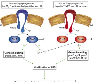
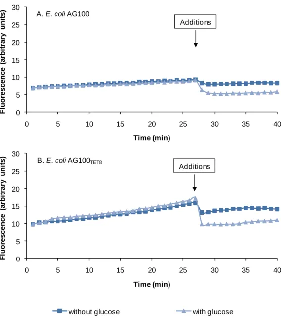
Figure 1 - Schematic representation of cell mechanisms of resistance. ... 7 Figure 2 – Schematic representation of the cell envelope of a bacterial cell. ... 10 Figure 3 - General structure of the cell membrane. ... 11 Figure 4 - Chemical structure of Hopanoid. ... 12 Figure 5 - Organization of the bacterial cell envelope. ... 13 Figure 6 - Lipopolysaccharide (LPS) composition ... 16 Figure 7 - Model of the activation and interaction of the PhoP/Q and PmrA/B two-component system in Salmonella spp. ... 18 Figure 8 – Structure of OmpF porin trimer. ... 23 Figure 9 – Schematic representation of the 5 families of membrane transporters in Gram-positive and Gram-negative bacteria. ... 26 Figure 10 - Schematic representation of each representative families belonging to the secondary membrane transporters. ... 31 Figure 11 - Distribution of the confirmed cases of XDRTB all over the world, until June 2008. ... 35 Figure 12 - Clinical and cellular outcomes of Mycobacterium tuberculosis and the role of the adaptive immune system. ... 37 Figure 13 – Membrane topology of the MDR-associated ABC transporters MDR1, MRP1. ... 43 Figure 14 – Proposed mechanism of action for antimicrobial peptides. ... 49 Figure 15 - Carpobrotus edulis. ... 56 Figure 16 –Protocol of purification of the compounds from the plant C. edulis. ... 77 Figure 17 - Gel SDS-PAGE 8,5% of S. enteritidis 104 and S. enteritidis 5048. ... 90 Figure 18 - The effect of glucose on the accumulation of EB by E. coli AG100 (A) and E. coli AG100TET8 (B). ... 98
Figure 19 - The effect of glucose on the efflux of EB by E. coli AG100 (A) and E. coli AG100TET8 (B). ... 99
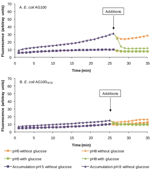
Figure 20 - The effect of pH and the need for metabolic energy for efflux of EB by E. coli AG100 (A) and E. coli AG100TET8 (B). ... 102
Figure 21 – Accumulation of EB in glucose and glucose free media pH 5, 7 and 8 by E. coli AG100. ... 103 Figure 22 – Effect of CCCP concentrations on efflux of EB by E. coli AG100 (A) and E. coli AG100TET8 (B) at pH 5 and 8. ... 105
Figure 23 - Effects of PAβN on efflux of EB by E. coli AG100 at pH 5 and 8. ... 108 Figure 24 - Effects of different concentration of PAβN on efflux of EB by E. coli AG100TET8 at pH 5 and 8. ... 109
Figure 25 - Competition between EB and PAβN: calculation of Km for PAβN relative to EB. ... 109 Figure 26 - The effects of concentrations of verapamil on the efflux of EB by E. coli AG100at pH 8. ... 110 Figure 27 - Accumulation of EB by E. coli AG100 (A) and E. coli AG100TET8 (B) at pH
7 and in the presence and absence of glucose and different concentrations of CPZ. ... 112 Figure 28 - Accumulation of EB by E. coli AG100 (A) and E. coli AG100TET8 (B) at pH
7 and in the presence and absence of glucose and different concentrations of TZ. ... 113 Figure 29 - Efflux of EB by E. coli AG100 (A) and E. coli AG100TET8 (B) at pH 7 and
in the presence and absence of glucose and different concentrations of CPZ. ... 114 Figure 30 - Efflux of EB by E. coli AG100 (A) and E. coli AG100TET8 (B) at pH 7 and
in the presence and absence of glucose and different concentrations of TZ. ... 115 Figure 31 - Efflux of EB by E. coli AG100 at pH 5 and 8, in the presence and absence of glucose and different concentrations of TZ. ... 116 Figure 32 – Calcium role on the efflux of EB by E. coli AG100 at pH 5. ... 118 Figure 33 – Calcium role on the efflux of EB by E. coli AG100 at pH 8. ... 119
Figure 34 – Effect of catechin on the accumulation of EB by E. coli AG100 in presence and absence of glucose. ... 133 Figure 35 – Effect of epicatechin on the accumulation of EB by E. coli AG100 in presence and absence of glucose. ... 133 Figure 36 – Effect of oleanolic acid on the accumulation of EB by E. coli AG100TET8 in
presence and absence of glucose. ... 134 Figure 37 - Effect of oleanolic acid on the efflux of EB by E. coli AG100 in presence and absence of glucose. ... 135 Figure 38 - Effect of epicatechin on the efflux of EB by E. coli AG100 in presence and absence of glucose. ... 135 Figure 39 - Effect of oleanolic acid on the efflux of EB by E. coli AG100TET8 in
presence and absence of glucose. ... 136 Figure 40 - Effect of epicatechin on the efflux of EB by E. coli AG100TET8 in presence
and absence of glucose. ... 136 Figure 41 - Effect of catechin on the efflux of EB by E. coli AG100TET8 in presence and
absence of glucose. ... 137 Figure 42 - Effect of uvaol on the accumulation of EB by MRSA COLOXA in presence
and absence of glucose. ... 139 Figure 43 - Effect of MGDG on the accumulation of EB by MRSA COLOXA in presence
and absence of glucose. ... 139 Figure 44 - Effect of the purified compounds from C. edulis on the increasing of the killing activity of macrophages infected with S. aureus strains. ... 140 Figure 45 - Histogram of amount of rhodamine accumulated in the MDR cell line and parental cell line (red) and in the MDR cell line treated with 4 mg/mL and 40 mg/L of uvaol (green). ... 144 Figure 46 - Accumulation of EB in MDR mouse lymphoma cells in the presence of uvaol. ... 146 Figure 47 - Accumulation of EB in MDR mouse lymphoma cells in the presence of oleanolic acid. ... 147
Figure 48 - Accumulation of EB in MDR mouse lymphoma cells in the presence of MGDG. ... 147 Figure 49 - The plasma membrane of Escherichia coli... 163 Figure 50 – Structures of the commonly used efflux modulators. ... 187 Figure 51 - Structures of the isolated compounds from the methanolic extract of C. edulis ... 187 Figure 52 - Representation of the proposed efflux mechanisms at pH 5. ... 192 Figure 53 - Representation of the proposed efflux mechanisms at pH 8. ... 193
TABLE INDEX
Table 1 - Representative classes and examples of antibiotics derived from natural products and their site of action. ... 52 Table 2 - Minimum Inhibitory concentration of erythromycin for MRSA COL strain during the adaptation passages in increasing concentrations of oxacillin. ... 92 Table 3 - Changes in sensitivity to other antibiotics as determined by the Kirby–Bauer susceptibility assay after adapting Staphylococcus aureus COL strain to 1600 mg/L oxacillin ... 92 Table 4 - The effect of serial exposure of the E. coli AG100TET8 strain to 10 mg/L of
tetracycline on the MIC of this antibiotic. ... 93 Table 5 - Phenotypic Array evaluation of E. coli AG100TET8 and E. coli AG100TET10
strains. ... 95 Table 6 – The slopes (rates) of EB accumulation by E. coli AG100 and E. coli AG100TET8. ... 106
Table 7 – Slopes of EB accumulation / Efflux after the addition of CCCP (Figure 22). ... 106 Table 8 - Slopes of EB accumulation / Efflux after the addition of TZ by E. coli AG100 (Figure 31) and E. coli AG100TET8. ... 116
Table 9 - Slopes of EB accumulation / efflux corresponding to the different conditions and influence of CPZ, Calcium and EDTA at pH 5. ... 120 Table 10 - Slopes of EB accumulation / efflux corresponding to the different conditions and influence of CPZ, Calcium and EDTA at pH 8. ... 121 Table 11 - Structures of the isolated compounds from the methanolic extract of C. edulis. ... 124 Table 12 – Minimum inhibitory concentration of C. edulis purified compounds on Gram-negative, Gram-positive and mycobacteria strains. ... 127 Table 13 – Effect of compounds isolated from C. edulis on the MIC of tetracycline on E. coli AG100 . ... 129
Table 14 – Effect of compounds isolated from C. edulis on the MIC of ciprofloxacin on S. enteritidis 5408CIP. ... 129
Table 15 – Effect of compounds isolated from C. edulis on the MIC of ciprofloxacin on S. enteritidis 104CIP. ... 130
Table 16 – Effect of compounds isolated from C. edulis on the MIC of oxacillin on MRSA COLOXA. ... 131
Table 17 – Effect of compounds isolated from C. edulis on the MIC of oxacillin on MRSA clinical strain. ... 131 Table 18 – Effect of the purified compounds from C. edulis on the increasing of the killing activity of macrophages infected with S. aureus strains. ... 141 Table 19 - Antiproliferative activity (IC50) of the compounds isolated from C. edulis 143
Table 20 - Fluorescence activity ratio (FAR) values for the isolated compounds at the two concentrations tested as well as the DMSO control. ... 145 Table 21 - Relative fluorescence factor (RFF) values for the isolated compounds at the two concentrations tested. ... 148
LIST OF ABBREVIATIONS
ABC ATP Binding Cassette
ABCB ATP Binding Cassette Superfamily type B ABCC ATP Binding Cassette Superfamily type C ABCG ATP Binding Cassette Superfamily type G
ADP Adenosine di-Phosphate
AIDS Acquired Immune Deficiency Syndrome AMC Amikacin
AMP Antimicrobial Peptide
ATCC American Type Culture Collection
ATP Adenosine tri-Phosphate
BCG Bacillus Calmette-Guérin
BCRP Breast Cancer Resistance Protein Ca Calcium ion – the same as Ca2+
CCCP m-chlorophenylhydrazone
CFU Colony Forming Units
CHL Chloramphenicol CIP Ciprofloxacin
CLSI Clinical and Laboratory Standards Institute CPZ Chlorpromazine
DMSO Dimethyl Sulfoxide
DNA Deoxyribonucleic Acid
EB Ethidium Bromide
EDTA Ethylenediaminetetraacetic acid
EGTA Ethyleneglycoltetraacetic Acid
EMB Ethambutol
EP Efflux Pump
EPI Efflux Pump Inhibitor ERY Erythromycin ETC Electron Transport Chain ETS Electron Transport Systems FAD Flavine Adenine Dinucleotide FAR Fluorescence Activity Ratio
FIX Fractional Inhibitory Concentration Index Fl Fluorescence
GI Growth Index
GSH Glutathione HBSS Hank’s Balanced Salts Solution HIV Human Immunodeficiency Virus
HMBC Heteronuclear Multiple Bond Correlation H-NMR Proton Nuclear Magnetic Resonance ESI-MS Electronspray-Impact Mass Spectrometry HSQC Heteronuclear Single Quantum Coherence
IC50 Concentration of a compound that inhibits cell proliferation in 50%
IFN Interferon IL Interleucin
IM Inner Membrane
INH Isoniazid
IUPAC International Union of Pure and Applied Chemistry
JMOD J-modulated spin-echo
KAN Kanamycin Kd Dissociation Constant KM Michaelis-Menten Constant LA Luria Agar LAM Lipoarabinomannans LB Luria Broth LM Lipomannans LPS Lipopolysaccharide LRP Lung Resistance-related Protein
MATE Multi-Antimicrobial and Toxin Extrusion MBC Minimum Bactericidal Concentration
MDR Multi-Drug Resistance
MDRTB Multi-drug Resistant Tuberculosis
MFP Membrane Fusion Protein
MFS Major Facilitator Superfamily
MHB Muller-Hinton broth MIC Minimum Inhibitory Concentration mRNA Messenger Ribonucleic Acid
MRP Multi-drug Resistance Associated Protein MRSA Methicillin Resistant Staphylococcus aureus
MS Mass Spectrometry
MTT 3-(4,5-dimethylthiazol-2-yl)-2,5-diphenyltetrazolium bromide
MW Molecular Weight
NADH Nicotinamide Adenine Dinucleotide (reduced form)
NBD Nucleotide Binding Domain
NMR Nuclear Magnetic Resonance
NOESY Nuclear Overhauser Effect Spectroscopy NOR Norfloxacin
NP-TLC Normal Phase – Thin Layer Chromatography
OD Optical Density
OFX Ofloxacin
OM Outer Membrane
OMP Outer Membrane Protein
OXA Oxacillin PAβN Phe-Arg-β-naphtylamide
PAGE Polyacrylamide Gel Electrophoresis
PAN the same as PAβN (use for legend of figures)
PANTA Antimicrobial mixture that contain Polymixin B, Amphotericin B, Nalidixic acid, Trimethropim and Azlocillin
PAR Parental mouse lymphoma cell line
PAS Para-aminosalicylic acid
PBP Penicillin-Binding Protein
PBS Phosphate Buffer Saline
PEP Phosphoenol Pyruvate
P-gp P-glycoprotein
PIM Phosphatidylinositol mannosides
PM Plasma Membrane
PTS Phosphotransferase System PZA Pyrazinamide
QSAR Quantitative Structure/Activity Relationship RES Reserpine
RFF Relative Final Fluorescence RIF Rifampin
RNA Ribonucleic Acid
RND Resistance Nodulation Division RPC Rotation Planar Chromatography RPM Rotations per minute
RPMI Roswell Park Memorial Institute Medium RP-TLC Reverse Phase – Thin Layer Chromatography
rRNA Ribossomal Ribonucleic Acid
rtRT-PCR real time Reverse Transcription Polymerase Chain Reaction
SDR Single-drug Resistance
SDS Sodium dodecyl sulfate SM Sulfonamide SMR Small Multi-drug Resistance Spp Species TB Tuberculosis
TCA Tricarboxylic Acid
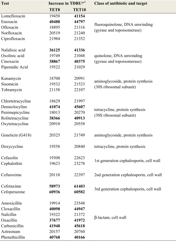
TDRU Tetrazolium Dye Reduction Units TET Tetracycline
TLC Thin Layer Chromatography
TMD Transmembrane Domain
TNF Tumor Necrosis Factor
TSA Tryptone Soya agar
TSB Tryptone Soya broth
TZ Thioridrazine v Volume VER Verapamil WHO World Health Organization
1. Chemotherapeutics and resistance
1.1 The new Era of the antibiotics
Antibacterial agents are derived either from natural sources (the antibiotics) or from total chemical synthesis. Antibiotics that are sufficiently nontoxic to the host are used as chemotherapeutic agents. Some antibacterial agents act as bactericidal, destroying bacteria, while others are bacteriostatic, inhibiting the growth of bacteria without destruction (1;2).
Since 1928, when Alexander Fleming discovered penicillin (3), and mainly after its commercialization in 1941 (4), the number of antibiotics used in medicine had increased as fast as it could not be imagined at that time. The discovery of penicillin was considered as a miracle of science by many people, and, in fact, it saved thousands of lives. After the success of penicillin other antibiotics, mainly β-lactams, were developed: streptomycin (discovered in 1944 (5)), tetracycline (derived from aureomycin (6) and patented in 1955 (7)) or chloramphenicol (first isolated in 1947 (8)). Within a few years resistance to an antibiotic was observed in the laboratory as well as clinically (3). A few years later the first cases of multi-drug resistance (MDR) were published (9). Due to mono-resistance and multi-drug resistance the need for additional effective antibiotics was evident and resulted in the creation of new antimicrobials derived from the few natural antibiotics known at that time. The “golden age of antibiotics” was born! However, it is amazing that, after thirty years of success in the search for new antibacterials, only three classes of antibiotics have entered the market since 1970 (10).
Nowadays, the wide range of antibiotics is grouped into different classes according to their mode of action, spectrum of activity or similarities in the chemical structure. The latter classification is the most common.
• Aminoglycosides: such as amikacin, gentamicin, kanamycin or streptomycin. This class of antibiotics derived from bacteria (order of Actinomycetales) interferes with protein synthesis by binding to the 30S component of the bacterial ribosome;
• Antimycobacterial agents: This group includes a wide range of antibacterials used against Mycobacterium spp. (tuberculosis, leprosy, etc) and includes, for example, rifamycin, isoniazid, pyrazinamide, ethambutol, streptomycin, amino salicylic acid, sulphones;
• Cephalosporins and related β-lactams: Cephalosporins are synthetic compounds derived for the natural antibiotic cephalosporin C. As the penicillins, members of this group inhibit bacterial cell wall synthesis. The first generation cephalosporins are active against Gram-positive bacteria but are not active against methicillin-resistant staphylococci. The second generation is highly resistant to β-lactamases and is active against Gram-negative bacteria. Third and fourth generations are broad range antibiotics because they are active against many Gram-positive and Gram-negative bacteria.
• Chloramphenicol: The first broad spectrum antibiotic whose activity is mainly bacteriostatic by interfering with protein synthesis.
• Glycopeptides: An example of this group is vancomycin that interferes with the cell wall synthesis. It is active against Gram-positive cocci.
• Macrolides: Large group of antibiotics that have a common macrocyclic lactone ring to which one or more sugars are attached. These antibiotics can be bacteriostatic or bactericidal, depending on the organism, and interfere with the protein synthesis. An example of this group is erythromycin.
• Penicillins: This group inhibits the cell wall synthesis and its action is, in general, bactericidal. Although penicillin is still in use, derivatives of this antibiotic, such as ampicillin, have been more widely used.
• Quinolones: Quinolones are synthetic antibiotics structurally related to nalidixic acid. Modifications of the structure of nalidixic acid yielded fluoroquinolones which include, for example, ciprofloxacin, ofloxacin or norfloxacin.
• Sulfonamides: They are usually bacteriostatic and interfere with the folic acid synthesis. Its use was greatly reduced because of the development of resistance. An example of a sulfonamide is the Prontosil.
• Tetracyclines: The compounds of this group are usually bacteriostatic and their mechanism of action is similar to that of aminoglycosides. Tetracyclines have a broad spectrum of activity. The increase of resistant strains and the adverse effects have reduced their use. However, they are the most commonly used antibiotics in animal husbandry.
The problem of resistance has been detected for all the classes of antibiotics described above (8). Development of antibiotic resistance results from the selection of bacterial populations whose antibiotic target has mutated during the time the population has been exposed to the antibiotic (antibiotic pressure). The frequency of resistance of a bacterial species to a given antibiotic is a product of antibiotic misuse and ineffective therapy. In economically disadvantaged countries, inadequate access to drugs contributes heavily to the frequency of antibiotic resistance (11). The problem of antibiotic resistance and its causes, are of extreme importance with respect to infections such as tuberculosis, and are the major motive for the work conducted in this thesis.
Antibiotic resistance is not restricted to bacteria. It is found in all infectious non-bacterial agents as well as in cells of an animal that is treated with a chemotherapeutic agent. Chemotherapeutically treated cancer that becomes refractory to the agent used for therapy and to any other chemotherapeutic agent, is an example of resistance that has been extensively described in the literature (12-15). A general overview of cancer, its therapy and resistance mechanisms will be discussed later on this thesis.
1.2 Fight against resistance
It is widely accepted that the problem of drug resistance, even in prokaryotic or eukaryotic cells, does not yet have solution (16-19).
Although, predictably, new resistance will emerge with time, there are ways to frustrate this possibility. Quality in education of health workers, easier access to a wide range of effective antibiotics, in economically disadvantaged countries, coupled to education of the patient population, the selection of appropriate therapy and patient compliance are the most important points according to this question.
Albeit, new drugs do not give the ultimate answer to this question, the discovery of new families of antibiotics, with new characteristics, still has high prospects.
On the other hand, to find new compounds that are able to potentiate or restore the decreased activity of existing antibiotics is, nowadays, commonly accepted as one of the best approaches to fight against multi-drug resistance (20-22).
The concept of adjuvant therapy has emerged (discussed in chapter 6 of this Introduction). In order to achieve practical benefits from this strategy, many studies have been performed leading to a better understanding of the mechanisms of resistance acquired by bacteria.
1.3 Mechanisms of resistance
As already mentioned, the understanding of the resistance mechanisms is one of the first steps leading to a possible solution against multi-drug resistance. Bacteria develop resistance against certain antibiotics or groups of antibiotics by several mechanisms. For the bacterial cells, these mechanisms can be intrinsic or acquired:
• Intrinsic resistance: some bacteria have low permeability to different classes of antibiotics/compounds (23). For example, Gram-negative bacteria and mycobacteria have thick and highly hydrophobic outer membranes, which act as a permeability barriers to hydrophilic compounds, such as macrolides (ex. erythromycin) (24).
• Acquired resistance: through mutation, acquisition of new genetic material (plasmids encoding for resistance mechanisms; foreign genetic elements; etc. (25;26)) affords survival under antibiotic pressure (19) - Figure 1.
Figure 1 - Schematic representation of cell mechanisms of resistance.
Some mechanisms can lead to resistance through different approaches, as for example the different ways by which the uptake of the antibiotic can be decreased or even forbidden. These differences are described in the text. Adapted from (27).
The most typical mechanisms of resistance, as presented by Figure 1, are:
• Modification of the antibiotic target / site of action, so that the antibiotic cannot bind to it and render it inactive (resistance to macrolides, vancomycin, β-lactams, fluoroquinolones and aminoglycosides) (28);
• Enzymatic inactivation of the antibiotic by secretions of enzymes that degrade the antibiotic (ex. β-lactamases) or modify the antibiotic so that it is ineffective (ex. the resistance to chloramphenicol (CHL) - In resistant strains,
CHL is acetylated at the C3 hydroxyl group, by a cytoplasmic enzyme, CHL acetyltransferase. CHL acetyltransferase enzymes can be plasmid or chromosomally encoded.) (29);
• Overproduction of the target such that the amount of antibiotic is not compatible with the amount of target (30) (ex. Resistance to vancomycin in staphylococci. The strain over-expresses the targets sites of vancomycin far from the key places for the cell survival, cell wall synthesis, so that it acts at that place without block the critical target sites) (31);
• The target to be inhibited is part of a multi-chemical pathway that has alternative pathways which lead to the needed product (Bypass of inhibitory steps) (ex. Sulfonamide resistance mediated by different plasmid encoded enzymes with the same activity as the antibiotic target but structurally different and not affected by the antibiotic) (32);
• Developing mechanisms that do not allow the accumulation of the antibiotic within the cell, preventing it from reaching the necessary concentration to be active (19):
Decreasing the permeability of the cell wall to an antibiotic. For instance by decreasing the expression of porins (for example OmpF and OmpX which are involved in the control of the penetration of antibiotics such as β-lactams and fluoroquinolones through the enterobacterial outer membrane (33)) or reducing the internal size of the pore through mutation of some amino acids (34);
Increasing efflux activity, which by extrusion of the antibiotic decreases its cellular concentration (19;35;36). Ex. resistance of Escherichia coli to tetracycline (8;35;37).
The combination of these two last systems leads to high level of resistance mainly in Gram-negative bacteria (38).
With respect to eukaryotic cells and, especially cancer, the main mechanisms of resistance are yet to be completely understood. However, overproduction and
modification of enzymatic targets, over-expressed efflux mechanisms, ability of the cells to avoid apoptosis, are well studied mechanisms (39). Resistance to anticancer drugs also results from other factors including individual variation among patients and somatic cell genetic differences in tumors, even those from the same tissue of origin. Frequently, resistance is intrinsic to the cancer, but with prolonged or ineffective therapy, acquired resistance is common. The most frequent cause for acquisition of resistance to a broad range of anticancer drugs is the over-expression of one or more energy-dependent transporters – efflux mechanisms. However, insensitivity to drug-induced apoptosis and induction of drug-detoxifying mechanisms probably play an important role in acquired anticancer drug resistance (12;15;39-41).
The next two chapters will focus on the mechanisms of resistance that are related with the bacterial cell envelope.
2. Cell envelope and resistance
The structure and ensuing characteristics of the cell envelope have an important role in the resistance of bacterial cells to antibiotics. The cell envelope of Gram-negative and Gram-positive bacteria, mycobacteria and the plasma membrane of eukaryotic cells will be described in this chapter.
Figure 2 – Schematic representation of the cell envelope of a bacterial cell.
The thickness and composition of the cell wall change between the Gram-positive and Gram-negative cells. Source: http://www.scq.ubc.ca/
2.1 Cell envelope
The cytoplasm of prokaryotes and eukaryotes is surrounded by a unit - the plasma membrane (PM). The plasma membrane is itself, a very important organelle of the cell that controls input and output of compounds. With respect to bacteria, an additional membrane is external to the plasma membrane and provides supplementary properties that contribute to its survival in the environment it normally inhabits. This outer membrane (OM) differs between Gram-negative and Gram-positive bacteria; the former having a more complex structure than that of the latter. Whereas intrinsic or acquired resistance of eukaryotes is determined by the characteristics of its plasma membrane, intrinsic and acquired resistance of prokaryotes is due to the structure of the cell wall and properties of the plasma membrane.
The cell envelope consists of different structures: 1) Plasma membrane
Electron microscopic examinations of cell membranes have led to the development of a lipid bilayer model of the PM. The bi-molecular leaflet of phospholipids is arranged as shown by Figure 3.
Figure 3 - General structure of the cell membrane.
Source: http://cellbiology.med.unsw.edu.au/units/images/cell_membrane.png
In general, membranes are highly conserved structures, similar for all living things (with exception of the virus). The bi-layer structure of the PM is stabilized by hydrophobic interactions - Wandervals forces, ionized groups of amino acids, lipid interactions. Membranes are very organized but asymmetric organelles, due to the difference of environments in both of their sides. Even if well organized organelles, membranes are dynamic in order to an easily adaptation to possible changes of the environmental conditions (42). Lipids and proteins are its main composition.
Attached to the surface and cytoplasmic side as well as within the leaflet layer of the PM are proteins. The majority of membrane proteins are responsible for structural functions, the moving of small molecules across the membrane
(transporters, ATPases, kinases, etc) or involved in energy generation (ATP synthase, enzymes involved in final steps of glycolysis, etc).
The lipids form a bilayer, with their hydrophilic portion facing the aqueous environment, and the hydrophobic parts clustering together inside the membrane. The majority of the lipid contents are phospholipids. The phospholipid bi-molecular leaflet with its functional transport units, bestow a semi-permeable barrier-like characteristic to the PM, allowing some molecules to enter under control and others are kept outside in a selective interaction manner (23). The fluidity of the lipid bilayer affects its permeability in an inversely proportional way. About 50% of the bacterial species also contain hopanoids (Figure 4), molecules with similar structure to sterols found in eukaryotic membranes that help to the stabilization of the membrane.
Figure 4 - Chemical structure of Hopanoid.
Hopanoid is a sterol molecule found in eukaryotic membranes that helps in its stabilization.
2) Cell wall
The cell wall is located outside the plasma membrane and protects the cell from osmotic lysis. It does not exist in all organisms. Some bacteria have cell walls containing peptidoglycan.
The structure of the cell envelope of Gram-negative and Gram-positive bacteria, as well as of mycobacteria, is different and provides them special characteristics and different levels of intrinsic resistance. The organization of their bacterial envelope is represented in Figure 5.
A
B
Figure 5 - Organization of the bacterial cell envelope.
A – Gram-negative and Gram-positive bacteria. Source: (43); B – Mycobacteria. Source: (44). LAM, lipoarabinomannans; LM, lipomannans; PIM, phosphatidylinositol mannosides
2.1.1 Gram-positive
In Gram-positive bacteria, the cytoplasmic membrane, that immediately surrounds the inside of the cell, is a thin structure with about 8 nm thick (Figure 5A). This structure is followed by a cell wall (15-80 nm thick), consisting of several layers of peptidoglycan complexed with teichoic acids. Some cell wall components protect against phagocytic engulfment or digestion. Examples of Gram-positive bacteria are the species belonging to the Staphylococcus genus. Staphylococcus aureus is an opportunistic pathogen and the major causative agent of numerous hospital and community acquired infections. The genome of S. aureus strains had been already sequenced (45) and became possible to identify many components of its cell envelope that interfere with its virulence, with its intrinsic resistance and components that are responsible to the acquisition of multi-drug resistance (discuss bellow on this section) (45;46).
2.1.2 Gram-negative
The cytoplasmic membrane is relatively fluid in order to allow the proper functioning of the cell membrane components (so then also permeable) and so, many bacteria developed other structures to protect themselves. Those include peptidoglycan cell wall, an outer membrane or layers of lipopolysaccharides (LPS) in which the lipid A possesses endotoxin activity (47).
The LPS, which are organized in several structures, according to the species, are, probably, the explanation of the unusual slow influx of lipophilic solutes through the outer membrane (23). The external membrane provides a very impermeable barrier and, so, bacteria developed other mechanisms that provide the influx of nutrients from the external medium. These uptake is done through proteins, called porins, which allow the influx of nutrients by providing non-specific and specific channels across the membrane (47). The hydrophobic (lipid) pathway, which is based on the interaction between the