DOI: 10.5935/2359-4802.20180097
ORIGINAL ARTICLE
Mailing Address: Matheus Lavorenti Rocha
Rua 240, s/n - Setor Leste Universitário. Postal Code: 74605-170, Goiânia, GO - Brazil. E-mail: [email protected], [email protected]
Antioxidant and Vasodilatory Action of Grape Juices Produced in Different Regions
of Brazil
José Britto Junior, Karla Carneiro Siqueira Leite, Eric de Souza Gil, Matheus Lavorenti Rocha Faculdade de Farmácia, Universidade Federal de Goiás (FF/UFG), GO - Brazil
Manuscript received on November 30, 2017, revised manuscript on July 20, 2018, accepted on August 07, 2018.
Abstract
Background: Grapes and its derivatives (wines and juices) are rich in polyphenols that have high antioxidant and vasodilator capacity. These biological activities may vary in the juices marketed and produced in different regions of Brazil.
Objectives: To determine the antioxidant and vasorelaxant effects of grape juice samples produced in different regions of Brazil.
Methods: The content of phenolic compounds and antioxidant capacity were evaluated by the methods of Folin-Ciocalteau, DPPH, ABTS and a new electroanalytical approach (differential pulse voltammetry - DPV). Vasodilator effects were analyzed in isolated aorta from rats in an organ bath.
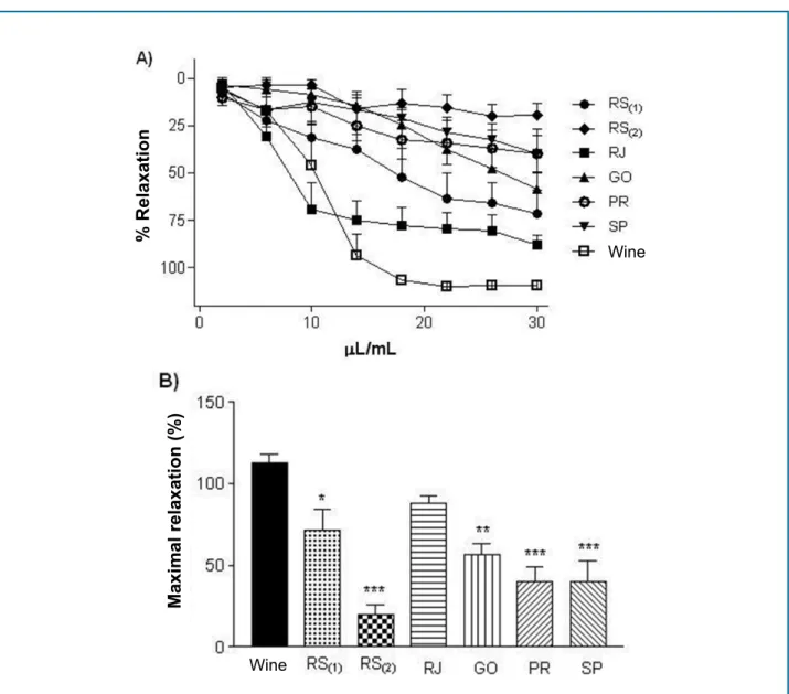
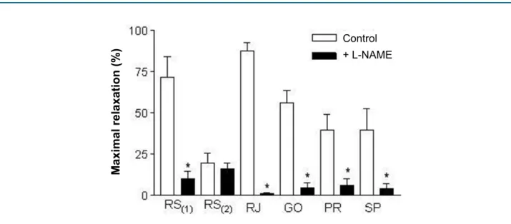
Results: The samples from RJ and SP presented respectively the higher and lower phenolic content and also antioxidant capacity by the methods used (ABTS and DPPH). The results of the electrochemical index corroborate to the other tests, with the best results to RJ (21.69 ± 3.15 µA/V) and worse to the SP sample (11.30 ± 0.52 µA/V). In the vascular reactivity studies, the relaxation induced by each sample presented more distinct differences, following the order: RJ (87.9 ± 4.8%) > RS1 (71.6 ± 8.6%) > GO (56.2 ± 7.2%) > SP (39.9 ± 7.8%) > PR (39.4 ± 9.5%) > RS2 (19.5 ± 6.2%). Inhibition of endothelial NO practically abolished (p < 0.001) the relaxation for all samples, except one.
Conclusion: The phenolic content and antioxidant capacity vary greatly among samples. The results obtained for the order of antioxidant activity were: RJ > RS1 > GO > RS2 > PR > SP. The juices were able to induce vascular relaxation at quite varied levels, and the RJ sample the most effective. The L-NAME practically blocked all samples except one (RS2). (Int J Cardiovasc Sci. 2019; [online].ahead print, PP.0-0)
Keywords: Antioxidants/pharmacology; Vasodilatation; Vasodilatador Agents/analysis; Vitis; Sucos; Fruit and Vegetable Juices/analysis; Epidemiology; Cardiovascular Diseases/prevention and control; Neoplasms/ prevention and control.
Introduction
Epidemiological evidence shows that the ingestion of foods rich in polyphenols (such as grapes and grape products) is associated with decreased mortality worldwide due to reduced cases of cancer and cardiovascular diseases.1
Polyphenols have beneficial antioxidant effects against oxidative stress, the main pathophysiological mechanism in the development of cardiovascular diseases, such as
hypertension, diabetes and atherosclerosis.2 They cause vasodilation by increasing the production of vasodilatory agents produced by the vascular endothelium (as NO) and decreasing vasoconstricting agents, such as endothelin-1.2,3 In addition, there is a direct correlation between the number of polyphenols in red wine and its capacity to induce endothelium-dependent vasodilation.4
the elderly, children and pregnant women. However, studies have found that the consumption of grape juice also has positive results, often comparable to wine consumption, such as an antioxidant and vascular relaxation action.5,6
Despite the existence of several studies with red wine from different parts of the world, including Brazil, a smaller number of information exists regarding the biological properties of red grape juice. Research on the functional properties of grapes was boosted by the benefits derived from wine consumption, encouraged by the “French paradox”, an expression that is known worldwide. Moreover, there is an increasing number of studies that demonstrate that grape juice has beneficial functional properties similar to wine, especially on the cardiovascular system.7,8
In Brazil, the consumption and production of whole-grape juice has increased by around 15% per year.9 However, the quantity and quality of the functional ingredients of grapes may be largely influenced by the climate, soil and geographic region of the crops.10,11 As Brazil is an enormous country, the juices produced in different regions may contain different antioxidant potentials and chemical compositions, which would certainly modify their functional properties. In this study, we evaluated the antioxidant properties and the vasodilatory capacity of grape juice of various brands produced in different regions across Brazil.
Methods
Sourcing of the juices analyzed
Samples of 6 whole-grape juices produced in a few regions in Brazil, marked with the acronym of the state where they were produced, have been purchased. The acronyms are the following: RS (2 samples: RS(1) and RS(2)), RJ, GO, PR and SP. The eligibility criteria were: i) whole red grape juice; (ii) no added preservatives, stabilizers or antioxidants; (iii) no added water; (iv) no added sugar; and v) samples whose ingredients were only “100% grape juice”. The juice samples were selected at random from the main producer states. We did not have access to any samples produced in the North and Northeast regions.
For the spectrophotometric and electroanalytical experiments, each sample was diluted in the concentration of 10% in an ethanolic solution. All tests were performed in triplicate.
Reagents
T h e r a d i c a l s A B T S . + ( 2 , 2 a z i n o b i s ( 3 -ethylbenzothiazoline-6-sulfonate), DPPH (2,2-diphenyl-1-picrylhydrazyl) and the reagents used in the analyzes were all analytically pure and sourced from Sigma Chemical Co. (St. Louis, MO, USA). All electrolytic solutions were prepared with purified water using the Millipore Milli-Q system, conductivity ≤ 0.1μS.cm-1 (Millipore S.A., Molsheim, France). Other reagents were purchased off-the-shelf.
Evaluation of antioxidant action by the DPPH radical sequestration method
The purple radical DPPH (2,2-diphenyl-1-picrylhydrazyl), when reduced by the antioxidant, is discolored and turns yellow. Reduction of radical DPPH is followed by a decrease in absorbance by 517 nm. The amount of analyte in µL required to reduce the DPPH absorbance by 50% (Efficient Concentration, EC50) is calculated to evaluate the antioxidant capacity of each sample and the free radical sequestering activity or percentage of discoloration. The analytes were solutions of 10% whole juices and the standard solution of gallic acid evaluated at different concentrations. Absorbance was monitored with a UV-Vis spectrophotometer (Jasco® V-530) and all tests were performed in triplicate.
Evaluation of the antioxidant activity by the ABTS radical method
T h e r a d i c a l A B T S . + ( 2 , 2 ’ a z i n o b i s ( 3 -ethylbenzthiazoline-6-sulfonate) is formed from the ABTS reaction (7 mM) with 88 µl of potassium persulfate (140 mM) in the absence of light. To perform the analyses, a volume of 300 µL of the ethanolic analyte solution was placed in a test tube with 2.7 mL of the ABTS radical, then the tubes were covered with Parafilm® and kept in the dark for 20 minutes and the absorbance was monitored at 734 nm using a UV-Vis spectrophotometer (Jasco® V-530) and all tests were performed in triplicate.
Determination of total phenols
with gallic acid. The total phenolic content was expressed as µg equivalents of gallic acid (GAE) per ml of sample. All analyses were done in triplicate.
Electrochemical analyses
To perform the electrochemical measurements, an µAutolab III potentiostat/galvanostat integrated with the software GPES 4.9 (Eco-Chemie, Utrecht, The Netherlands) was used. The analyses were carried out in an electrochemical cell with capacity for 3 mL of a solution operated with a three-electrode system, an Ag/ AgCl reference electrode (KCl 3 mol.L-1), an auxiliary platinum electrode and, as a carbon paste electrode prepared with 0.075 g of carbon and 0.035 g of mineral oil was used as a working electrode.
The tests using differential pulse voltammetry (DPV) were performed with pulse amplitude of 50 mV and scanning velocity of ʋ = 10mVs-1. The analyses were carried out in an electrochemical cell containing 1.75 ml of phosphate buffer pH 7.0 (0.1 M), adding 25 µL of the ethanolic solution of the analyte. The electrochemical index (EI) of the samples was calculated by summing up the result of each division of the current by the potential (I/E) for each anodic peak observed on the differential pulse voltammograms.
Animals used and preparation of isolated arteries
Male Wistar rats (200-230 g) from the UFG central vivarium were used. All experimental protocols respected the protocols approved by the UFG Research Ethics Committee (protocol: 044/17). This study is in line with the European Union Guide for the Care and Use of Experimental Animals (2010/63/EU).
The rats were euthanized by exsanguination under inhalational anesthesia (n = 5-6). The thoracic aorta was isolated, separated from the connective and adipose tissues and cut into rings (± 4 mm), which were mounted between two metal hooks, one of which was connected to a power transducer to record the isometric voltage (DATAQ Instruments, Akron, OH, USA) and the other was attached to the vial for the isolated organ containing modified Krebs solution [composition in mM: NaCl, 130.0; KCl, 4.7; KH 2 PO 4, 1.2; CaCl2, 1.6; MgSO4, 1.2; NaHCO3, 14.9; glucose, 5.5], pH 7.4, under gasification with carbon dioxide (95% O2 + 5% of CO2) at 37°C and maintained at baseline at 1.5 g (optimal rest tension, as previously standardized in our laboratory).
Experimental protocols
After 60 minutes of basal tension stabilization, the arteries were pre-contracted with phenylephrine (0.1 µM) and the presence of endothelium was determined using ACh (1 µM). The rings were disposed of when the relaxation for ACh was smaller than 80%. The whole grape juices analyzed were filtered on a filter paper under light and immediately divided and frozen at -20°C for future experiments.
The arteries with intact precontracted endothelium (phenylephrine, 0.1 µM) were stimulated to relax by increasing the concentration of juice directly in the bath solution (0 to 30 µL/mL). As wine is popularly known throughout the world as a vasodilator, we also perform relaxation curves stimulated with Cabernet Sauvignon red wine, produced in France in 2015, in order to compare the vascular effect.
In another experiment series, the vasodilatory effect of the juice samples was repeated after treatment with the NOS, L-NAME (100 µM, 30 min) inhibitor in order to determine the participation of NO in the mediated juice-stimulated vasodilation.
Statistical analysis
The charts were made and analyzed by the software GraphPad Prism (GraphPad Software Corporation, version 5.0) by ANOVA plus Bonferroni post-test. In the analyses, the continuous variables showed normal distribution and the results were expressed as the mean ± standard deviation of the mean of at least five experiments (n = 5-6) obtained from different animals. All analyses considered a statistical significance level of 5% (p < 0.05).
Results
Evaluation of antioxidant activity and determination of total phenols
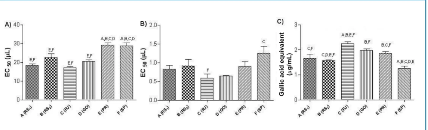
Figure 1 - Graphical representation of the results of antioxidant tests DPPH (A), ABTS (B) and total content of phenolic compounds by Folin-Ciocalteu reaction (C), expressed in μg gallic acid equivalent (GAE/mL). The letters on the bars represent the statistical difference (p < 0.05) between the indicated groups.
Gallic acid equivalent
(
µ
g/mL)
The ABTS technique (Figure 1B) confirmed the best antioxidant activity of the RJ sample (CE50: 0.59 ± 0.11 µL). The SP sample had a lower antioxidant potential (CE50: 1.25 ± 0.19 µL), being statistically different (p < 0.05) from the best sample (RJ). In turn, the other samples presented similar results (p > 0.05). The order of activity for the ABTS TEST was RJ > RS(1) (0.83 ± 0.1 µL) > RS(2) (0.91 ± 0.18 µL) > GO (0.65 ± 0.01 µL) > PR (0.90 ± 0.13 µL) > SP.
In Figure 1C, we can see the number of phenolic compounds (expressed in µg equivalents of gallic acid/ mL of the sample) present in the samples. The results are according to the antioxidant activity (Figures 1A, B), and the sample with the highest antioxidant potential presented a higher concentration of phenolic compounds (RJ: 2.25 ± 0.06 µg/mL). The lowest concentration of phenolic compounds was observed in the SP sample (1.26 ± 0.08 µg/mL), being different (p < 0.05) from all others.
Electrochemical analyses
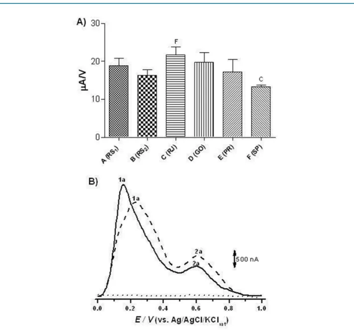
Figure 2A shows the results of the electroanalytical analyses of the samples. Although the electrochemical indices showed a higher activity for the RJ sample (21.7 ± 2.1 µA/V), the samples from RS(1), RS(2), GO and PR presented equivalent electrochemical indices (p > 0.05) (18.8 ± 1.9; 16.3 ± 1.5; 19.8 ± 2.5; 17.2 ± 3.3 µA/V, respectively). Only the SP sample (11.3 ± 0.5 µA/V) showed a statistical difference (p < 0.05) compared to most effective sample (RJ).
The IE values are calculated from the main electrochemical parameters, that is, an anode peak potential — Epa. The lower the anode peak potential the
higher the thermodynamic capacity of the species to reduce others, and anodic peak current intensity — Ipa — which in addition to being related to concentration, also concerns the speed with which the reduction process occurs. The presence of electroactive compounds in both the most effective sample (RJ) and in the least effective sample (SP), which presented two oxidation peaks (1a and 2a). The oxidation peak of the RJ sample occurs at a lower voltage than the SP sample, showing its greater reducing potential. (Figure 2B).
Vasodilator effect
Figure 2 - Electrochemical Index (EI) chart representing the sum of the Ipa/Epa (A) rates and differential pulse voltammograms (B)
obtained using carbon paste electrode for analysis of C (RJ) (─) and F (--) (SP), which presented the highest and the lowest potential of antioxidant activity, respectively. The white (...) was achieved in 0.01 M pH 7 sodium phosphate buffer.
Regarding the mechanism of action, all juice samples tested revealed a major participation in the endothelial NO in the induction of relaxation, as L-NAME nearly completely inhibited relaxation (Figure 4). Only the RS(2) sample showed no difference (p > 0.05) after inhibition of NO with L-NAME, revealing an action mechanism unrelated to NO release by the vascular endothelium.
Discussion
The greatest finding of this study is that the grape juices marketed in Brazil do not have the same functional
properties. Its antioxidant capacities, phenolic compound concentration and vasodilator activity are different among samples tested from different regions. Moreover, the vasodilatory capacity of grape juice is not equivalent to the red wine-induced dilation, except for one sample (obtained from RJ).
Figure 3 - Vascular relaxation curve induced by different juice samples in aortas of rats precontracted with phenylephrine (A) and maximal vascular relaxation (B). Red wine was used for comparison. The dots represent means ± SEM of the relaxing effect expressed in % relaxation. * Significant difference (p < 0.05) vs. red wine.
% Relaxation
Maximal relaxation (%)
Wine
Wine
as red wines, which have different properties depending on the brand and place of manufacture.12 In fact, the juice sample obtained from RJ was the most powerful in the tests of antioxidant activity and the one from SP was the worst one. With regard to the DPPH radical elimination activity, it was found that the juice from RJ had the highest antioxidant potential, with similar results for the RS(1), RS(2) and GO samples. The samples produced in SP and PR were the worst ones, presenting the same potential compared to each other, and lower than the others.
Similar results were obtained with the ABTS technique, where the best and the worst antioxidant activity was
found in the RJ and SP samples, respectively. Grape juices are sources of different antioxidant substances, mainly polyphenols, which includes them in the range of foods with high antioxidant potential.1,5,13 As expected, the sample of highest antioxidant capacity (RJ) also presented the highest concentration of polyphenols. Likewise, the sample of lowest antioxidant activity also had the lowest concentration of polyphenols (SP). This different concentration of polyphenols may be responsible for the antioxidant capacity of the samples, as it was also found in other foods and beverages.13,14
Figure 4 - Maximum vascular relaxation induced by grape juice samples in aortas of rats pre-contracted with phenylephrine. The bars represent the maximal relaxation percentage (Emax) with or without treatment with L-NAME (100 μM, 30 min). * Significant difference (p < 0.05) vs. Control (with no L-NAME).
Maximal relaxation (%)
Control
+ L-NAME
one of the main tools to determine antioxidant activity. In fact, good electron-donating agents (antioxidants) can reversibly oxidize at lower peak potentials (Epa < 0.5 mV, pH = 7). Following this idea, the concept of electrochemical index (EI) was previously proposed to qualify compounds with antioxidant capacity.15
The evaluation of antioxidant activity using spectrophotometric and electrochemical methods shows that the analyses conducted with the classical methods (DPPH, ABTS and FC) agree with the electrochemical index, which allows the comparison of the antioxidant activity measured by electroanalytical methods. The small differences presented in the analyses are due to the color of the analyte, which generates interference in the spectrophotometric tests. In these analyses, the RJ sample also presented the best antioxidant activity, while the SP sample was the worst of them. The decreasing order of antioxidant activity obtained by EI calculations was: RJ ≥ GO ≥ RS(1) ≥ PR ≥ RS(2) > SP.
Differential pulse voltammetry (Figure 2B) of the best (RJ) and worst (SP) sample had two anode peaks, one with Epa approximately at 0.1V and the other at 0.6V. Therefore, it can be stated that the electroactive species found in the juice are oxidizing at a low potential and this behavior is observed in samples that are rich in antioxidants.16 Resveratrol, a phenolic compound that is very present in the grapes, oxidizes at a potential close
to 0.6V; this finding justifies the presence of the second peak (2a) presented in the voltammogram.17
Several studies report that the ingestion foods rich in polyphenols (mainly vegetables, fruits, wines and teas) are associated with a protective effect on the cardiovascular system in humans and animals.18 In most published studies, red wine was highlighted as a functional food that is used to test the benefits of intake of antioxidants. Its effects include vascular dilatation in laboratory animals or patients with or without hypertension.19,20 However, not only red wines can induce protection against cardiovascular risk factors. Many researchers have shown that the consumption of other beverages such as grape juice and teas can bring important benefits, often comparable to those of wine.5,21
Our results showed that the different juice samples produced different cardiovascular results, having different levels of efficacy in the induction of vascular relaxation. The RJ sample was the one that presented the best result, being the only one that induced vascular relaxation at a similar level to the red wine tested. This result may be associated with a greater quantity of phenolic compounds, whose vasodilatory and cardioprotective action has been well documented.22-24
1. Chong MF, MacDonald R, Lovegrove JA. Fruit polyphenols and CVD risk: a review of human intervention studies. Br J Nutr. 2010;104(S3):S28-39.
2. Mudnic I, Budimir D, Modun D, Gunjaca G, Generalic I, Skroza D, et al. Antioxidant and vasodilatory effects of blackberry and grape wines. J Med Food. 2012;15(3):315-21.
3. Stoclet JC, Chataigneau T, Ndiaye M, Oak MH, El Bedoui J, Chataigneau M, et al. Vascular protection by dietary polyphenols. Eur J Pharmacol. 2004;500(1-3):299-313.
4. Burns J, Gardner PT, O’Neil J, Crawford S, Morecroft I, McPhail DB, et al. Relationship among antioxidantactivity, vasodilation capacity, and phenolic content ofred wines. J Agric Food Chem. 2000;48:220-30.
5. Stein JH, Keevil JG, Wiebe DA, Aeschlimann S, Folts JD. Purple grape juice improves endothelial function and reduces the susceptibility of LDL cholesterol to oxidation in patients with coronary artery disease. Circulation. 1999;100(10):1050-5.
6. Anselm E, Chataigneau M, Ndiaye M, Chataigneau T, Schini-Kerth VB. Grape juice causes endothelium-dependent relaxation via a
redox-References
endothelial cells.25 In the blood vessels, endothelial cells play a critical role in maintaining local homeostasis by producing various autacoids that act on neighboring vascular cells. Among these endothelial-derived factors, NO, which is synthesized by the endothelial NO synthase, is the main one and produces a potent vasodilator action. Some studies have shown that NO can be stimulated by red wine in isolated arteries.18,26 In contrast, other studies have shown that inhibition of NO synthase has no effect on red wine-induced relaxation.25
In this study, the inhibition of NO synthase with L-NAME significantly inhibited the relaxation of almost all samples, revealing the important role of endothelial NO in the vasodilator effect of grape juices. Only the RS(2) sample showed no difference in the vascular relaxation effect when NO production was inhibited. This shows that the mechanism of induction of vasodilation in this sample is little effective and is not related to the endothelial NO release stimulus, like most of the samples tested.
Conclusion
This study shows that the grape juices analyzed have different levels of antioxidant activity. Besides, the vasodilatory effect also presented a varying intensity from one sample to another. The mechanism of vasodilatory action seems to be related to the release of endothelial NO, except for one of the samples (RS(2)). Only one sample (RJ) has a vasodilator activity comparable to that of red wine. Finally, these findings help understanding that the functional qualities of grape juice can vary greatly across different regions of Brazil. Even though all the samples meet the eligibility criteria, not all of them have functional effects as an appreciable antioxidant and vasodilator activity, some of which
are probably incapable of providing protection against cardiovascular diseases.
Acknowledgements
CAPES (Coordenação de Aperfeiçoamento de Pessoal de Nível Superior) awarded a Masters’ Grant to José Britto Junior. Matheus L. Rocha receives financial support for research productivity (PQ2) from CNPq.
Author contributions
Conception and design of the research:Britto Junior J, Rocha ML. Acquisition of data: Britto Junior J, Leite KCS. Analysis and interpretation of the data: Britto Junior J, Leite KCS, Gil ES, Rocha ML. Statistical analysis: Rocha ML. Writing of the manuscript: Gil ES, Rocha ML. Critical revision of the manuscript for intellectual content: Gil ES, Rocha ML.
Potential Conflict of Interest
No potential conflict of interest relevant to this article was reported.
Sources of Funding
There were no external funding sources for this study.
Study Association
This article is part of the thesis of master submitted by José Britto Junior, from Universidade Federal de Goiás.
Ethics approval and consent to participate This study was approved by the Ethics Committee on Animal Experiments of the Universidade Federal de Goiás
sensitive Src-and Akt-dependent activation of eNOS. Cardiovasc Res. 2007;73(2):404-13.
7. Vinson JA, Teufel K, Wu N. Red wine, dealcoholized red wine, and especially grape juice, inhibit atherosclerosis in a hamster model. Atherosclerosis. 2001;156(1):67-72.
8. Coimbra SR, Lage SH, Brandizzi L, Yoshida V, Luz PL. The action of red wine and purple grape juice on vascular reactivity is independent of plasma lipids in hypercholesterolemic patients. Braz J Med Biol Res. 2005;38(9):1339-47.
9. Carmo MCL, Dantas MIS, Ribeiro SMR. Caracterização do mercado consumidor de sucos prontos para o consumo. Braz J Fod Technol. 2015;17(4):305-9.
10. Rizzon LA, Link M. Composição do suco de uva caseiro de diferentes cultivares. Revista Ciência Rural. 2016;36(2):689-92.
11. Pepi S, Coletta A, Crupi P, Leis M, Russo S, Sansone L, et al. Geochemical characterization of elements in Vitis vinifera cv. Negroamaro grape berries grown under different soil managements. Environ Monit Assess. 2016;188(4):211.
12. Cetó X, Gutiérrez JM, Gutiérrez M, Céspedes F, Capdevila J, Mínguez S, et al. Determination of total polyphenol index in wines employing a voltammetric electronic tongue. Anal Chim Acta. 2012;732:172-9.
13. Dani C, Oliboni LS, Vanderlinde R, Bonatto D, Salvador M, Henriquesa JA. Phenolic content and antioxidant activities of white and purple juices manufactured with organically-or conventionally-produced grapes. Food Chem Toxicol. 2007;45(12):2574-80.
14. Lino FM, Sá LZ, Torres IM, Rocha ML, Dinis TC, Ghedini PC, et al. Voltammetric and spectrometric determination of antioxidantcapacity of selected wines. Electrochim Acta. 2014 May 10;128:25-31.
15. Blasco AJ, González MC, Escarpa A. Electrochemical approach for discriminating and measuring predominant flavonoids and phenolic acids using differential pulse voltammetry: towards an electrochemical index of natural antioxidants. Anal Chim Acta. 2004;511(1):71-81.
16. Kilmartin PA, Zou H, Waterhouse AL. A cyclic voltammetry method suitablefot characterizing antioxidant properties of wine and wine phenolics. J Agric Food Chem. 2001;49(4):1957-65.
17. Burin VM, Falcão DL, GonzagaI LV, Fett R, Rosier JP, Bordignon-Luiz MT. Colour, phenolic content and antioxidant activity of grape juice. Ciênc Tecnol Aliment. 2010 out-dez 2010;30(4):1027-32.
18. Luciano MN, Ribeiro TP, França-Silva MS, Nascimento RJ, Oliveira EJ, França KC, et al. Uncovering the vasorelaxant effect induced by Vale do São Francisco red wine: a role for nitric oxide. J Cardiovasc Pharmacol. 2011;57(6):696–701.
19. Brizic I, Modun D, Vukovic J, Budimir D, Katalinic V, Boban M. Differences in vasodilatory response to red wine in rat and guinea pig aorta. J Cardiovasc Pharmacol. 2009;53(2);116-20.
20. Porteri E, Rizzoni D, Ciuceis C, Boari GE, Platto C, Pilu A, et al. Vasodilator effects of red wines in subcutaneous small resistance artery of patients with essential hypertension. Am J Hypertens. 2010;23(4),373-8.
21. Kawada T. Green tea consumption and risk of cardiovascular disease or stroke. Int J Cardiol. 2016 Oct 15;221:831.
22. Hugel HM, Jackson N, May B, Zhang AL, Xue CC. Polyphenol protection and treatment of hypertension. Phytomedicine. 2016;23(2):220-31.
23. Leifert WR, Abeywardena MY. Cardioprotective actions of grape polyphenols. Nutrit Res. 2008;28(11):729-37.
24. Barona J, Aristizabal JC, Blesso CN, Volek JS, Fernandez ML. Grape polyphenols reduce blood pressure and increase flow-mediated vasodilation in men with metabolic syndrome. J Nutrit. 2012;142(9):1626-32.
25. Leblais V, Krisa S, Valls J, Courtois A, Abdelouhab S, Vila AM, et al. Relaxation induced by red wine polyphenolic compounds in rat pulmonary arteries: lack of inhibition by NO-synthase inhibitor. Fundam Clin Pharmacol. 2008;22(1):25-35.
26. Li H, Xia N, Förstermann U. Cardiovascular effects and molecular targets of resveratrol. Nitric Oxide. 2012;26(2):102-10.