Nádia Filipa Marques Grilo
Licenciada em Biologia
Protein adducts from the anti-HIV drug
abacavir – possible biomarkers of drug
toxicity
Dissertação para obtenção do Grau de Mestre em
Genética Molecular e Biomedicina
Orientador: Sofia de Azeredo Pereira, Professora Auxiliar
Convidada, Faculdade de Ciências Médicas, UNL
Co-orientador: Alexandra Moita Antunes, Investigadora
Auxiliar, Instituto Superior Técnico, UTL
Jurí:
Presidente: Prof. Doutora Ilda Sanches
Arguente: Prof. Doutor Frederico Pereira
Vogal: Prof. Doutora Sofia Pereira
Nádia Filipa Marques Grilo
Licenciada em Biologia
Protein adducts from the anti-HIV drug
abacavir – possible biomarkers of drug
toxicity
Dissertação para obtenção do Grau de Mestre em
Genética Molecular e Biomedicina
Orientador: Sofia de Azeredo Pereira, Professora Auxiliar
Convidada, Faculdade de Ciências Médicas, UNL
Co-orientador: Alexandra Moita Antunes, Investigadora
Auxiliar, Instituto Superior Técnico, UTL
Júri:
Presidente: Prof. Doutora Ilda Sanches
Arguente: Prof. Doutor Frederico Pereira
Vogal: Prof. Doutora Sofia Pereira
iii Protein adducts from the anti-HIV drug abacavir - possible biomarkers of drug toxicity
Copyright Nádia Filipa Marques Grilo, FCT/UNL, UNL
v The results discussed in this thesis originated:
Publications in international scientific journals:
Grilo NM, Antunes AMM, Antunes, Caixas U, Marinho AT, Charneira C, Gouveia S, Oliveira MC, Marques MM, Pereira SA. 2012. Abacavir bioactivation in man yields an aldehyde capable of reacting with proteins. Chem Res Toxicol. (Submitted)
Charneira C, Grilo NM, Pereira SA, Godinho ALA, Monteiro EC, Marques MM, Antunes AMM. 2012. N-terminal valine adduct from the anti-HIV drug abacavir in rat hemoglobin as evidence for abacavir metabolism to a reactive aldehyde in vivo. Br J Pharmacol. doi: 10.1111/j.1476-5381.2012.02079.x.
Oral comunications in national meetings:
Grilo NM, Pereira SA, Charneira C, Monteiro EC, Marques MM, Antunes, AMM. N-terminal valine adduct from the anti-HIV drug abacavir in rat hemoglobin as evidence of bioactivation to a reactive aldehyde in vivo. XLII Reunião Anual da Sociedade Portuguesa de Farmacologia, XXX Reunião de Farmacologia Clinica, XI Reunião de Toxicologia, Lisboa, Portugal, 2012.
Grilo NM, Pereira SA, Antunes AMM. Factors influencing Abacavir hemoglobin adducts in HIV-infected patients. Jornadas Intercalares das Dissertações Anuais dos Mestrados. Faculdade de Ciências e Tecnologia - Universidade Nova de Lisboa, Lisboa, Portugal, 2012.
Oral comunications in international meetings:
Grilo NM, Antunes AMM, Caixas U, Gouveia S, Oliveira C, Marinho AT, Monteiro EC, Marques MM, Pereira SA. Hemoglobin adducts from the antiretroviral abacavir in HIV-infected patients as evidence of bioactivation. 244th ACS National Meeting & Exposition, Philadelphia, Pennsylvania, USA, 2012.
Poster comunication in international meetings:
vi
International conference proceedings:
Grilo NM, Antunes AMM, Caixas U, Gouveia S, Oliveira C, Charneira C, Marinho AT, Monteiro EC, Marques MM, Pereira SA. Hemoglobin adducts from the antiretroviral abacavir in HIV-infected patients as evidence of bioactivation. 244th ACS National Meeting & Exposition, Philadelphia, Pennsylvania, USA, 2012. Abstracts of Papers of the American Chemical Society (in press)
Grilo NM, Antunes AM, Caixas U, Gouveia S, Charneira C, Marinho AT, Oliveira C, Marques MM, Monteiro EC, Pereira SA. Evidence of haptenation upon bioactivation of the anti-HIV drug abacavir to a reactive aldehyde in vivo. 6th European Congress of Pharmacology. Granada, Spain, 2012. MEDIMOND International Proceedings (in press)
The participation in other ongoing projects of the research team originated the follow publications in international scientific journals:
Caixas U, Antunes AMM, Marinho AT, Godinho ALA, Grilo NM, Marques MM, Oliveira MC, Branco T, Monteiro EC, Pereira SA. 2012. Evidence for nevirapine bioactivation in man: Searching for the first step in the mechanism of nevirapine toxicity. Toxicology 301: 33-39.
vii
Acknowledgments
Em primeiro lugar, gostaria de agradecer à Prof. Doutora Sofia Pereira e à Doutora Alexandra Antunes a oportunidade de realizar este projecto, dando-me a conhecer o mundo da investigação, e pela confiança que depositaram em mim. Agradeço toda a disponibilidade e simpatia em me transmitirem preciosos conhecimentos. À Prof. Doutora Sofia Pereira agradeço ainda o entusiasmo contagiante, a motivação e todas as oportunidades que me deu ao longo deste ano. À Doutora Alexandra Antunes agradeço a enorme paciência e simpatia que sempre mostrou ao me apresentar o mundo da Química.
Gostaria de agradecer também à Prof. Doutora Emília Monteiro, por ter tornado possível a minha integração no laboratório de Farmacologia.
Agradeço também à Prof. Doutora Matilde Marques e à Prof. Doutora Conceição Oliveira a disponibilidade e a transmissão de conhecimentos.
Agradeço à Dra. Umbelina Caixas e à Dra. Sandra Gouveia por me ajudarem com a “Clínica”. Agradeço especialmente à Dra. Umbelina pela simpatia e disponibilidade em ajudar-me ao longo do meu projecto.
Gostaria de agradecer à Aline Marinho, à Ana Godinho e à Inês Faustino pela disponibilidade em me ajudarem sempre que precisei, pelos conhecimentos que me transmitiram, pela paciência, por me terem acolhido tão bem e pelo companheirismo. Em especial, à Aline pelos dias longos, pelos passeios à máquina dos chocolates e por estar “sempre lá”, e à Ana por muitas vezes me mostrar o caminho entre a Bio e a Química e por me guiar por um laboratório de Química.
Gostaria de agradecer aos restantes elementos do laboratório de Farmacologia pela simpatia e boa disposição sempre demonstradas, à Prof. Doutora Sílvia Conde, à Dra. Joana Batuca e em especial à Joana Sacramento e à Maria João Ribeiro, pelas risadas, pelos almoços tão “interessantes” e por terem “enriquecido” a minha cultura musical!!
Agradeço a disponibilidade, que sempre tiveram para me ajudar, aos meus colegas da Química, Shrika, Catarina, Inês, Riccardo e em especial ao David e ao Pedro, não só pela ajuda mas essencialmente por animarem os dias do “Batman” no laboratório.
viii
Quero agradecer à minha mãe, por tornar mais esta etapa possível, por acreditar sempre em mim, por aceitar as minhas escolhas e por nunca desistir!
Agradeço ao meu pai a motivação constante e por me relembrar que “sem esforço não se vai a lado nenhum”!
Agradeço aos meus avós, Clementina e Filipe, pelo exemplo que são e por tudo o que fizeram e continuam a fazer por mim!
ix
Abstract
While the benefits of combined antiretroviral therapy have revolutionized the life expectancy of patients infected with the human immunodeficiency virus, treatment-associated toxicity is frequently observed. Abacavir is a nucleoside reverse transcriptase inhibitor associated with acute toxic events, such as hypersensitivity reactions. In addition, its long-term use has increasingly been recognized as associated with an increased risk of myocardial infarction. While the mechanisms underlying abacavir-induced hypersensitivity and cardiotoxicity are not fully understood, abacavir bioactivation to a reactive aldehyde metabolite is thought to play a crucial role in this context. However, as a short lived specie,
in vivo, its formation has so far eluded detection. Our initial hypothesis was that it could be efficiently
trapped by N-terminal valine of hemoglobin, forming N-terminal-valine-abacavir adducts.
With the ultimate goal of gain insight into the role of abacavir metabolism in abacavir-induced toxicity, the present work was focused on obtaining evidence for the abacavir bioactivation to a reactive aldehyde metabolite, in human immunodeficiency virus - infected patients, and on the evaluation of this reactive metabolite ability to undergo protein modification. To address these issues: 1) abacavir-Edman standards were prepared to monitor the presence of these adducts in vivo; 2) the
presence of abacavir-Edman adduct was screened in Wistar rats, for optimization of analytical
method; and 3) in patients infected with the human immunodeficiency virus. The experimental approach used, involving N-Alkyl Edman degradation, for specific detachment of N
-terminal-valine-abacavir adducts from protein, followed by liquid chromatography-electrospray ionization tandem mass spectrometry analysis of detached abacavir-Edman adducts, upon comparison with standards
prepared in vitro, allowed the unequivocal identification of these adducts in animal and human
samples. These results represent the first report on the involvement of a conjugated aldehyde intermediate in the metabolic activation of abacavir in man. Whereas this evidence does not imply an exclusive relation between abacavir-Edman adducts and abacavir toxicity, the search for causal
relationships between the formation of abacabir-derived protein adducts and the occurrence of abacavir-induced toxic events in patients is worth pursuing, and is currently underway, towards the clarification of mechanism(s) of abacavir-induced toxicity.
xi
Resumo
Apesar da terapêutica antiretroviral combinada ter revolucionado a esperança de vida de doentes infectados pelo vírus da imunodeficiência humano, a toxicidade associada ao tratamento é frequentemente observada. O abacavir é um nucleósido inibidor da transcriptase reversa que está relacionado com eventos tóxicos agudos, tais como reacções de hipersensibilidade. A sua utilização a longo prazo tem sido também associada a um aumento do risco de enfarte do miocárdio. Apesar dos mecanismos subjacentes à hipersensibilidade e à cardiotoxicidade induzidas pelo abacavir não serem completamente compreendidos. A bioactivação do fármaco a um metabolito aldeídico reactivo tem sido frequentemente apontada como tendo um papel crucial neste contexto. No entanto, a detecção deste metabolito in vivo ainda não foi conseguida, provavelmente devido ao curto tempo de vida
deste tipo de metabolitos em condições fisiológicas. Como hipótese inicial, equacionou-se que este intermediário poderia ser armadilhado pela valina N-terminal da hemoglobina, formando um aduto
covalente passível ser identificado/quantificado.
Tendo como objectivo global a elucidação do papel do metabolismo do abacavir na toxicidade induzida por este fármaco, o presente estudo visou a obtenção de evidencias para a ativação do abacavir a um aldeído reativo, em doentes infectados pelo vírus da imunodeficiência humano, e a avaliação do potencial deste metabolito reactivo para modificar covalentemente proteínas. Para alcançar estes objectivos:1) foram preparados sinteticamente padrões dos adutos de abacavir para monitorar a presença destes in vivo; 2) foi investigada a presença destes adutos em ratos Wistar
expostos ao abacavir, com o vista à optimização da técnica analítica para a identificação/quanticação de adutos; 3) foi investigada a presença dos adutos em doentes. Através de um procedimento experimental que envolveu a libertação selectiva do aduto, por degradação de Edman da valina N
-terminal, seguida da análise do aduto libertado por cromatografia líquida acoplada a espectrometria de massa com ionização por electrospray, e comparando com os padrões, foi possível identificar inequivocamente estes adutos in vivo. Estes resultados representam a primeira evidência do
envolvimento de um aldeído conjugado na activação metabólica do abacavir no homem. Embora estes resultados não impliquem uma relação exclusiva entre a formação destes adutos e a toxicidade induzida pelo abacavir, sublinham a importância do estabelecimento de correlações entre a presença/concentração destes adutos e a ocorrência de eventos tóxicos. Atualmente, mais trabalho está a ser desenvolvido com o objectivo de esclarecer os mecanismos subjacentes à toxicidade do fármaco.
xiii
Table of Contents
Acknowledgments ... vii
Abstract ... ix
Resumo... xi
Table of Contents ... xiii
Index of Figures ... xv
Index of tables ... xvii
Abbreviations ... xix
1 Introduction ... 1
1.1
Human Immunodeficiency Virus Infection and the combined antiretroviral therapy ... 3
1.2
Antiretroviral drugs ... 4
1.3
A focus on abacavir pharmacology ... 5
1.4
A briefly overview on abacavir toxicity mechanism ... 8
1.4.1
Hypersensitivity reactions ... 9
1.4.1
Heart toxicity mechanism ... 11
1.5
Reactive metabolites, protein-adducts and drug toxicity ... 12
1.6
Objectives ... 13
2 Materials and Methods... 15
2.1
Materials ... 17
2.1.1
Reagents ... 17
2.1.2
Enzymes ... 17
2.1.3
Consumables ... 17
2.2
Equipment ... 17
2.2.1
High-performance liquid chromatography ... 17
2.2.2
Liquid Chomatography-Electrospay Ionization-Tandem Mass Spectrometry ... 18
2.3
Methods ... 18
2.3.1
Preparation of abacavir adduct standards ... 18
2.3.1.1 Abacavir generation from abacavir sulphate...18
2.3.1.2 Swern Oxidation - preparation of abacavir-conjugated aldehyde...18
2.3.1.3 Reaction of ethyl valinate with abacavir-conjugated aldehyde...19
2.3.1.4 Abacavir-Edman adduct standard...19
2.3.2
Identification of abacavir-adducts in Wistar rats ... 20
2.3.2.1 Animal treatment...20
2.3.2.2 Isolation of hemoglobin...20
xiv
2.3.2.4 Detachment of N-terminal valine adducts from hemoglobin - N Alkyl Edman
degradation...21
2.3.2.5 Hydrolysis of serum albumin to amino acids...21
2.3.2.6 Quantification of abacavir concentration in Wistar rats...22
2.3.2.6.1 Preparation of stock solution and calibration solutions……...22
2.3.2.6.2 Samples extration procedure and HPLC analysis………..22
2.3.2.7 Identification and quantification of abacavir-Edman adducts in HIV-infected patients………..22
2.3.2.7.1 Enrollment of patients and, demographic and clinical data collection..………..22
2.3.2.7.2 Abacavir-Edman adduct analysis in HIV-infected patients……….………...23
2.3.2.7.3 Quantification of abacavir plasma concentration in HIV-infected patients………...24
3 Results ... 25
3.1
Preparation of abacavir adducts standards ... 27
3.2
Identification of abacavir-adducts in Wistar rats ... 28
3.2.1
Identification of abacavir-Edman adducts with N-terminal valine of hemoglobin ... 28
3.2.2
Identification of abacavir adducts with other amino acids from serum albumin ... 29
3.2.3
Quantification of abacavir in plasma from Wistar rats ... 30
3.1
Identification and quantification of abacavir-adducts in HIV-infected patients ... 31
3.1.1
Patients demographic and clinical data ... 31
3.1.2
Abacavir-adduct analysis in HIV-infected patients... 33
3.1.3
Quantification of abacavir in plasma of HIV-infected patients ... 34
4 Discussion ... 35
5 References ... 45
xv
Index of Figures
Figure 1.1. Schematic overview of the human immunodefiency virus (HIV)-life cycle main steps and
the targets for antiretroviral drugs.. ... 5
Figure 1.2. Structure of abacavir. ... 6
Figure 1.3. Intracellular conversion of abacavir (ABC) to its pharmacologically active metabolite carbovir triphosphate.. ... 7
Figure 1.4. Scheme of abacavir bioactivation and elimination pathways ... 8
Figure 1.5. Hapten/prohapten hypothesis ... 10
Figure 1.6. Pharmacological interaction (P-i) hypothesis. ... 10
Figure 1.7. Danger hypothesis. ... 10
Figure 1.8. Hapten hypothesis for a restricted generation of immunogenic complexes in abacavir hypersensitivity syndrome on HLA-B*57:01 positive patients ... 11
Figure 1.9. Drug bioactivation and toxicity ... 13
Figure 1.10. Graphic summary of study work plan. ... 14
Figure 3.1. Formation of abacavir-Edman adduct ... 27
Figure 3.2. LC-ESI-MS/MS chromatogram of ion m/z 503 from ABC-Edman adduct standard. ... 28
Figure 3.3. LC-ESI-MS/MS fragmentation pattern for the ABC-Edman adduct ... 28
Figure 3.4 LC-ESI-MS/MS analysis from hemoglobin of the Wistar rats exposed to abacavir. ... 29
Figure 3.5. Structures of the cysteine and lysine covalent adducts expeted. ... 30
Figure 3.6. Quantification of abacavir concentration in Wistar rats exposed to abacavir ... 31
Figure 3.7. Ionic chromatogram obtained following LC-ESI-MS/MS analysis of the MS3 transition for the protonated molecule of the abacavir-valine Edman adduct ... 33
Figure 3.8. Quantification of abacavir concentration in HIV-infected patients ... 34
xvii
Index of tables
Table 1.1. Examples of drugs activated via aldehyde metabolites and their associated toxicities ... 13
Table 2.1. Chromatographic conditions for HPLC analysis - ABC-Edman adduct standard. ... 19
Table 2.2. Chromatographic conditions for LC-ESI-MS analysis of ABC-Edman adduct standard ... 20
Table 2.3. Chromatographic conditions used for LC-ESI-MS analysis of rat serum albumin ... 21
xix
Abbreviations
ABC Abacavir
ABC-MP Abacavir monophosphate
ADH Alcohol dehydrogenase
AIDS Acquired Immunodeficiency Syndrome
APC Antigen-presenting cell
BID Twice daily
cART Combination antiretroviral therapy
CBV-DP Carbovir diphosphate
CBV-MP Carbovir monophosphate
CBV-TP Carbovir triphosphate
dGTP Deoxyguanosine triphosphate
DMSO Dimethyl sulfoxide
DMF N,N-dimethylformamide
DNA Deoxyribonucleic acid
FDA Food and Drug Administration
Hb Hemoglobin
HIV Human immunodeficiency virus
HPLC High-performance liquid chromatography
HSR Hypersensitivity reactions
INI Integrase Inhibitor
LC-ESI-MS/MS Liquid Chomatography - Electrospay Ionization-Tandem Mass Spectrometry
MHC Major histocompatibility complex
MI Myocardial infarction
NNRTI Non-nucleoside reverse transcriptase inhibitors
NRTI Nucleoside reverse transcriptase inhibitors
NtRTI Nucleotide reverse transcriptase inhibitors
NVP Nevirapine
OD Once daily
PI Protease inhibitor
PTIC Phenyl isothiocyanate
RNA Ribonucleic acid
TAP Transporter associated with antigen processing
TCR T-cell receptors
xx
Abbreviations
TLC Thin layer chromatography
1
1
Introdução
Protein adducts from the anti-HIV drug abacavir – possible biomarkers of drug toxicity 1. Introduction
3
1.1
Human Immunodeficiency Virus Infection and the combined antiretroviral
therapy
More than thirty years have passed since human immunodeficiency virus (HIV)-infection epidemic began. However, this infection is still a serious worldwide public health problem. Nevertheless, the success of the combined antiretroviral therapy (cART) changed dramatically the prognosis of this infection, which is currently perceived as a chronic disease, particularly in the developed countries (Mehellou and De Clercq, 2010). At the end of 2010, 6.65 million people were on cART and this number is still increasing (WHO et al., 2011). Despite its indisputable benefits, cART life-long use
raises concerns about its adverse effects, especially the long-term toxic effects. This potential toxicity of antiretroviral drugs can have a negative impact on clinical outcomes, ultimately affecting the quality of life and life expectancy of the patients. Moreover, the management of toxicity outcomes may require additional hospital visits and admissions, increasing the economic burden on already strained medical care systems. Thus, an understanding of the mechanisms underlying drug toxicity is essential for establishing dependable risk-benefit relationships that can guide decisions on treatment options.
The HIV-infection can be considered as having four main stages. Following the primary infection, there is a clinical latent period, asymptomatic, with active viral replication associated with a progressive destruction of the CD4 cells. This stage can last an average of 8-11 years, depending on several factors including the person´s health status and life style. In this period of latency, there are enough immune cells to afford an immune response against the infection. But, eventually, a significant number of T cells are destroyed and the rate of production of these cells cannot follow the rate of its destruction, and the patient comes to a symptomatic stage. The cART is recommended to begin in this stage, according to the national (DGS, 2012) and international guidelines (EACS, 2011), when CD4 count is bellow 350 cell/mm3. CD4 counts should be obtained every 3-6 months during periods of clinical stability, and more frequently should symptomatic disease occur. If CD4 count drops to baseline or below 50% of increase from pre-treatment (aprox. 30 cells.mm-3) then the cART should be changed. The maintenance of sequential viral load values < 150,000 copies/ml is associated with higher short-term survival rates, thus higher viral load is also a criteria to start the cART. Viral load is currently the preferred method for monitoring the response to cART. There should be at least 1 log reduction in viral load, preferably to less than 10,000 copies/ml HIV-RNA within 2-4 weeks after the beginning of treatment. If a lower reduction in viral load is observed or it stays above 100,000 cps/mL, then the treatment should be adjusted by either adding or switching drugs. After six months it should be maintained lower 20 cps/mL. Viral load measurement should be repeated every 4-6 months if patient is clinically stable. If viral load returns to 0.3-0.5 log of pre-treatment levels, or there are two consecutive blips in its value, then the cART is no longer working and should be changed. The appearance of AIDS-defining conditions and the deterioration of the immune system causes the progression of the HIV-infection to AIDS.
1. Introduction Protein adducts from the anti-HIV drug abacavir – possible biomarkers of drug toxicity
4
inhibitors (NtRTI; tenofovir/emtricitabine or abacavir/lamivudine) plus one of the following options a non-nucleoside reverse transcriptase inhibitor (NNRTI; efavirenz or nevirapine); one protease inhibitor (PI; atazanavir or darunavir) boosted with ritonavir; one integrase inhibitor (INI, raltegravir). (EACS, 2011; DGS, 2012). Occasionally, fist-line regimens needs to be altered - for example in case of virological failure, side effects, pregnancy, co-infections - and alternative regiments are adopted (EACS, 2011; DGS, 2012).
As a cure to the HIV infection seems not likely to occur in a near future, chronic treatment with anti-HIV drugs to control the infection is presently unavoidable. Therefore, the greater the knowledge about the risks associated with these drugs; greater will be the support for the selection of the best drug for the right patient.
1.2
Antiretroviral drugs
Antiretroviral drugs are distributed by seven classes, according to their mechanism of action in the several steps of HIV life cycle (Fig. 1.1). The NRTIs, as abacavir (ABC), act by inhibiting the HIV reverse transcriptase enzyme. They are analogues of the naturally occurring deoxynucleotides and must be phosphorylated to their pharmacologicaly triphosphate derivates, to later compete with the natural deoxyribonucleotides for the incorporation into the growing DNA by cell DNA polymerases. Unlike the natural deoxyribonucleotides, NRTI do not have a 3’-hydroxyl group on the deoxyribose moiety and, as consequence, following incorporation, the next incoming deoxyribonucleotide cannot form the 5’-3’ phosphodiester bond, which is needed to extend the DNA chain, leading to chain termination. The nucleotide reverse transcriptase inhibitors (NtRTIs), as tenofovir, are also pro-drugs and share with NRTIs the same mechanism of action. The difference between both classes is that NtRTs already have one phosphate group and only two additional phosphorylation steps are required to be converted into their pharmacological active form (Anderson et al., 2004; Piliero, 2004). The
NNRTIs, like efavirenz, also inhibit the reverse transcriptase, but in a non-competitive manner. They bind to an allosteric site, near the active site of reverse transcriptase. This binding, changes the conformation of reverse transcriptase, distorting the position of the residues which bind DNA,inhibiting its polymerization (De Clercq, 2004). The protease inhibitors (PIs) prevent viral replication by inhibiting HIV protease, which is the responsible for cleavage precursor polyproteins to structural proteins (eg.
p24) and functional proteins (eg. reverse transcriptase), preventing the formation of infectious virus
Protein adducts from the anti-HIV drug abacavir – possible biomarkers of drug toxicity 1. Introduction
5 Figure 1.1. Schematic overview of the human immunodefiency virus (HIV)-life cycle main steps and the targets for antiretroviral drugs. Mechanism of action for the different classes of antiviral drugs nucleoside reverse transcriptase inhibitors (NRTIs), nucleotide reverse transcriptase inhibitors (NtRTIs), non-nucleoside reverse transcriptase inhibitors (NNRTIs), fusion inhibitors, protease inhibitors, co-receptor antagonists and integrase inhibitors throughout the different phases of HIV-life cycle. In: De Clercq (2007).
Despite the irrefutable benefits brought by cART, it is associated with several complications, as the increased prevalence of resistant virus strains, the increased incidence of transmission of drug-resistant virus and the drug toxicity. For these reasons, the research for new compounds, new drug targets and novel therapeutic strategies, such as minimizing antiretroviral toxicity, are essential towards successful and safer treatment.
1.3
A focus on abacavir pharmacology
1. Introduction Protein adducts from the anti-HIV drug abacavir – possible biomarkers of drug toxicity
6
2000). The recommended ABC dosage for adults is 300 mg twice daily (BID) or 600 mg once daily (OD). In Kivexa® formulation, ABC is co-formulated with 3TC in a dose of 600 mg for ABC and 300 mg for 3TC allowing an OD schedule. However, in those patients with renal insufficiency 3TC dose adjustment is required and a BID schedule is recommended.
Figure 1.2.Structure of abacavir.
ABC enters cells by non-facilitated diffusion and binds o cytosolic proteins. The pharmacologic effect of ABC is achieved after its intracellular conversion to the carbovir thriphosphate (CBV-TP) metabolite via a stepwise anabolism, involving enzymes that are not implicated in the phosphorylation of the other NRTIs (Faletto et al., 1997). Firstly, ABC is phosphorylated to abacavir 5’-monophosphate
(ABC-MP) by adenosine phosphotransferase, followed by deamination via acytosolic enzyme to form
Protein adducts from the anti-HIV drug abacavir – possible biomarkers of drug toxicity 1. Introduction
7 Figure 1.3. Intracellular conversion of abacavir (ABC) to its pharmacologically active metabolite
carbovir triphosphate. Phosphorylation steps involve adenosine phosphotransferase, guanylate kinase
nucleoside diphosphate kinase activities to generate abacavir monophosphate (ABC-MP), carbovir monophosphate (CBV-MP) and carbovir triphosphate (CBV-TP). This pharmacologically active metabolite, CBV-TP, competes with the endogenous 2’-deoxyguanosine triphosphate for the incorporation into the nucleic acid chain.
ABC absolute bioavailability in adults is about 83% (EMEA, 2010; Sivasubramanian et al., 2010).
Maximum plasma concentration is reached in approximately 0.8 to 1 hour post-dose and ranges from 4.10 to 5.46 ìg/mL after single 600 mg dose and 2.39 to 11 ìg/mL after a single 300 mg dose (Chittick
et al., 1999; McDowell et al., 1999; Yuen et al., 2001). This NRTI has a widespread penetration into
body tissues, including the blood-brain-barrier, reaching the brain, which is a reservoir of HIV-infection. This NRTI can also cross the human placenta, allowing concentrations in the newborn at birth, which provides protection against HIV-transmission (Best et al., 2006). Unlikely the other drugs of the same
class, ABC is extensively biotransformed by the liver, with less than 2% being eliminated unchanged in the urine (Chittick et al., 1999; McDowell et al., 1999). ABC is biotransformed via two pathways
comprising a Phase II glucuronidation, mediated by uridine diphosphate glucuronyltransferase (UGT), yielding an inactive glucuronide metabolite; and a Phase I oxidation, mediated by alcohol dehydrogenase (ADH), yielding an inactive carboxylate metabolite. These metabolites are excreted by the kidney, where in combination they account for approximately 66% of the net dose; an additional 15% of the dose is converted into a number of minor metabolites (McDowell et al., 1999; Hervey and
Guanylate Kinase
Nucleoside Diphosphate Kinase
1. Introduction Protein adducts from the anti-HIV drug abacavir – possible biomarkers of drug toxicity
8
Perry, 2000; Yuen et al., 2008). ABC metabolites and unchanged ABC account for 83% of the
administrated dose eliminated in the urine and the remainder is eliminated in the faeces.
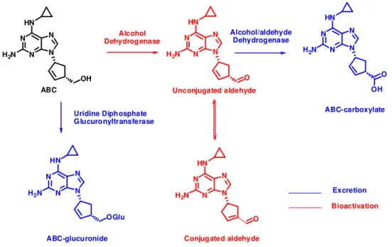
It is well established that ABC can be bioactivated during the formation of the carboxylate metabolite (Walsh et al., 2002). The formation of this metabolite involves a two-step oxidation
process, via a reactive aldehyde intermediate. The formation of isomers the acid metabolite and the ABC in itself through ADH activity led Walsh et al.(2002) to propose a metabolic pathway involving
double bond migration and epimerization processes upon formation of two putative aldehyde intermediates: an unconjugated aldehyde, which rapidly isomerizes into a thermodynamically more stable conjugated aldehyde (ABC aldehyde; Fig. 1.4). This conjugated ABC aldehyde is capable of reacting with proteinsin vitro (Charneira et al., 2011), generating adducts; which potentially can act as
antigens and be recognized by the immune system, triggering an immune response (Park et al.,
2011a). However, so far this reactive metabolite has eluded detection in humans.
Figure 1.4. Scheme of abacavir bioactivation and elimination pathways. Metabolic pathway of
abacavir (ABC) involving the formation of two putative aldehyde intermediates: the unconjugated aldehyde, which rapidly isomerizes into the conjugated aldehyde. This conjugated aldehyde is capable of reacting with proteinsforming protein adducts (Charneira et al., 2011).
1.4
A briefly overview on abacavir toxicity mechanism
Despite the therapeutic benefits of ABC in HIV-infection treatment, this antiretroviral has been associated with several adverse reactions. Nausea is the most frequently reported. In addition, vomiting, malaise and fatigue, headache, diarrhea, sleep disorders, cough, anorexia and rash have also been described, but in lower frequency. These adverse events tend to be more frequent soon after the starting of ABC and are mild or moderate in intensity and transient. The most concerning
OH N N N N HN
H2N
ABC O N N N N HN
H2N
ABC-carboxylate OH OGlu N N N N HN
H2N
ABC-glucuronide O N N N N HN
H2N
Unconjugated aldehyde O N N N N HN
H2N
Protein adducts from the anti-HIV drug abacavir – possible biomarkers of drug toxicity 1. Introduction
9 factor of ABC use is its association with hypersensitivity reactions (HSR) and with myocardial infarction (MI).
1.4.1 Hypersensitivity reactions
HSR has been reported in 3 to 5% of patients, usually occurring within the first 6 weeks of ABC use (Clay, 2002) and can lead to drug discontinuation. This adverse reaction is characterized by symptoms representative of a multi-organ involvement. It is diagnosed by the manifestation of, at least, two of the following symptoms: rash, fever, vomiting, headache, respiratory and gastrointestinal symptoms, lethargy, myalgia or arthralgia (Mallal et al., 2002). Usually, these symptoms rapidly
disappear, after ABC discontinuation. Some patients can be misdiagnosed with respiratory diseases, gastroenteritis or reactions to other medications, which may result in a more severe reaction, multi-organ failure and death. The discontinuation of ABC must be immediate and cannot be re-challenged. A strong association between ABC-induced HSR risk and a HLA-B*5701 allele has been described in several clinical trials. In the Clinical Trial 1 (PREDICT-1) trial (Mallal et al., 2008) and the Study of Hypersensitivity to Abacavir and Pharmacogenetic Evaluation (SHAPE) study (Saag et al., 2008) it
has been showed that those patients (especially among Caucasians) who carry the HLA-B*5701 allele were at higher risk for experiencing an ABC-induced HSR, with a positive predictive value of 47.9% and a negative predictive value of 100%. Therefore, current guidelines recommended the performance of a prospective test to detect the allele-positive patients, to identify to whom ABC should not be prescribe (Nolan, 2009). This prospective test is one of the major successes in the area of personalized medicine by mean of pharmacogenetics. However, this test does not predict which patients will definitely develop HSR (Mallal et al., 2008), allowing the possibility of different
mechanisms at the onset of HSR.
Currently, three major complementary models are considered for these immune-mediated adverse reactions: 1) the hapten/prohapten hypothesis (Fig. 1.5) (Uetrecht, 2007); 2) the pharmacologic interaction with immune receptors, the p-i hypothesis (Fig. 1.6) (Pichler et al., 2006); and 3) the
danger model, which is complementar of the hapten hypothesis, adding a secondary signal (eg. an infection) (Fig. 1.7) (Pirmohamed et al., 2002). The hapten hypothesis considers that drugs or their
metabolites are too small to be immunogenic. Instead, they can bind irreversibly to proteins, generating covalent adducts, which act as antigens (Uetrecht, 2007). These drug-modified proteins are seen as foreign by the immune system, leading to an immune response. Possibly, theses adducts are processed by antigen presenting cells (APCs) into peptide fragments, which associate with the major histocompatibility complex (MHC) from class I (genes HLA-A, -B and –C) and are presented to
T-cell receptors (TCR) (Uetrecht, 2007; Bharadwaj et al., 2010; Park et al., 2011b). The p-i hypothesis
1. Introduction Protein adducts from the anti-HIV drug abacavir – possible biomarkers of drug toxicity
10
Figure 1.5. Hapten/prohapten hypothesis. The drug or reactive metabolite covalently binds to protein.
The modified protein is taken up by antigen presenting cells, processed, and presented through major histocompatibility complex (MHC) to helper (CD4) T cells. In: Uetrecht (2007)
Figure 1.6. Pharmacological interaction (P-i)
hypothesis. The drug binds directly to the major
histocompatibility complex leading to an immune response to the parent drug.In:Uetrecht(2007).
Figure 1.7. Danger hypothesis. Together with
Protein adducts from the anti-HIV drug abacavir – possible biomarkers of drug toxicity 1. Introduction
11 Additionally, a specific model for restricted generation of immunogenic complexes in ABC-induced HSR, through the hapten hypothesis, was recently suggested (Bharadwaj et al., 2012), taking into
account the link to the HLA-B*57:01. Briefly, the covalent modification of a protein, by ABC or a metabolite, generates a hapten. This hapten is processed in the cytoplasm by proteasome to produce peptide fragments, which are transferred into the endoplasmic reticulum by the transporter associated with antigen processing (TAP) (McCluskey et al., 2004), where peptide loading is optimized by tapasin
(Williams et al., 2002b). Then the peptides are loaded onto HLA-B*5701. The antigen-specific T cells
recognizing the HLA-B*5701-restricted viral peptide, causing an immune response (Fig. 1.8). Functional loss of either TAP or tapasin leads to defective peptide loading and impaired antigen presentation (Williams et al., 2002a).
Figure 1.8. Hapten hypothesis for a restricted generation of immunogenic complexes in abacavir
hypersensitivity syndrome on HLA-B*57:01 positive patients. Adapted from Bharadwaj el al. (2012).
1.4.2 Heart toxicity mechanism
In addition to the adverse effects described above, long-term ABC exposure has been associated with an increased risk of MI (Costagliola et al., 2010; Worm et al., 2010; Islam et al., 2012).
This association is still controversial, but a recent meta-analysis (Islam et al., 2012) report that
together with the fact that HIV-patients are at higher risk of devolving cardiovascular disease, ABC is the antiretroviral drug associated with greater risk of MI. Also, current guidelines recommend caution with ABC administration in patients at higher risk of cardiovascular disease (Thompson et al., 2010)
1. Introduction Protein adducts from the anti-HIV drug abacavir – possible biomarkers of drug toxicity
12
Based on well documented knowledge of the link between drug aldheyde metabolites and cardiotoxicity (Guo and Ren, 2010a), it can be anticipated that ABC aldehyde might play a significant role at the onset of ABC-induced heart toxic effects. Nevertheless, this ABC aldehyde has never been identified in man, probably due to the lack of suitable methodological approaches.
1.5
Reactive metabolites, protein-adducts and drug toxicity
The exact mechanism for both cardiotoxicity and HSR associated ABC is not clear. However, ABC bioactivation can be involved. The reactive metabolites generated from drug biotransformation are usually electrophile species, which react easily with macromolecules (eg. proteins), generating adducts. The nucleophilic sites of proteins such as cysteine thiols, lysine amines, histidine imidazoles side chains and protein N-terminal amines (Guengerich et al., 2001; Casini et al., 2002) are the
expected targets of reaction with the electrophilic reactive metabolites.
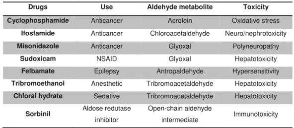
Whereas solid proof for the involvement of ABC bioactivation in toxic events induced by this antiretroviral has not been found yet, some evidence support this hypothesis: 1) the formation of adducts has been implicated in several drug-induced toxic events (Levine and Ovary, 1961; Padovan
et al., 1996); 2) aldehydes are short-lived and extremely difficult to detect in vivo, but can form stable
covalent adducts with proteins; 3) aldehydes are often implicated in HSR conditions due to their ability to undergo protein modification (Table 1.1) (O'Brien et al., 2005); 4) ADH is present in epithelial
tissues, including the skin (Lockley et al., 2005) and heart (Estonius et al., 1996); 5) aldehydes are
often implicated in cardiotoxicity (Carvalho et al., 2004; Luo et al., 2007; Guo and Ren, 2010b). As
Protein adducts from the anti-HIV drug abacavir – possible biomarkers of drug toxicity 1. Introduction
13
Table 1.1. Examples of drugs activated via aldehyde metabolites and their associated toxicities
Drugs Use Aldehyde metabolite Toxicity
Cyclophosphamide Anticancer Acrolein Oxidative stress
Ifosfamide Anticancer Chloroacetaldehyde Neuro/nephrotoxicity
Misonidazole Anticancer Glyoxal Polyneuropathy
Sudoxicam NSAID Glyoxal Hepatotoxicity
Felbamate Epilepsy Antropaldehyde Hypersensitivity
Tribromoethanol Anesthetic Tribromoacetaldehyde Hepatotoxicity
Chloral hydrate Sedative Tribromoacetaldehyde Hepatotoxicity
Sorbinil Aldose redutase
inhibitor
Open-chain aldehyde
intermediate Immunotoxicity
Figure 1.9. Drug bioactivation and toxicity. Toxicity may occur through the parent drug accumulation or by
drug bioactivation through formation of reactive (toxic) metabolites, which, if not detoxified can bind covalently to macromolecules.
1.6
Objectives
The fact that ABC-induced HSR and cardiotoxicity mechanisms are relevant and unclear subjects and the necessity for a prospective biomarker of ABC toxicity, especially for preventing chronic events, has established the objectives of the present work. ABC bioactivation to an aldheyde metabolite has never been identified in man, mainly due to the absence of suitable methodological approach. Our initial hypothesis was that this reactive metabolite could be efficiently trapped by N-terminal valine of
hemoglobin, forming N-terminal-valine-abacavir adducts that could be used to quantify ABC
HIV-1. Introduction Protein adducts from the anti-HIV drug abacavir – possible biomarkers of drug toxicity
14
infected patients represented the second objective (Fig. 1.10). Finally, the identification of factors which could influence the formation of ABC adducts were also projected.
Specific aims:
1) Develop a methodology to access ABC adducts 2) Test the methodology and optimize in an animal model 3) Identify ABC adducts in HIV-infected patients
4) Search for factors that may influence the formation of adducts
Figure 1.10. Graphic summary of study work plan. Preparation of
ABC-adduct standards
Wistar rats treated
with ABC
Identification of ABC-adducts in Wistar rats
Enrolment of patients
15
2
Materials and Methods
Protein adducts from the anti-HIV drug abacavir – possible biomarkers of drug toxicity 2. Materials and Methods....
17
2.1
Materials
2.1.1 Reagents
All reagents were acquired from Sigma-Aldrich Co. (St. Louis, MO, USA). The solvents were purified and dried when applicable.
Abacavir (ABC) reference was supplied through the program NIH AIDS Research and Reference Reagent Program (abacavir, Cat 4680). ABC sulfate was kindly offered by Dr. Frederick Beland
(NCTR, Arkansas, USA).
2.1.2 Enzymes
Pronase E (EC 3.4.24.31) and leucine aminopeptidase M (EC 3.4.11.2) have been both acquired from Sigma-Aldrich Co. (St. Louis, MO, USA). Pronase E solution (0.53 mg/mL) was prepared in phosphate buffered saline 10mM (PBS) and leucine aminopeptidase M solution (0.13 mg/mL) was prepared in distilled water.
2.1.3 Consumables
Silica gel plates 60 F254 from Merck (Darmstadt, Germany) with a thickness of 0.5 mm were
purchased for performing preparative thin layer chromatography (TLC). The plates were observed by ultraviolet light at a wavelength of 254 nm.
Amicon® Ultra-4 Centrifugal Filter Devices, 30,000 MWCO from Millipore (Billerica, MA, USA) were
used for isolation of albumin from plasma.
C-18 Sep-Pak cartridges from Waters (Milford, MA, USA) were used when performing enzymatic
hydrolysis, for sample concentration.
2.2
Equipment
2.2.1 High-performance liquid chromatography (HPLC)
The semi-preparative and analytical HPLC analysis of ABC-adducts standards was conducted on an Ultimate 3000 Dionex (Sunnyvale, CA, USA) system, consisting of a LPG-3400A quaternary gradient
pump, a diode array spectrophotometric detector and an automatic 8125 Rheodyne (Rohnert Park,
CA, USA) injector. The semi-preparative analyses were performed on a Luna C18 column (250 × 10 mm; 5 m) and the analytical were in a Luna C18 column (250 mm × 4.6 mm; 5 m), both from
Phenomenex (Torrance, CA, USA). Data acquisition and processing was performed on the
2. Materials and Protein adducts from the anti-HIV drug abacavir – possible biomarkers of drug toxicity Methods
18
The quantification of ABC in plasma by HPLC was performed in a Schimadzu (Kyoto, Japan)
system, consisting of a LC 9-A solvent delivery pump, a 7725i injector, a SPD-6 AV UV-VIS detector and a CTO-10AS VP column oven. Was used a LiChrospher 100 RP-18 (250 x 4 mm; 5 m) column
protected by a LiChrospher 100 RP-18e (4 x 4mm; 5 m) guard-column, both from Merck (New
Jersey, USA.). Data acquisition and processing were performed on the Shimadzu Class VP 7.X software. Isocratic elution was with a mixture of methanol:acetonitrile:potassium dihydrogen phosphate 3.7 mM at pH 5.65 (35:10:55; v/v/v), a flow rate of 0.6 mL min at 25 ºC. The UV detector was set at a wavelength of 275 nm.
2.2.2 Liquid Chomatography-Electrospay Ionization-Tandem Mass Spectrometry
(LC-ESI-MS)
LC-ESI-MS/MS analyses were performed using a ProStar 410 autosampler, two 210-LC pumps, a ProStar 335 diode array detector and a 500-MS ion trap mass spectrometer with an ESI ion source
from Varian, Inc. (Palo Alto, CA, USA). Data acquisition and processing were performed using the Varian MS Control 6.9.3 software. The samples were injected via an injector with a 20 µL loop from Rheodyne (Rohnert Park, CA, USA). A Luna C18 column (150 mm x 2 mm, 3 µm) from Phenomenex
(Torrance, CA, USA) was used. The MS was operated in the positive ESI mode. The optimized operating parameters were: ion spray voltage, +5.2 kV; capillary voltage, 80 V and RF loading, 70%. Nitrogen was used as the nebulizing and drying gas, at pressures of 45 and 10 psi, respectively. The drying gas temperature was 350 °C. MS/MS spectra were obtained with an isolation window of 2 Da, excitation energy of 1.7 V and excitation time of 10 ms. The MSn spectra were obtained with an isolation window of 1.0 Da, excitation energy values of 1.5 and 1.2 V and an excitation time of 10 ms (CID up to MS3).
2.3
Methods
2.3.1 Preparation of abacavir adduct standards
2.3.1.1 Abacavir generation from abacavir sulphate
ABC sulphate powder (600 mg) was dissolved in water (150 mL), followed by the addition of potassium hydroxide 1M, to reach pH 8. This solution was then extracted with dichloromethane (4 x 200 mL). Subsequently, the organic phase was dried with anhydrous magnesium sulfate and filtered. The solvent was removed under reduced pressure, affording a white solid and the purity of this compound was confirmed by HPLC analysis upon comparison with ABC reference.
2.3.1.2 Swern Oxidation – Preparation of abacavir conjugated aldehyde
Protein adducts from the anti-HIV drug abacavir – possible biomarkers of drug toxicity 2. Materials and Methods....
19 added and stirred at -78 oC, for 1 h. At this stage, anhydrous triethylamine (507 L) was added to the mixture, the temperature was allowed to rise to 0 oC and stirred for 1 h. Afterwards, distilled water (9 mL) was added and the mixture was extracted with diethyl ether (3 x 100 mL). Anhydrous magnesium sulfate was added to the organic phase to remove water and the solvent was removed under reduced pressure. The mixture was purified by preparative TLC [eluted in dichloromethane/methanol (9:1)], affording a yellow oil. Identification of the obtained compound was confirmed by analytical HPLC analysis upon comparison with a previously prepared standard (Charneira et al., 2011).
2.3.1.3 Reaction of ethyl valinate with abacavir-conjugated aldehyde
A solution of ethyl valinate hydrochloride (40 mg) was prepared in phosphate buffer 50 mM at pH 7.4 (800 L) and subsequently treated with sodium hydrogen carbonate (18 mg) for 30 min at 37 oC. Following the addition of ABC-conjugated aldehyde solution in THF (0.5 mL) the mixture was incubated for 30 min, at 37 oC. Sodium cyanoborohydride (150 mg) was then added and the resulting mixture was re-incubated at this temperature overnight. The solvent was removed under reduced pressure.
2.3.1.4 Abacavir-
Edman
adduct standard
The mixture afforded at section 2.3.1.3 was dissolved in N,N-dimethylformamide (DMF; 1 mL).
sodium hydroxide 1M (40 L) and phenyl isothiocyanate (PTIC; 6 L) were both added. The solution was subsequently stirred for 2 h at 37 oC and then for 1.5 h at 45 oC. Upon cooling to room temperature, water (2.5 mL) was added and the solution was extracted with ethyl acetate (2 x 2.5 mL). The organic phase was dried under reduced pressure. The ABC-Edman adduct was purified by
semi-preparative HPLC, using the chromatographic conditions described in Table 2.1 and its purity was subsequently confirmed upon LC-ESI-MS analysis (Table 2.2).
Table 2.1. Chromatographic conditions for HPLC analysis and isolation of ABC-Edman adduct
standard.
Time (min) Formic acid
0.1% (%) Acetonitrile (%) Flow (mL/min) UV absorbance (nm) Temperature
(oC)
0 100 0
3 254 25 oC
15 0 100
17 0 100
25 100 0
2. Materials and Protein adducts from the anti-HIV drug abacavir – possible biomarkers of drug toxicity Methods
20
Table 2.2. Chromatographic conditions for LC-ESI-MS/MS analysis of ABC-Edman adduct standard.
2.3.2 Identification of abacavir-adducts in
Wistar
rats
2.3.2.1 Animal treatment
The animal handling protocol was approved by the Institutional Animal Care and Use Committee.
A group of Wistar rats (10-13 weeks old; three males and three females; 274-388 g) were obtained from the vivarium of the Faculty of Medical Sciences, New University of Lisbon, where the animals
were kept under controlled temperature with 12/12h light/dark cycles. They received a standard rodent diet and tap water ad libitum.
The Wistar rats were exposed to ABC, which was suspended in methyl cellulose (0.2% in water) with 5% of methanol. The rats were administered eight daily intraperitoneal doses of 120 mg ABC/kg body weight. An extra group of each gender (two males and two females) received the vehicle alone. Two hours after the last treatment, the rats were anesthetized (60 mg/kg of pentobarbital), the chest cavity was opened and blood was collected by cardiac puncture into EDTA tubes. The blood samples were centrifuged at 3,000 g for 10 min, to separate plasma from blood cells. The samples were aliquoted
and stored at -80 oC until Hb isolation1.
2.3.2.2 Isolation of hemoglobin (Tornqvist
et al.
, 2002)
Aliquots of the red blood cells (400 µL) were washed with sodium chloride 0.9% (3 x 400 µL). The saline solution was discarded and Milli-Q water (600 µL) was added to each sample, to promote cell lysis. Next, a hydrochloric acid 50 mM in 2-propanol solution (6mL) was added to 1 mL of the lysate and the mixture was centrifuged at 3000 g for 10 min, to remove cell membranes. To the supernatant
was added iced ethyl acetate (40 mL) to precipitate Hb and the pellet was washed with 20 mL of
n-pentane and finally dried under reduced pressure.
1The blood samples were already available at the Pharmacology lab.
Time (min) Formic acid 0.1
% (%) Acetonitrile (%) Flow (mL /min) Temperature
(oC)
0 100 0
0.2 30
15 0 100
17 0 100
Protein adducts from the anti-HIV drug abacavir – possible biomarkers of drug toxicity 2. Materials and Methods....
21
2.3.2.3 Serum albumin isolation (Lindstrom
et al.
, 1998)
To the rat plasma (400 µL) was added a saturated ammonium sulfate solution (400 µL). The solution was centrifuged at 900 g, 4 oC, for 30 min to remove the immunoglobulins. The supernatant was
filtered through an Amicon®centrifugal filter with centrifugation at 3800 g, 4 oC, for 20 min and then the
resulted solution was dried under reduced pressure.
2.3.2.4 Detachment of
N
-terminal valine adducts from hemoglobin -
N
-Alkyl
Edman
degradation (Charneira
et al.
, 2012)
Each Hb sample (50 mg) from section 2.3.2.2 was dissolved in DMF (1.5 mL), followed the addition of sodium hydroxide 1M (65 µL) and PTIC (10 µL). The samples were subsequently stirred for 2 h at 37 oC and for 1.5 h at 45 oC. Upon cooling to room temperature, water (2 mL) was added and the resulted mixtures were extracted with ethyl acetate (2 x 1 mL). The organic phases were dried under reduced pressure, resuspended in methanol (50 µL) and were analyzed by LC-ESI-MS/MS using the chromatographic conditions described in Table 2.2.
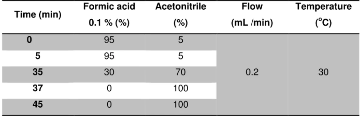
2.3.2.5 Hydrolysis of serum albumin to amino acids (Tsao and Otter, 1999)
Serum albumin samples (10 mg) from section 2.3.2.3 were dissolved in PBS 10mM (3.5 mL) and solutions of Pronase E (190 µL, 0.53 mg/mL) and leucine aminopeptidase M (80 µL, 0.13 mg/mL) were added. The solution was stirred at 37 oC overnight. The enzymatic hydrolysate was concentrated in a C-18 Sep-Pak cartridge. The cartridge was conditioned with methanol (3 mL), followed by water (6 mL). The sample was then loaded, and the cartridge was rinsed with water (1 mL) and methanol (2 mL). The methanolic eluate was dried under reduced pressure, reconstituted in 60 µL acetonitrile/0.1% aqueous formic acid (1:1) and analyzed by LC- ESI-MS/MS through the chromatographic conditions described in Table 2.3.
Table 2.3. Chromatographic conditions used for LC-ESI-MS/MS analysis of rat serum albumin hydrolisates.
Time (min) Formic acid 0.1 % (%)
Acetonitrile (%)
Flow (mL /min)
Temperature (oC)
0 95 5
0.2 30
5 95 5
35 30 70
37 0 100
2. Materials and Protein adducts from the anti-HIV drug abacavir – possible biomarkers of drug toxicity Methods
22
2.3.2.6 Quantification of abacavir concentration in
Wistar
rats
2.3.2.6.1 Preparation of stock and calibration solutions.
Stock solutions of ABC (1 mg/mL) were prepared in methanol and were stored at -80o C until handling.
Different calibration solutions, to obtain a calibration curve covering the range from 0.005 to 20 mg/L, were prepared by dilution of the ABC solution in plasma from rats not exposed to ABC.
All calibration solutions were heated for 60 minutes at 60 ºC before being submitted to the extraction procedure.
2.3.2.6.2 Samples extraction procedure and HPLC analyses.
A liquid-liquid extraction procedure was performed. Briefly, dichloromethane was added to human plasma (500 µL) in a proportion of 1:10, in a 10 mL glass tube. The solution was mixed on a vortex mixer and centrifuged at 2 000 g for 5 min, at 4ºC. The organic phase was recovered, and the aqueous phase was re-extracted. The two organic phases were combined. The solvent was dried under reduced pressure, reconstituted in potassium dihydrogen phosphate 3.7 mM/methanol (1:1; 150
L) and 100 L where injected in the HPLC.
2.3.2.7 Identification and quantification of abacavir-adducts in HIV-infected patients
2.3.2.7.1 Enrollment of patients and, demographic and clinical data collection.
The protocol received prior approval from the Ethics Committee of Centro Hospitalar de Lisboa Central, EPE. The patients gave their written informed consent and adherence was controlled through
a questionnaire. All eligible patients (5 men and 5 women) were adults with documented HIV-infection who had received continuous treatment with ABC-containing cART regimens (300 mg twice daily or 600 mg once daily) for more than 1 month, regardless of past therapeutic history. Exclusion criteria were being under 18 years of age, having AIDS-defining conditions or compliance issues. A control group (2 healthy volunteers) was also included in the study. From all the patients relevant data were collected: age, sex, weight, height, ethnicity, alcohol consumption, injection drug use, smoking habits, opportunistic infections, co-morbidities, adverse events, antiretroviral medication, and other analysis consider relevant for the study (see Annex I – Case Report Form).
Blood samples (2 mL) were collected in EDTA tubes. The samples were centrifuged at 3 000 g for
10 min, 4 oC. The samples of plasma and cells were aliquoted and store at -80 oC until use.
Protein adducts from the anti-HIV drug abacavir – possible biomarkers of drug toxicity 2. Materials and Methods....
23
2.3.2.7.2 Abacavir-
Edman
adduct analysis in HIV-infected patients
Identification and quantification of the ABC-Edman adduct was conducted in three steps: a.
hemoglobin isolation and purification from blood samples (see section 2.3.2.2); b. detachment of N
-terminal valine adducts from haemoglobin (see section 2.3.2.4); and c. analysis of the detached adducts by LC-ESI-MS/MS (see section2.3.2.4).
For ABC-Edman adducts quantification in patients a stock solution of ABC-adduct standard (1
mg/mL) was prepared in methanol. Solutions were stored at -20 oC until analyze. Afterwards, different calibration solutions, to perform a calibration curve covering the range from 0.25 to 2.5 ng/L, were prepared by dilution from the ABC-adduct standard solutions in methanol.
2.3.2.7.3 Quantification of abacavir plasma concentration in HIV-infected patients
25
3
Results
Protein adducts from the anti-HIV drug abacavir - possible biomarkers of drug toxicity 3. Results
27
3.1
Preparation of abacavir adducts standards
ABC-conjugated aldehyde was prepared upon ABC Swern oxidation, using trietilamine, as base. The resulting mixture from Swern reaction was purified by preparative TLC, which allowed the isolation of the ABC-conjugated aldehyde with 18% yield. For the preparation of the stabilized adduct (Fig. 3.1), ABC-conjugated aldehyde in THF was initially incubated with a solution of ethyl valinate in a phosphate buffer 50 mM and then subjected to reduction with sodium cyanoborohydride to stabilize the Shciff base formed. The reaction mixture was then submitted to N-Alkyl Edman degradation, using
the phenyl isothiocyanate as the derivatizing agent (Chevolleau et al., 2007), and following purification
by semi-preparative HPLC the ABC-Edman adduct was obtained.
Figure 3.1. Formation of abacavir-Edman adduct. Swern oxidation of abacavir into its conjugated
aldehyde derivative followed by formation of the Schiff base upon reaction with ethyl valinate, which is then stabilized by reduction and derivatizated with phenyl isothiocyanate in a basic medium.
The purified compound was then analyzed by LC-ESI-MS/MS. The ionic chromatogram exhibited only one signal at 9.7 min, confirming the purity of the obtained product, the ABC-Edman adduct
standard. The identification of this adduct was based upon undistinguishable mass spectra and identical retention time, when compared with previously prepared synthetic standard (Fig. 3.2). The mass spectra exhibited a protonated molecule at m/z 503 that upon tandem mass fragmentation
afforded three fragment ions (Fig. 3.3), stemming from loss of the purine moiety (m/z 313), loss of the
3. Results Protein adducts from the anti-HIV drug abacavir – possible biomarkers of drug toxicity
28
moiety (m/z 191), respectively. Taken together, all data are in accordance the ones described by
Charneira et al. (2011), confirming that the isolated product is the ABC-Edman adduct (Charneira et al., 2011).
Figure 3.2. LC-ESI-MS/MS chromatogram of ion m/z 503 from ABC-Edman adduct standard.
Figure 3.3. LC-ESI-MS/MS fragmentation pattern for the ABC-Edman adduct (protonated molecule
m/z 503).
3.2
Identification of abacavir-adducts in
Wistar
rats
3.2.1 Identification of abacavir-
Edman
adducts with
N
-terminal valine of hemoglobin
To obtain evidence for ABC-conjugated aldehyde ability to modify proteins in vivo, three male and
three female Wistar rats were treated with eight daily doses of ABC (120 mg/kg body weight) and the formation of covalent adducts with the N-terminal valine of Hb was investigated by LC-ESI-MS/MS. The LC-ESI-MS/MS analysis of the ion at m/z 503 (corresponding to the protonated molecule of the expected adduct) allowed the identification of the N-terminal valine adduct from two (one male and
Protein adducts from the anti-HIV drug abacavir - possible biomarkers of drug toxicity 3. Results
29 503 consistently presented three fragment ions, stemming from loss of the cyclopropylaminopurine moiety (m/z 313), loss of the abacavir moiety (m/z 235), and cleavage of the purine-cyclopentene bond, with protonation on the purine moiety (m/z 191). These signals were absent from all the control rat samples.
Figure 3.4 LC-ESI-MS/MS analysis from hemoglobin of the Wistar rats exposed to abacavir. A.
female Wistar rat; B. male Wistar rat and, C. abacavir-Edman adduct standard.
3.2.2 Identification of abacavir adducts with other amino acids from serum albumin
3. Results Protein adducts from the anti-HIV drug abacavir – possible biomarkers of drug toxicity
30
Figure 3.5 Structures of the covalent adducts expected upon A) Michael addition of cysteine to the
double bound of abacavir-conjugated aldehyde, B) Shiff base formation, upon lysine addition to the abacavir carbonyl group, followed by reductive stabilization.
3.2.3 Quantification of abacavir in plasma from
Wistar
rats
Thought analytical HPLC analysis was possible the quantification of ABC concentration in plasma from the Wistar rats treated with ABC. Based on identical retention times (8.3 min) when compared with the standards solutions was possible the identification of ABC peak, and by a calibration curve ABC plasma concentrations were estimated to range between 7.5 and 14.4 mg/L. For rats with ABC-
Edman adducts the mean ± standard deviation (SD) of ABC concentration was 10.8 ± 1.73 mg/L. In
those rats which were not identified with adducts, the mean ± SD of ABC concentration was 13.5 ± 5.7 mg/L. No differences were found (unpaired T-test) between ABC concentration in rats with
Protein adducts from the anti-HIV drug abacavir - possible biomarkers of drug toxicity 3. Results
31 Figure 3.6. Quantification of abacavir concentration in Wistar rats exposed to abacavir. A, Chromatogram obtained following analytical HPLC analysis of a rat exposed to abacavir. The peak identified with an arrow corresponds to abacavir; B, Chromatogram from a rat not exposed to abacavir; C, Chromatogram from a calibration solution in a concentration of 1.5 mg/L (retention time 8.3 min).