The use of bedside echocardiography for
measuring cardiac index and systemic vascular
resistance in pediatric patients with septic shock
INTRODUCTION
Early aggressive fluid resuscitation, antibiotics, and vasoactive therapies are recommended by the American College of Critical Care Medicine as clinical practice parameters for hemodynamic support of septic shock in children and neonates; they also recommend echocardiography to guide fluid and vasoactive regimens as well as to rule out pericardial effusion.(1,2)
The hemodynamic monitoring of shock in the intensive care unit (ICU) is of great value for diagnosis of septic shock as one of the differential diagnoses of
Faten A. Abdalaziz1, Hebat Allah Fadel Algebaly1, Reem Ibrahim Ismail1, Seham Awad El-Sherbini1, Ahmed Behairy1
1. Department of Pediatrics, Faculty of Medicine, Cairo university - Cairo, Egypt.
Objective: Follow-up of cardiac index and systemic vascular resistance index by bedside echocardiography until resuscitation.
Methods: A set of hemodynamic parameters was obtained, including cardiac output, stroke volume, cardiac index, systemic vascular resistance index, velocity time integral, myocardial performance index, capillary refill time, and heart rate at 0 hours after fluid boluses before the start of inotropes, and followed up after 6 hours and 24 hours.
Results: Included were 45 patients with community-acquired septic shock. Septic foci were gastroenteritis (24%), intestinal perforation requiring emergency surgery (24%), pneumonia (20%), central nervous system infection (22%) and soft tissue infection (8%). Klebsiella and Enterobacter were the most frequent isolates. We estimated the factors affecting the cardiac index: high central venous pressure at zero time (r = 0.33, p = 0.024) and persistently
Conflicts of interest: None.
Submitted on April 15, 2018 Accepted on July 26, 2018
Corresponding author:
Hebat Allah Fadel Algebaly
Department of Pediatrics of the Faculty of Medicine
Cairo University 11 Ali Basha Street 11352 Cairo, Egito
E-mail: [email protected]
Responsible editor: Jefferson Pedro Piva
Uso de ecocardiografia junto ao leito para medir índices cardíaco
e de resistência vascular sistêmica em pacientes pediátricos com
choque séptico
ABSTRACT
Keywords: Echocardiography; Hemodynamic; Septic shock; Child
high heart rate at hour 6 (r = 0.33, p = 0.03). The systemic vascular resistance index was high in most patients at 0 and 24 hours and at the time of resuscitation and inversely affected the cardiac index as well as affecting the velocity time integral (r = -0.416, -0.61, 0.55 and -0.295). Prolonged capillary refill time was a clinical predictor of the low velocity time integral at 24 hours (r = -0.4). The mortality was 27%. Lower systemic vascular resistance index and higher cardiac output were observed in nonsurviving patients.
Conclusion: There was a persistently high systemic vascular resistance index in cold shock patients that influenced the stroke volume index, cardiac index, and velocity time integral. The use of echocardiograms for hemodynamic measurements is important in pediatric septic shock patients to adjust dilators, and vasopressor doses and achieve resuscitation targets in a timely manner.
circulatory shock, evaluating the hemodynamic condition involving the detection of therapeutic conflicts, and directing the treatment regimen.(3)
Septic shock in children is classically differentiated into either cold shock (low cardiac index, often high systemic vascular resistance index - SVRI), and warm shock (high cardiac index, low SVRI).(4)
In applying any hemodynamic monitoring technique, cardiac index is considered the reference standard parameter for targeting organ perfusion and oxygen delivery in shock.(5)
Cardiac output is classically evaluated by insertion of a pulmonary artery catheter (Swan-Ganz) using the thermodilution technique.(6)There is an increased interest
in replacing this invasive method with noninvasive or minimally invasive technologies to measure cardiac index, including pulse contour analysis, lithium dye dilution, electrical bioimpedance, and transesophageal and transthoracic echocardiography with pulsed or continuous
wave Doppler ultrasound.(7) A focused ultrasound cardiac
examination to evaluate cardiac index, SVRI, and other echo parameters have been discussed in recent studies as a possible method used by emergency room (ER) physicians for the management of critically ill patients.(8,9) It was
found that increased cardiac index was associated with better renal function and better renal outcome in patients with acute renal injury.(10)
Systemic vascular resistance index was found to be a powerful predictor of mortality in children with septic shock because low values of SVRI denote endothelial layer injuries that are one of the important pathophysiologies of sepsis.(11)
Left ventricular outflow tract-velocity time integral (LVOT-VTI) (used to calculate stroke volume) is thought to be a better indicator of LV systolic function than stroke volume, without the confounding factor of LVOT-area measurements, which are difficult to obtain in ICU patients.(12)
We aimed to study the role of bedside echocardiography as a noninvasive tool in assessing the hemodynamic changes in critically ill patients with septic shock with the hope of helping manage such patients.
METHODS
This prospective observational cohort study was conducted between June 2014 and July 2015, in an academic pediatric intensive care unit (PICU) of Cairo University Specialized Hospital with a 14-bed capacity and approximately 300 yearly admissions. The study
was approved by the institutional review board at our institution and was deemed to pose minimal risk to the subjects. Before study enrollment, a PICU physician was trained to measure the echocardiographic parameters using the ultrasound system echocardiographic equipment (GE, Logic P3). Training of the PICU physician included a total of 48 hours of hands-on instruction by a certified cardiac sonographer to obtain the LVOT-VTI and the Doppler mitral inflow signal to be able to calculate cardiac index, SVRI, myocardial performance index (Tei index) and fractional shortening (FS).
Patients with strong clinical suspicion of septic shock at the time of admission to the PICU were included. Septic shock was diagnosed according to the American College of Critical Care Medicine Clinical Practice Parameters for Hemodynamic Support of Pediatric and Neonatal Septic Shock 2012 by clinical signs, including hypothermia or hyperthermia, altered mental status, and peripheral vasodilation (warm shock) or vasoconstriction with capillary refill time (CRT) greater than 2 seconds
(cold shock) before hypotension occurs.(13) All cases
included were considered to be in a state of fluid refractory septic shock when they received at least 40mL/kg shock fluids (normal saline or Ringer’s) and required inotropic/ vasopressor support for hemodynamic resuscitation, whether ventilated or not.
Patients with congenital heart diseases, cardiomyopathy, and valvular disorders were excluded. After informed consent was obtained from the caregiver, transthoracic two-dimensional, M-mode, and Doppler echocardiography was performed on commercially available echocardiographic equipment (GE, Logic P3) using a 6S probe. Echocardiography was performed to measure the LVOT diameter measured in the long-axis parasternal view, and the time-velocity integral of the flow wave across the aortic valve (VTI) was obtained by pulsed wave Doppler. All ultrasound images obtained by the PICU physician and sonographer were stored and then verified by a cardiologist using a scale to rate the acceptability of the ultrasound measurements.
to the aortic annulus.(4) Stroke volume was calculated
by the equation: π × VTI × (LVOT diameter/2)2. Stroke
volume index (SVI) = stroke volume/body surface area. Cardiac output = stroke volume × heart rate. Cardiac index = cardiac output/ body surface area. M-mode echocardiography was done to measure left ventricular end systolic diameter (LVESD), left ventricular end diastolic diameter (LVEDD), FS is measured by the equation: LVEDD - LVESD/LVEDD × 100. Myocardial performance index (Tei Index) was calculated by the equation: the sum of isovolumic contraction time and isovolumic relaxation time (total systolic time- ejection time) divided by the ejection time obtained by imaging the Doppler mitral inflow signal. Myocardial performance index - MPI (Tei index) = Total systolic time- ejection time/ ejection time. Systemic vascular resistance was calculated by the equation: 80 (mean blood pressure-central venous pressure)/cardiac index.(14)
All cases were subjected to full clinical and echocardiographic examination at the time of PICU admission before inotropic support (Time 0), after 6 hours, after 24 hours and then at the time of stabilization (resuscitation point), which was identified by normalization of heart rate (resolved tachycardia), blood pressure (resolved hypotension if present), CRT (less than 2 seconds), good urine output (≥ 1mL/kg per hour), and regaining of consciousness with the start of vasoactive drug withdrawal.
Different vasoactive agents were used in management of the study cases with fluid refractory septic shock. These agents included adrenaline, noradrenaline, dobutamine, milrinone, and dopamine; overt vasodilator agents such as nitroglycerin were also used.
Inotropic score (IS) was calculated by the Wernowsky equation:
Wernowsky IS = dopamine dose + dobutamine dose +100 × epinephrine dose(15)
Vasoactive IS (VIS) was calculated by the following equation:
VIS = IS + 10 × milrinone dose + 10,000 × vasopressin
dose + 100 × norepinephrine dose(16)
Statistical analysis
Data were analyzed using the Statistical Package of Social Science Software (SPSS) program, version 21.
Data were summarized using mean and standard deviation for quantitative variables and frequency and percentage for qualitative variables.
Comparison between groups was performed using independent sample t-tests or one-way ANOVA with post hoc Tukey’s tests for quantitative variables and chi square or Fisher’s exact tests for qualitative variables.
Paired t-tests were conducted to compare quantitative data.
Pearson or Spearman correlation coefficients were calculated to test the association between parametric and nonparametric variables, respectively.
P values < 0.05 were considered statistically significant, and < 0.01 were considered highly significant.
RESULTS
In this study, 45 patients with fluid refractory septic shock were included. There were 26 (57.8%) males and 19 (42.2%) females. Their mean age was 20.1 ± 29.1 months (2 - 120 months) and mean body weight was 9.8 ± 6.2kg. Their mean CRT at time of admission was 5 ± 0.9 seconds, and mean heart rate at the time of admission was 153 ± 24bpm. The most common causes of sepsis were gastroenteritis and postoperative sepsis followed by CNS infections. The most common isolated organism in blood culture was Klebsiella followed by Enterobacter. No growth was detected in 66.7% of cases, as shown in table 1. Of the 45 cases, 33 (73%) patients improved and were discharged from the PICU, and 12 (27%) patients died.
Duration of resuscitation from septic shock was assessed. Thirty (66.7%) of 45 cases were resuscitated before 24 hours, and the median resuscitation time of all survivors was 34 hours.
Thirty-seven (82%) of 45 cases had echocardiographic criteria of cold septic shock with low normal or low
cardiac index (≤ 3.3L/m/m2) and normal or high SVRI
(≥ 1,600dyn-sec/cm5/m2), whereas 8 (18%) cases had
echocardiographic criteria of warm septic shock (high
cardiac index > 6L/m/m2) with low normal or low SVRI
(≤ 800dyn-sec/cm5/m2).
admission versus 0.4 ± 0.1 by mean time of resuscitation, with p value 0.008), MPI showed a negative correlation with the central venous pressure (r = -0.3; p = 0.04).
It was interesting to find that the initial SVRI was
1,514 ± 550 dyn-sec/cm5/m2. SVRI declined to 1,398 ±
535dyn-sec/cm5/m2 at 6 hours, and then had a rebound
elevation at the mean time of resuscitation (34 hours) to
1,402 ± 400dyn-sec/cm5/m2 (Table 2). The SVRI was
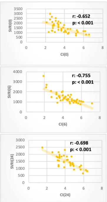
the only measured index that showed a second rise at the time point of resuscitation in cases with cold shock, and we intended to see its effect as afterload on the other hemodynamic parameters. We found that VTI, SVI, and cardiac index were negatively affected by the SVRI at various time intervals. The heart rate and the myocardial performance index were not affected by the SVRI (Table 3, Figure 2).
The cardiac index was found to have a linear positive correlation with SVI and VTI assessed at different time intervals and had a negative correlation with SVRI (Table 4).
Velocity-time integral was negatively affected by the rising SVRI at different time intervals and with CRT at 24 hours (Table 5).
Because all the cases included in this study had fluid-resistant septic shock, all required inotropic support for management, and many (62.2%) required more than two inotropic agents.
- Adrenaline was the starting inotrope in 11 (24%)
cases; 6 of 11 had low cardiac index (< 3.3L/
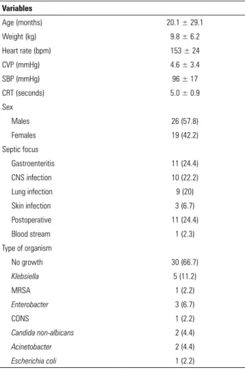
Table 1 - Clinical and laboratory characteristics of the study population
Variables
Age (months) 20.1 ± 29.1
Weight (kg) 9.8 ± 6.2
Heart rate (bpm) 153 ± 24
CVP (mmHg) 4.6 ± 3.4
SBP (mmHg) 96 ± 17
CRT (seconds) 5.0 ± 0.9
Sex
Males 26 (57.8)
Females 19 (42.2)
Septic focus
Gastroenteritis 11 (24.4)
CNS infection 10 (22.2)
Lung infection 9 (20)
Skin infection 3 (6.7)
Postoperative 11 (24.4)
Blood stream 1 (2.3)
Type of organism
No growth 30 (66.7)
Klebsiella 5 (11.2)
MRSA 1 (2.2)
Enterobacter 3 (6.7)
CONS 1 (2.2)
Candida non-albicans 2 (4.4)
Acinetobacter 2 (4.4)
Escherichia coli 1 (2.2)
CVP - central venous pressure; SBP - systolic blood pressure; CRT - capillary refill time; MRSA - Methicillin-resistant Staphylococcus aureus; CONS - Coagulase Negative
Staphylococci. Results expressed as mean ± standard deviation or n (%).
Table 2 - The echocardiographic data assessed at time of admission, after 6 hours and at the time of resuscitation
Clinical/echocardiography data Admission
data After 6 hours After 24 hours Resuscitation point
Data on admission versus time of resuscitation
p value
Heart rate (beats/minute) 153 ± 24 154 ± 24 142.7 ± 25.2 138 ± 22 0.003
CRT (seconds) 5 ± 0.9 3.2 ± 1 2.3 ± 1.3 1.8 ± 1.1 < 0.001
TVI 13.5 ± 3.7 15.5 ± 3.6 15.9 ± 4.1 16.4 ± 3.9 < 0.001
SV (mL) 11.3 ± 7.1 13.1 ± .7 14.1 ± 8.7 14.6 ± 8.1 0.001
SVI (mL/beat) 24.9 ± 8.3 29 ± 7.9 30 ± 8.4 32.1 ± 8.1 0.001
CO (mL/minute) 1.653 ± 0.888 1.938 ± 0.919 1.908 ± 0.917 1.914 ± 0.832 0.06
CI (L/m/m2) 3.7 ± 1.2 4.4 ± 1.1 4.2 ± 1.1 4.4 ± 1.1 < 0.001
FS (%) 35.0 ± 8.4 36.2 ± 7.0 37.4 ± 6.8 37.4 ± 6.2 0.13
SVRI (dyn sec/cm5/m2) 1.514 ± 550 1.398 ± 535 1.487.9 ± 430.6 1.402 ± 400 0.218
Tei index 0.3 ± 0.1 0.4 ± 0.1 0.4 ± 0.1 0.4 ± 0.1 0.043
Figure 1 - Line curve showing pattern of time velocity integral and stroke volume index measured at different time intervals. TVI - time velocity integral; SVI - stroke volume index.
min/m2), and 5 had normal cardiac index (3.3 -
6L/m/m2).
- Noradrenaline was used as an add-on vasopressor
in 4 (9%) cases (these 4 cases were on adrenaline or dobutamine before adding noradrenaline).
- Dobutamine was the starting inotrope in 32 cases
(71%); 20 of 32 cases had normal cardiac index
(3.3 - 5.5L/min/m2) but with high normal or high
SVRI (≥ 1,600dyn-sec/cm5/m2), and 12 cases had
a low cardiac index (< 3.3L/min/m2) with high
normal or high SVRI.
- Dobutamine was used as an add-on inotrope (inodilator) in 8 cases (these cases were on adrenaline or dopamine before adding dobutamine).
- Milrinone was used as an add-on inotrope (inodilator) in 6 (13%) cases; it is used as an alternative to dobutamine.
- Dopamine was used as the starting inotrope in 2
patients only and as add-on inotrope in 1 patient after dobutamine.
- Nitroglycerin drip was proven to have a good rule
in resuscitation of septic shock (cold type) because it was given in 14 (31%) cases with normal or low cardiac index and a high SVRI; it helped in titrated decreases in the SVRI with improvement of the cardiac index (Figure 3).
Nonsurvivors had a higher cardiac output and lower SVRI (they had warm septic shock type on admission). We found that the nonsurvivors had higher inotropes and vasopressor scores at T6 hours (Table 6).
DISCUSSION
Shock is considered the main leading cause of morbidity and mortality in the PICU. Septic shock is a serious condition with high mortality rates ranging from 18% to 50%. To improve outcomes, it is important to upgrade hemodynamic monitoring parameters, not just the traditional clinical signs to guide management.(11)
Bedside echocardiographic examination was performed to assess the hemodynamic parameters for patients with fluid refractory septic shock. The heart rate, CRT, and base deficit were the clinical and biochemical parameters taken as endpoints of resuscitation. Titration of inotropes and dilators until therapeutic-end points was preceded by measuring the hemodynamic parameters via echocardiographic assessment at fixed time intervals.
The main findings of this study were that VTI, SVI, and cardiac index were compromised at the time of PICU admission (T0) and progressively improved, matching clinical therapeutic target achievement, inotrope and dilator modulation. Fractional shortening was within the normal range and remained there, with no remarkable changes throughout the follow-up period. Furthermore, the Tei index became prolonged (indicating impaired global myocardial performance) at T6 hours and then remained almost unchanged afterward.
echocardiography measuring FS and ejection fraction. They stated that these standard parameters were load-dependent and often revealed cardiac dysfunction only after clinical deterioration had occurred.(17)
This impairment of MPI detected in our study can be explained by the fact that global myocardial dysfunction can occur in septic shock and goal-directed therapy helps in preventing worsening of myocardial performance. This
is consistent with an adult study done by Nizamuddin et al., which concluded that MPI used to assess subtle changes in myocardial function not detected by routine hemodynamic monitoring, strongly correlated with mortality among patients with severe sepsis and septic shock; changes in MPI may be a prognostic tool in patients with septic shock and may raise the possibility
Table 3 - Correlations between systemic vascular resistance index with other variables at different time intervals
Systemic vascular resistance index
Admission After 6 hours After 24 hours Resuscitation point
TVI
r -0.416 -0.617 -0.559 -0.295
p value 0.006 < 0.001 < 0.001 0.049
SVI
r -0.509 -0.736 -0.637 -0.533
p value < 0.001 < 0.001 < 0.001 < 0.001
HR
r -0.124 -0.160 -0.050 -0.159
p value 0.427 0.294 0.765 0.298
CI
r -0.652 -0.755 -0.698 -0.717
p value < 0.001 < 0.001 < 0.001 < 0.001
LVESD
r -0.227 -0.048 0.039 -0.202
p value 0.143 0.755 0.818 0.182
LVEDD
r -0.263 -0.049 -0.026 -0.245
p value 0.088 0.747 0.877 0.105
FS
r -0.029 -0.219 -0.165 -0.003
p value 0.854 0.149 0.323 0.986
CVP
r -0.364 -0.033 0.019 -0.166
p value 0.016 0.828 0.911 0.276
TEI
r -0.082 0.002 -0.144 0.117
p value 0.621 0.990 0.389 0.443
CRT
r -0.277 0.087 0.385 0.242
p value 0.072 0.569 0.017 0.109
Figure 2 - Showing the significant negative correlation between the cardiac index and systemic vascular resistance index at different time intervals. SVRI - systemic vascular resistance index; CI - cardiac index.
that myocardial dysfunction plays a mechanistic role in sepsis-related mortality.(18)
Optimization of cardiac output and SVRI was associated with improved outcomes in fluid-refractory septic shock in children. Similar observations have been made in adults as well, using goal-directed therapy.(19)
In this study, the significant increase of the cardiac index was observed by the end of resuscitation of septic shock, consistent with findings of other studies that
considered cardiac index to be the standard parameter in resuscitation of septic shock, because attainment of the therapeutic goal of a cardiac index between 3.3 and 6.0L/min/m2 may result in improved survival in patients with septic shock.(1,2) The cardiac index was found to be
negatively correlated with SVRI because excessively high SVRI has detrimental effects on the cardiac index; this is consistent with the results of the study done by Deep et
al., which found a strong negative correlation between
cardiac index and SVRI.(20)
In this study, we also observed that SVRI was significantly decreased after 6 hours of resuscitation in comparison to Time 0, and then showed mild rebound elevation at T24 hours with no significant difference. This may be explained by sympathomimetic withdrawal lagging behind cardiac index optimization despite the use of dilators such as nitroglycerine and milrinone. The theory of ischemia/reperfusion should be considered, because the bioavailability of nitric oxide, an important mediator of vasodilation, is profoundly decreased during the reperfusion period, resulting in impaired vasodilation of arterioles. Release of inflammatory mediators and increased expression of adhesion molecules initiate inflammatory and coagulation cascades that culminate in the occlusion of capillaries, known as the “no-reflow” phenomenon.
(19) Such a rebound increase in resistance can lead to a
decrease in cardiac output, and vice versa. Furthermore, SVRI was observed to be negatively correlated with VTI. Despite the systemic vascular resistance being critical in the infants’ compensatory mechanisms, its persistent or rebound elevation has a poor impact on hemodynamic resuscitation parameters.(21)
In our study, echocardiographic assessment was helpful in differentiating the type of septic shock (cold and warm). Cold septic shock predominated in our study (82%), whereas warm septic shock represented 12%; nevertheless, it had the highest case fatality (34.4%), and this is consistent with the results of the study conducted by Deep et al., which concluded that the cold type of
septic shock is far more common than the warm type.(20)
In essence, this provides a strong argument for quantification and consideration of all three circulatory components when treating septic shock: flow, pressure and resistance.(19)
Table 4 - Correlations between cardiac index and other variables at different time intervals
Cardiac index
Admission After 6 hours After 24 hours Resuscitation point
LVOT
r 0.050 0.118 0.175 0.257
p value 0.744 0.440 0.293 0.089
TVI
r 0.775 0.613 0.526 0.371
p value < 0.001 < 0.001 0.001 0.012
SVI
r 0.858 0.842 0.719 0.776
p value < 0.001 < 0.001 < 0.001 < 0.001
HR
r 0.094 0.337 0.257 0.204
p value 0.539 0.023 0.118 0.178
LVESD
r 0.136 0.082 -0.011 0.162
p value 0.374 0.591 0.947 0.289
LVEDD
r 0.171 0.020 -0.009 0.150
p value 0.261 0.898 0.959 0.326
FS
r 0.105 0.173 0.002 -0.062
p value 0.494 0.255 0.990 0.684
CVP
r 0.341 0.011 -0.142 -0.003
p value 0.024 0.941 0.395 0.984
MBP
r 0.133 0.154 0.042 0.104
p value 0.391 0.312 0.801 0.496
SVRI
r -0.652 -0.755 -0.698 -0.717
p value < 0.001 < 0.001 < 0.001 < 0.001
TEI
r -0.030 0.057 -0.014 -0.028
p value 0.852 0.717 0.936 0.855
CRT
r 0.078 -0.320 -0.262 -0.237
p value 0.613 0.032 0.112 0.117
Table 5 - Correlations between time velocity integral and other variables at different time intervals
Time velocity integral of aortic outflow signal
Admission After 6 hours After 24 hours Resuscitation point
SVRI
r -0.416 -0.617 -0.559 -0.295
p value 0.006 <0.001 <0.001 0.049
LVOT
r 0.035 0.118 0.157 0.100
p value 0.819 0.442 0.345 0.512
LVESD
r 0.116 0.202 0.240 0.400
p value 0.450 0.184 0.146 0.006
LVEDD
r 0.221 0.261 0.293 0.370
p value 0.144 0.084 0.074 0.012
FS
r 0.109 0.277 0.028 -0.142
p value 0.476 0.066 0.869 0.352
CVP
r 0.278 0.091 -0.056 0.051
p value 0.068 0.552 0.737 0.742
TEI
r 0.025 -0.117 -0.196 -0.213
p value 0.878 0.454 0.238 0.160
CRT
r 0.116 -0.243 -0.393 -0.197
p value 0.448 0.107 0.015 0.195
SVRI - systemic vascular resistance index; LVOT- left ventricular outflow tract diameter; LVESD - left ventricular end systolic diameter; LVEDD - left ventricular end diastolic diameter; FS - fraction shortening; CVP - central venous pressure; TEI index - myocardial performance index; CRT - capillary refill time.
Figure 3 - Bar chart describing the different vasoactive agents used in the study.
Table 6 - Comparing cardiac output, systemic vascular resistance index and treatment score between the survivor and nonsurvivor groups
Mortality (long term)
Nonsurvivors (n = 12) Survivors (n = 33) p value
CO T0 (mL/min) 1900 ± 741 1564 ± 931 0.021
SVRI T0 (dyn-sec/cm5/m2) 1158 ± 396 1623 ± 550 0.01
Inotropic score T6 22.9 ± 12.5 14.2 ± 6.9 0.012
Vasopressor score T6 26.7 ± 14.8 14.6 ± 6.9 0.008
COP - cardiac output; T0 - time of pediatric intensive care unit admission before inotropic support; SVRI - systemic vascular resistance index; T6 - six hours after inotropic support. Results expressed as mean ± standard deviation.
on 36 children with fluid refractory septic shock, the resuscitation time was 42 hours.(20)
The mortality rate in our study was 27%, which was higher than a similar study conducted on patients of the same age group.(21)
Objetivo: Acompanhar o índice cardíaco e o índice de resistência vascular sistêmica até a ressuscitação.
Métodos: Por meio de ecocardiografia junto ao leito, obteve-se um conjunto de parâmetros hemodinâmicos, inclusive débito cardíaco, volume sistólico, índice cardíaco, índice de resistência vascular sistêmica, integral velocidade-tempo, índice de desempenho miocárdico, tempo de reenchimento capilar e frequência cardíaca no momento zero após infusão de fluidos em bolo, e início e utilização de fármacos inotrópicos, com seguimento até 6 horas e 24 horas.
Resultados: Incluíram-se 45 pacientes com choque séptico adquirido na comunidade. Os focos de infecção foram gastrenterite (24%), perfuração intestinal com necessidade de cirurgia emergencial (24%), pneumonia (20%), infecção do sistema nervoso central (22%) e infecção de tecidos moles (8%). Os isolados mais frequentes foram de Klebsiella e Enterobacter. Estimamos os fatores que afetaram o índice cardíaco: pressão venosa central elevada no momento zero (r = 0,33; p = 0,024) e persistência de frequência cardíaca elevada após 6 horas
(r = 0,33; p = 0,03). O índice de resistência vascular sistêmica foi alto na maioria dos pacientes no momento zero e após 24 horas, e por ocasião da ressuscitação, afetando inversamente o índice cardíaco, assim como a integral velocidade-tempo (r = -0,416; -0,61; 0,55 e -0,295). O tempo de reenchimento capilar aumentado foi preditor clínico de valores baixos de integral velocidade-tempo após 24 horas (r = -0,4). O índice de mortalidade foi de 27%. Nos pacientes que não sobreviveram, observaram-se índices de resistência vascular sistêmica mais baixos e débitos cardíacos mais altos.
Conclusão: O índice de resistência vascular sistêmica esteve persistentemente elevado em pacientes com choque frio, o que influenciou no índice de volume sistólico, no índice cardíaco e na integral velocidade-tempo. O uso de ecocardiografia para obtenção de mensurações hemodinâmicas é importante em pacientes pediátricos com choque séptico, para que se possam ajustar as doses de vasodilatadores e vasopressores, e obter os objetivos da ressuscitação em tempo apropriado.
RESUMO
Descritores: Ecocardiografia; Hemodinâmica; Choque séptico; Criança
REFERENCES
1. Carcillo JA, Fields AI; American College of Critical Care Medicine Task Force Committee Members. Clinical practice parameters for hemodynamic support of pediatric and neonatal patients in septic shock. Crit Care Med. 2002;30(6):1365-78.
2. Brierley J, Carcillo JA, Choong K, Cornell T, Decaen A, Deymann A, et al. Clinical practice parameters for hemodynamic support of pediatric and neonatal septic shock: 2007 update from the American College of Critical Care Medicine. Crit Care Med. 2009;37(2):666-88. Erratum in: Crit Care Med. 2009;37(4):1536. Skache, Sara [corrected to Kache, Saraswati]; Irazusta, Jose [corrected to Irazuzta, Jose].
3. Saugel B, Huber W, Nierhaus A, Kluge S, Reuter DA, Wagner JY. Advanced hemodynamic management in patients with septic shock. Biomed Res Int. 2016;2016:8268569.
4. Han YY, Carcillo JA, Dragotta MA, Bills DM, Watson RS, Westerman ME, et al. Early reversal of pediatric-neonatal septic shock by community physicians is associated with improved outcome. Pediatrics. 2003;112(4):793-9.
5. Antonelli M, Levy M, Andrews PJ, Chastre J, Hudson LD, Manthous C, et al. Hemodynamic monitoring in shock and implications for management. International Consensus Conference, Paris, France, 27-28 April 2006. Intensive Care Med. 2007;33(4):575-90.
6. Swan HJ, Ganz W, Forrester J, Marcus H, Diamond G, Chonette D. Catheterization of the heart in man with use of a flow-directed balloon-tipped catheter. N Engl J Med. 1970;283(9):447-51.
7. Hofer CK, Ganter MT, Zollinger A. What technique should I use to measure cardiac output? Curr Opin Crit Care 2007;13(3):308-17.
Our nonsurvivors had significantly lower SVRI than
did survivors with a mean of 1,158 ± 396dyn-sec/cm5/
m2; this value was even lower than the value identified
by Lee et al., who stated that SVRI of 1,167dyn-sec/cm5/
m2 was a convenient point to predict mortality (measured
by pulmonary thermodilution method).(11) This may be
explained by the fact that sepsis causes circulatory failure, secondary to vasodilation (especially caused by endothelial injuries).(23)
CONCLUSION
8. Dinh VA, Ko HS, Rao R, Bansal RC, Smith DD, Kim TE, et al. Measuring cardiac index with a focused cardiac ultrasound examination in the ED. Am J Emerg Med. 2012;30(9):1845-51.
9. Ranjit S, Kissoon N. Bedside echocardiography is useful in assessing children with fluid and inotrope resistant septic shock. Indian J Crit Care Med. 2013;17(4):224-30.
10. Luo JC, Qiu XH, Pan C, Xie JF, Yu T, Liu L, et al. Increased cardiac index attenuates septic acute kidney injury: a prospective observational study. BMC Anesthesiol. 2015;15:22.
11. Lee EP, Hsia SH, Lin JJ, Chan OW, Lee J, Lin CY, et al. Hemodynamic analysis of pediatric septic shock and cardiogenic shock using transpulmonary thermodilution. Biomed Res Int. 2017;2017:3613475.
12. Jensen MB, Sloth E, Larsen KM, Schmidt MB. Transthoracic echocardiography for cardiopulmonary monitoring in intensive care. Eur J Anaesthesiol. 2004;21(9):700-7.
13. Dellinger RP, Levy MM, Rhodes A, Annane D, Gerlach H, Opal SM, Sevransky JE, Sprung CL, Douglas IS, Jaeschke R, Osborn TM, Nunnally ME, Townsend SR, Reinhart K, Kleinpell RM, Angus DC, Deutschman CS, Machado FR, Rubenfeld GD, Webb SA, Beale RJ, Vincent JL, Moreno R; Surviving SepsisCampaign Guidelines Committee including the Pediatric Subgroup. Surviving sepsis campaign: international guidelines for management of severe sepsis and septic shock: 2012. Crit Care Med. 2013;41(2):580-637.
14. O’Rourke RA, Fuster V, Alexander RW. Hurst’s the heart. Manual of cardiology. 11th ed. New York: McGraw-Hill; 2005. p. 513.
15. Wernovsky G, Wypij D, Jonas RA, Mayer JE Jr, Hanley FL, Hickey PR, et al. Postoperative course and hemodynamic profile after the
arterial switch operation in neonates and infants. A comparison of low-flow cardiopulmonary bypass and circulatory arrest. Circulation. 1995;92(8):2226-35.
16. Gaies MG, Gurney JG, Yen AH, Napoli ML, Gajarski RJ, Ohye RG, et al. Vasoactive-inotropic score as a predictor of morbidity and mortality in infants after cardiopulmonary bypass. Pediatr Crit Care Med. 2010;11(2):234-8.
17. Basu S, Frank LH, Fenton KE, Sable CA, Levy RJ, Berger JT. Two-dimensional speckle tracking imaging detects impaired myocardial performance in children with septic shock, not recognized by conventional echocardiography. Pediatr Crit Care Med. 2012;13(3):259-64.
18. Nizamuddin J, Mahmood F, Tung A, Mueller A, Brown SM, Shaefi S, et al. Interval changes in myocardial performance index predict outcome in severe sepsis. J Cardiothorac Vasc Anesth. 2017;31(3):957-64. 19. Seal JB, Gewertz BL. Vascular dysfunction in ischemia-reperfusion injury.
Ann Vasc Surg. 2005;19(4):572-84.
20. Deep A, Goonasekera CD, Wang Y, Brierley J. Evolution of haemodynamics and outcome of fluid-refractory septic shock in children. Intensive Care Med. 2013;39(9):1602-9.
21. Pedro TC, Morcillo AM, Baracat EC. Etiology and prognostic factors of sepsis among children and adolescents admitted to the intensive care unit. Rev Bras Ter Intensiva. 2015;27(3):240-6.
22. Peters MJ, Brierley J. No representation without taxation: declaration of (load) independence in septic cardiomyopathy. Pediatr Crit Care Med. 2012;13(3):349-50.