A R T I G O D E R E V I S Ã O
T H E G E N E T I C S O F O S T E O P O R O S I S
Frances MK Williams*,Tim D Spector*
tores genéticos. Do mesmo modo, a densidade mi-neral óssea (DMO) e outros factores de risco para fracturas, como a baixa qualidade óssea, a geome-tria do colo do fémur e o turnover ósseo são gran-demente hereditários. A susceptibilidade para a os-teoporose é, com grande probabilidade, mediada por múltiplos genes, com um pequeno contributo cada. Várias abordagens têm sido utilizadas para identificar os genes envolvidos, incluindo estudos de linkage em humanos e em modelos animais, es-tudo de genes candidatos e de expressão de genes. Os estudos de linkage identificaram múltiplos quantitative trait loci (QTL) para a regulação da DMO e, juntamente com estudos em gémeos, in-dicaram que o efeito destes loci na DMO é depen-dente do local e específica para o sexo. No global, os genes responsáveis pela regulação da DMO ntes QTL não foram identificados. A maioria dos es-tudos utilizou genes candidatos, baseando-se no metabolismo ósseo. O gene do receptor da vitami-na D (RVD), do colagénio tipo I alfa1 (COLIA1) e do receptor dos esterogénios (RE) alfa têm sido ampla-mente investigados e concluiu-se que têm um pa-pel na regulação da DMO. Uma meta-análise recen-te sugere contudo que o RVD não desempenha um papel relevante e que o efeito dos outros 2 genes é modesto – provavelmente contribuindo em menos de 3% para a componente genética da DMO. Os testes genéticos em larga escala mais acessíveis permitirão grandes estudos populacionais multi-nacionais de análise de genes candidatos, meta--análises, pooling de DNA e estudos de expressão de genes.
Palavras-chave: Osteoporose; Gene; QTL; Densida-de Mineral Óssea
Osteoporosis Genes and their Identification
Osteoporosis is characterized by diminished bone mineral density and deterioration in bone micro-architecture. The main clinical endpoint is fractu-re. It is common and costly, both financially and in *Twin Research & Genetic Epidemiology Unit,St Thomas’ Hospital, London
Summary
Osteoporosis is highly influenced by genetic factors. Bone mineral density (BMD) has also been shown to be highly heritable, as are other risk factors for oste-oporotic fracture such as bone quality, femoral neck geometry and bone turnover. Susceptibility to osteo-porosis is mediated, in all likelihood, by multiple ge-nes each having small effect and a number of diffe-rent approaches are being employed to identify the genes involved. Study methods include linkage stu-dies in both humans and experimental animals as well as candidate gene and gene expression studies. Linkage studies have identified multiple quantitati-ve trait loci (QTL) for regulation of BMD and, along with twin studies, have indicated that the effects of these loci on BMD are site-dependent and sex-spe-cific. On the whole, the genes responsible for BMD regulation at these QTL have not been identified. Most studies have used the candidate gene appro-ach, based on what is known of bone metabolism. The vitamin D receptor gene (VDR), the collagen type I alpha I gene (COLIA1) and estrogen receptor gene (ER) alpha have been widely investigated and found to play a role in regulating BMD. A recent meta-analysis suggests, however, that VDR plays no significant role, and the effects of the other 2 genes are modest – probably accounting for less than 3% of the genetic contribution to BMD. Cost-effective large scale genetic testing is becoming available and lends itself to combining large multi-national popu-lations for candidate gene analysis, meta-analyses, DNA pooling studies and gene expression studies. Keywords: Osteoporosis; Gene; QTL; Bone Mineral Density,
Resumo
fac-be inherited together. Evidence suggests that LD varies greatly and variably according to both chro-mosomal region and human population studied, but can extend to 350kb or further.20Linkage stu-dies are a well validated method for the identifica-tion of genes responsible for monogenic diseases and have been applied to the identification of chro-mosomal regions which harbour genes regulating quantitative traits such as bone mass, in so-called quantitative trait loci (QTL). An advantage of linka-ge-based studies is that they offer the prospect of identifying new molecular pathways that regulate bone metabolism. In addition they are not influen-ced by population admixture. A major disadvanta-ge is their low statistical power to detect disadvanta-genes ha-ving modest effects and so they require family sam-ples of considerable size (several thousand). An in-dependent validation group is now recognised as providing important confirmatory replication data – many previous findings have not been replicated. This type of study is waning in popularity as the ge-nome-wide association scans become available (see below).
Linkage studies in Animals
Linkage studies in experimental animals have also been used in the identification of genes responsi-ble for complex traits. This approach has several advantages: optimal control over the animals’ envi-ronment minimises the influence of confounding factors; and large numbers of progeny may be ge-nerated,providing excellent statistical power. In ad-dition, fine mapping of loci identified may be achi-eved using a technique known as ‘back crossing’. The most obvious drawback of the approach is that genes/loci regulating BMD in mice may not be in-fluential in regulating BMD in man.
A recent study has combined genetic and geno-mic approaches in geno-mice to provide evidence of a role for the Alox15 gene. Earlier work had identifi-ed a region on mouse chromosome 11 as influen-cing peak BMD.21A congenic mouse model was then constructed using the area of interest on chro-mosome 11 and shown to have increased BMD.22 Microarray analysis identified Alox15 as the diffe-rentially expressed gene which encodes 12/15 li-poxygenase (12/15-LO), and other studies confir-med that this overexpression had biological im-pact (increased expression of CD36 and reduced osteocalcin). A 12/15-LO knock out mouse model also confirmed the findings, as did pharmacologi-cal inhibitors of 15-lipoxygenase.22Work in humans
T H E G E N E T I C S O F O S T E O P O R O S I S
social terms. Genetic factors have long been recog-nised to play an important role in both osteoporo-sis and its associated phenotypes, including bone mineral density (BMD), bone mass, broadband ul-trasound attenuation (BUA), velocity of sound (VOS). Twin and family studies have estimated that 50-85% of the variance in bone mass is genetically determined.1-4Similar studies have shown eviden-ce of significant genetic effects on other determi-nants of fracture risk, including quantitative ul-trasound,5several aspects of femoral neck geo-metry,5muscle strength,6bone turnover markers,7;8 body mass index9and age at menopause.10
Unfortunately, there are few data describing the heritability of osteoporotic fracture, mainly becau-se recruiting adequate numbers of study subjects with fracture is difficult and expensive. Several stu-dies have shown that a family history of fracture is a risk factor for fracture, and importantly, this is in-dependent of BMD.11-14Two studies (one of sib-pairs, one of twins) have shown wrist fracture to be clearly heritable15;16and suggest that the genes in-volved may be separate to those influencing BMD.17This illustrates the important difference between associated phenotypes, osteoporosis and fracture: associated phenotypes have been found to be highly heritable but finding the genes res-ponsible for them does not necessarily identify ge-nes causative for other phenotypes or fracture. Another such example is that of genes influencing bone density and speed of sound in bone. A UK twin study reported both wrist ultrasound and BMD to be heritable. However only a modest ge-netic overlap was found between genes influen-cing BMD/VOS properties of bone and genes in-fluencing fracture.18
Several approaches are being employed cur-rently in the search for genes which contribute to osteoporosis in the general population (reviewed by Huang et al.).19Rare monogenic conditions af-fecting bone have already been used to cast light on genes which may influence population osteo-porosis. Going forward, the most important ap-proaches include association studies and, to a les-ser extent, linkage studies.
Methods of Identifying Genes in Osteoporosis
Linkage Studies
Linkage disequilibrium (LD) refers to the pheno-menon whereby genes lying close together tend to
has also shown linkage to a region on human chro-mosome 17 containing the genes for 12 and 15 li-poxygenase, suggesting that the findings in mice may be of direct relevance to human BMD regula-tion23and further evidence for both forms of li-poxygenase is emerging, if somewhat conflic-ting.24;25
Linkage Studies in Humans
Linkage studies in sib-pairs and extended families having osteoporosis have also been used to iden-tify loci linked to BMD. Early studies identified loci on chromosomes 1p36, 2p23-p24 and 4q32-34,23 with subsequent work in a second sample confir-ming linkage to the 1p36 locus.26A genome wide se-arch in a Chinese sample for loci regulating fore-arm BMD27also revealed the highest LOD score at 2p23-24. Koller and co-workers conducted a who-le genome search in a series of 595 healthy Cauca-sian and African-American female sib-pairs, fin-ding LOD = +3.86 at chromosome 1q21-2328and an area suggestive of linkage at 5q33-35. Linkage stu-dies in the same population identified multiple loci for regulation of femoral neck geometry on chro-mosome 5q and 4q and 17q.29Karasik and collea-gues30have reported a genome scan on 330 fami-lies (Framingham study) and identified QTL sug-gestive of linkage on chromosome 6 and 20. Of
in-terest, a subsequent analysis using the same popu-lation suggested that QTL regupopu-lation of BMD dif-fers between men and women, and different QTL were found for the phenotypes peak bone mass and bone loss.31Wilson et al. have performed one of the largest linkage studies with 1100 dizygous UK twin pairs, defining two regions of suggestive linkage on chromosomes 1p36 and 3p21. Linkage to the 3p21 region was confirmed in a validation sample of 254 extreme discordant or concordant affected sib pairs having low BMD.32
Most linkage studies have examined BMD as the associated phenotype of interest. However in a study of Icelandic families, Styrkasdottir and col-leagues detected significant linkage of osteoporo-sis to chromosome 20p12 (LOD = 5.1) using a no-vel classification system.33In this study, subjects were scored as “affected” if they had reduced BMD (Z-score less than –1.0 at spine or hip) or if they had a history of fragility fractures, or if they were under-going bisphosphonate treatment for osteoporosis. The Icelandic study also suggested linkage of spi-ne and hip BMD to chromosome 20p12, with LOD scores of around +3.0 on the genome wide scan and LOD scores of between +3.4 and +4.0 on fine mapping. Further analysis showed that part of the linkage signal was due to an association between osteoporosis and a polymorphism in the BMP2 gene which results in a serine-alanine amino acid change at codon 37, but of note this was not replicated in the large Rotterdam cohort.34
Associated osteoporosis risk phe-notypes other than BMD have also been examined. Using ultrasound to genera-te two associagenera-ted phenotypes, BUA and VOS, Wilson et al. have performed a ge-nome-wide screen of dizygous twin pairs using 737 highly polymorphic mi-crosatellite markers. Evidence was found of linkage to chromosome 2q33-37 (BUA, LOD 2.1-5.1) and 4q12-21 (VOS, LOD2.2-3.4). LOD scores >2 were also identified on chromosomes 1,2,13,14 and X.35 Similar work on the Framingham study sample showed quantitative ultrasound to be linked to chromosomal regions 1p36.1.36 Subse-quently, this group has used combined bone phenotypes to see if more infor-mation may be obtained. Using BMD and quantitative ultrasound they
per-F R A N C E S M K W I L L I A M S E C O L.
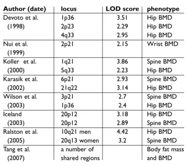
Table I. Summary of main Quantitative Trait Loci findings for BMD in humans
Author (date) locus LOD score phenotype
Devoto et al. 1p36 3.51 Hip BMD
(1998) 2p23 2.29 Hip BMD
4q33 2.95 Hip BMD
Nui et al. 2p21 2.15 Wrist BMD
(1999)
Koller et al. 1q21 3.86 Spine BMD
(2000) 5q33 2.23 Hip BMD
Karasik et al. 6p21 2.93 Spine BMD
(2002) 21q22 3.14 Hip BMD
Wilson et al. 3p21 2.7 Spine BMD
(2003) 1p36 2.4 Hip BMD
Iceland 20p12 3.18 Hip BMD
(2003) 20p12 2.89 Spine BMD
Ralston et al. 10q21 men 4.42 Hip BMD
(2005) 20q13 women 3.2 Spine BMD
Tang et al. a number of Body fat mass
formed principal components analysis: linkage to 1q21.3 and 8q24.3 was found with the first prinici-pal component (LODs 2.5, 2.4) while 1p36 was found with the second (LOD 2.1).37
Given the polygenic nature of BMD regulation and osteoporosis susceptibility, most linkage stu-dies performed to date have probably been under-powered, although the two large studies mentio-ned included over 1000 subjects in each.32;33 Ove-rall, results from different linkage studies show more discrepancy than agreement, probably be-cause of differing study populations and differing criteria for subject enrolment. It is partly for these reasons and easier access to clinical samples that association studies are becoming more widely used.
Candidate Gene Studies
Candidate gene studies have been widely used. Candidate gene association studies are relatively easy to perform and may have sufficient power to detect small allelic effects. They may be disadvan-taged, however, by the effects of confounding fac-tors, genetic heterogeneity and population strati-fication. Furthermore, demonstration of an asso-ciation between a candidate gene and BMD does not necessarily mean that the gene is causally res-ponsible for the effect observed, as there may be linkage disequilibrium with a nearby causal gene. The transmission desequilibrum test (TDT) can help by testing candidate genes for both associa-tion and linkage.
Candidate genes investigated thus far have in-cluded genes influencing cytokines and growth factors which regulate bone turnover; genes that encode components of bone matrix; and genes that encode receptors for calciotropic hormones. Individual candidate genes that have been impli-cated in the regulation of bone mass or osteopo-rotic fractures have been reviewed elsewhere.38 Re-cent advances in knowledge are discussed in more detail below. Candidate genes have been sugges-ted on the basis on what is known about bone me-tabolism. In future, novel genes may be identified by genome-wide association scans (see below).
Vitamin D Receptor (VDR)
Vitamin D interacts with its receptor to play an im-portant role in calcium homeostasis by regulating bone cell growth and differentiation, intestinal cal-cium absorption and parathyroid hormone secre-tion. The VDR was therefore a natural place to
be-gin looking for genetic variation that might ac-count for osteoporosis. The original finding that VDR alleles played a role in BMD is over 10 years old.39Other studies of VDR in relation to bone mass have since been conflicting, and it is likely that the VDR genotype is associated with relatively modest effects on bone mass. Various different polymor-phisms have been described, in different popula-tions,40;41although the mechanisms by which the-se polymorphisms modulate VDR function remain unclear: some 3’ polymorphisms may influence RNA stability, and isoforms of VDR encoded by dif-ferent alleles may possess difdif-ferent functions.40In addition there are data to suggest that an interac-tion between 5’ and 3’ polymorphisms is involved in regulating VDR function, and that the risk alle-le involving such polymorphisms may result in lower levels of VDR mRNA.42However, a recent gi-ant study of 26,000 participgi-ants could not find a re-lationship with either BMD or fracture, which se-riously calls into question the results of the smal-ler studies.43
Type I collagen
The genes encoding type I collagen (COLIA1 and COLIA2) are important, well studied candidates for the pathogenesis of osteoporosis. A common polymorphism affecting the transcription factor Sp1 binding site has been shown to have increased prevalence in osteoporosis patients.48Positive as-sociations between the COLIA1 Sp1 polymorphism and bone mass or osteoporotic fractures were sub-sequently reported in several populations, and meta-analysis also supported the COLIA1 ge-notype conferring differences in BMD.44Ethnic dif-ferences have been reported in population preva-lence of COLIA1 Sp1 alleles with the polymorphism being common in Caucasian populations, but rare in Africans and Chinese.45Overall the data suggest that the COLIA1 Sp1 polymorphism is a functional variant which has adverse effects on bone compo-sition and mechanical strength. Haplotype analy-sis has shown that susceptibility to fracture is dri-ven by the Sp1 polymorphism rather than other known polymorphisms at the COLIA1 locus,46 al-though it remains possible that hitherto unidenti-fied polymorphisms in linkage disequilibrium with the Sp1 polymorphism exist and contribute to the observed effects. From a clinical viewpoint, the COLIA1 polymorphism may be of value not as a therapeutic target but as a marker of osteoporotic fracture risk, since it predicts fractures
indepen-dent of BMD and interacts with BMD to enhance fracture prediction.47
Estrogen Receptors and Aromatase Genes
In view of the strong relationship between estro-gen deficiency and bone loss, the estroestro-gen recep-tor alpha (ER) gene has long been a strong candi-date gene for osteoporosis. An association has been reported between a TA repeat polymorphism in the ER promoter and bone mass in both Japa-nese and U.S. populations. Other investigators have reported positive associations between ha-plotypes defined by PvuII and XbaI polymor-phisms in intron 1 of the ER gene and bone mass48 as well as age at menopause.49The molecular me-chanisms by which these polymorphisms influen-ce bone mass are as yet unclear, but a meta-anal-ysis of the intron 1 polymorphisms indicated that the association with BMD and fracture is attributa-ble mainly to variation at the XbaI site.50More re-cently, a large-scale study comprising 8 European centres has attempted to answer the question more definitively using almost 19,000 subjects. Three common ER gene polymorphisms were studied and none of the polymorphisms was shown to be associated with BMD. The absence of a Xba1 poly-morphism recognition site conferred a risk reduc-tion in all fractures of 19% while the risk reducreduc-tion for vertebral fractures was 35%. The effects on frac-ture were independent of BMD (but may be asso-ciated with BUA).51Polymorphisms in PvuII and TA repeats did not appear to have any influence.52 Aromatase is the enzyme which converts andro-gens into estroandro-gens so is likely to be of importan-ce in bone metabolism in men and post-meno-pausal women. It is encoded by the CYP19 gene. A recent study from Australia has shown the TTTA re-peat polymorphism of CYP19 to be associated with higher circulating estradiol, higher BMD at hip and lumbar spine and lower markers of bone turnover, in over 1200 women age 70 years or older.53 Simi-lar findings have also been reported in elderly Ita-lian men.54
Other Genes
Polymorphisms in several other candidate genes have been associated with bone mass and/or oste-oporotic fracture including TGFb-1 and the IL-6 lo-cus. The effects of these polymorphisms on IL-6 function are yet to be determined. Two studies have looked at the possible associations between apoli-poprotein E (APOE) alleles and osteoporosis but
again the mechanisms by which APOE alleles influ-ence susceptibility to osteoporosis remain unclear. Two groups have reported an association between a coding polymorphism of the calcitonin receptor gene and BMD. The osteocalcin gene has been found to be associated and linked to BMD and bone quality.55Other candidate genes which have been studied in relation to BMD include; parathyroid hormone; the androgen receptor, aromatase, oste-oprotegerin, Klotho and the interleukin-1 receptor antagonist (IL-1ra). Most of the original findings, however, have not been consistently replicated.
In addition to the study of single genes or poly-morphisms in isolation, it has been realized that both gene-gene and gene-environment interac-tions play an important role in influencing the va-riation of expression of complex traits such as os-teoporosis within populations. Such interactions are discussed below.
Gene-Gene and Gene-Environment Interactions
A Dutch study of 1000 postmenopausal women lo-oked at the effects of a combination of both the G to A polymorphism in the COL1A1 Sp1 binding site and the ‘baT’ haplotype of VDR. They found that there was a significant interaction between the ge-notypes, both being independent of the effect of BMD.56The Danish Osteoporosis Prevention Study has recently reported the influence of polymor-phisms within the CYP19 and androgen receptor genes in almost 1800 newly postmenopausal wo-men who were randomized to receive estrogen re-placement therapy or no treatment.57While peri-menopausal bone loss was not associated with ei-ther genes’ polymorphisms, the BMD response over 5 years to estrogen was influenced by ge-notype: one CYP19 allele was associated with sig-nificantly greater response. While the androgen re-ceptor genotype was not related to BMD, a mo-difying effect of sex hormone-binding globulin (SHBG) was observed. Thus in the highest quarti-le of SHBG, androgen receptor genotype was asso-ciated with baseline BMD.
These types of study emphasize the importan-ce of both gene-gene and gene-environment inte-ractions and highlight once again the need for lar-ge, usually multi-center, studies to recruit suffi-cient subjects to enable well-powered studies to be performed. They also create new difficulties of their own, particularly problems associated with multiple testing of subgroups – which increase the likelihood of spurious positive findings unless they
are taken into account in the analysis.
Gene Expression Studies
A novel approach to the question of osteoporosis genes is that involving gene expression studies. In this type of study, differences in gene expression are explored in tissues derived from subjects ex-pressing and not exex-pressing the trait of interest. Very much greater power is obtained if the genetic background of the trait-discordant subjects is simi-lar or the same, as in the case of identical twins. One such small study has used osteoblast-like cul-ture from 2 pairs of monozygotic twins discordant for BMD and one concordant pair. Genome-wide gene expression of the cell culture derived from bone marrow aspirates suggests the following ge-nes were differentially expressed: chondroitin beta 1,4 N-acetylgalactosaminyltransferase, inhibin beta A, interleukin 1 beta and colony stimulating factor 1 macrophage.58These genes are known to play a part in bone physiology. Although the num-bers studied were small this study highlights both the potential of the emerging new technology for examining gene expression and the further bene-fits that may be derived from the twin registers around the world in providing informative willing subjects for intensive study .
Pooling studies
Another newer method being used for increasing the power and cost-effectiveness of studies to de-tect genes associated with osteoporosis is that of pooling. This type of association study contrasts DNA pools from 200-300 subjects with and without the trait of interest, for example BMD. One such study has used 25,000 SNPs in 16,000 genes from women divided into study groups by expression of the traits high and low BMD. Because of the loss of power with multiple testing, the findings were verified by individual genotyping in two further case control groups. The differences in allele fre-quency between the two trait expression groups suggested a candidate locus in the phosphodies-terase 4D (PDE4D) gene on chromosome 5q12. This was fine mapped using 80 SNPs within 50 kB of the marker SNP.59This study also produced evi-dence in support of the association with the Ser37Val polymorphism in BMP2, a gene known to interact with PDE4D (and implicated in Icelandic studies). These data illustrate the potential of the-se methods but also highlight the need for the-several replication groups.
Overlapping phenotypes
In addition to the associated phenotypes and traits which may be used as surrogates of the main cli-nical outcome of interest, other bone diseases may also shed light on genes of importance in osteopo-rosis. Studies have shown that perhaps 30% of ge-nes involved in bone metabolism overlap with tho-se influencing osteoarthritis – a ditho-seatho-se of bone as well as cartilage. Genes believed to be common to both include the VDR, the COLIA1 and possibly the ER genes.60 A recent example of an associati-on study of OA progressiassociati-on by Valdes et al implica-ted several bone genes such as BMP-261and genes involved in inflammation and cytokines have been found, somewhat surprisingly, to be associated with chronic diseases such as disc degeneration.62 With the finding that the LRP-5 gene is associated with osteoporosis comes the realization that genes controlling pathways such as lipid metabolism and inflammation may be important in what were con-sidered non-inflammatory bone conditions. Thus the choice of potential candidate genes is getting considerably larger and genetic researchers have increasingly to cross the traditional disease boun-daries.
In addition to the overlap with osteoarthritis, osteoporosis is also associated with obesity. The tools are now available to us to dissect this rela-tionship using a variety of methods. It would ap-pear, however, that the negative association with body mass index is not independent of the loading expected on bone,63thus advising people to gain weight will not be appropriate. It is likely that sha-red genes operate to account for this association.64
Genes of Rare Monogenic Diseases
Osteoporosis and fragility fractures are features of several rare monogenic diseases, and provide an obvious place to start the search for genes influen-cing osteoporosis in the general population. Such conditions are not always informative, however. They include osteogenesis imperfecta (OI), the osteoporosis-pseudoglioma syndrome (OPS) and syndromes associated with inactivating mutations of the oestrogen receptor alpha and aromatase genes.
OI describes a heterogeneous group of mono-genic disorders characterised by multiple bone fractures which, in most forms, is caused by
mu-tations in the type I collagen genes COLIA1 and COLIA2. The genes which encode type I collagen possess mutations in many different places, ac-counting for the heterogenous nature of the disor-der – from mild to extremely severe. OPS is a rare, autosomal recessive disorder characterised by ju-venile onset osteoporosis and blindness due to persistent vascularisation of the eye. Initial linka-ge studies mapped OPS to chromosome 11q12--13.65Subsequent work showed the disease to be caused by inactivating mutations in the low den-sity lipoprotein-related receptor-5 (LRP-5).66 Ano-ther phenotype, autosomal dominant high-bone-mass, maps to the same region67and indepen-dently was reported to be caused by an activating mutation of the same receptor.68Osteoporosis has been reported in association with homozygous inactivating mutations of the estrogen receptor and aromatase genes, emphasising the importan-ce of estrogen in the attainment and maintenanimportan-ce of peak bone mass. Mutations in the latency-acti-vating peptide (LAP) domain of the TGF beta 1 gene are associated with Camurati-Engelmann disease – a condition characterised by increased BMD in the diaphysis of long bones.69Mutations of the TCIRG1 gene, which encodes a subunit of the os-teoclast proton pump, have been shown responsi-ble for the autosomal recessive condition osteope-trosis.70
The important question is do the genetic clues obtained from rare diseases cast any light on the osteoporosis and fractures seen in the normal po-pulation? There is evidence that some of these ge-nes do contribute to regulation of ‘normal’ BMD. For example, LRP gene polymorphisms have re-cently been shown to be associated with bone mi-neral content, bone area and stature particularly in males.71;72Several groups have reported polymor-phisms in the TGF beta gene to be associated with BMD and osteoporotic fracture14;73and polymor-phisms of the TCIRG1 genes (subunit of osteoclast proton pump) have been found to be associated with BMD in normal subjects.74Finally, the SOST gene causative in the slerosteosis/van Buchem di-sease phenotype has been shown to be associated with BMD in elderly Dutch white volunteers.75
Genome wide association scans
At present, some argue, lines of investigation are driven by technology and the availability of new as-say techniques handling ever larger numbers of polymorphisms. Although the estimated number
of human genes continues to fall (currently around 23,000) the number of recognized SNPs increases – with over 30,000 known non-synonymous SNPs and the possibilty of testing samples with over 250,000 validated SNPs at a cost of less than 1 US cent per SNP. The use of genome wide scans is al-ready beginning to yield exciting new genes in oth-er complex genetic traits, such as diabetes.76The new technology will enable increasingly large pa-nels of polymorphisms, as well as gene expression levels and, eventually, proteins and metabolic pro-files to be studied simultaneously. Funding for fu-ture work should be prioritised for those study pro-posals demonstrating sufficient power to answer the question being addressed, although the increa-sing problem of multiple testing and the difficulti-es in having large numbers of replicate clinical co-horts will make the task no less challenging.
In conclusion, osteoporosis is a perfect example of a complex genetic trait. The associated phe-notypes studied thus far have heritabilities of 50--80% and a large number of genes are likely to be involved in its pathogenesis. Several candidate ge-nes have been identified but their individual ef-fects are small. Many genome-wide linkage scans have been performed but the results are inconsis-tent - underlining some of the difficulties in pin-pointing the genes - and suggest that to maximise the chances of gene discovery a full range of phe-notypes and methods will need to be utilised. Re-gardless of the methods employed, combining da-tasets will be essential to obtain sufficient power. This means national and international collabora-tion will play a vital role in taking forward the work done so far.
Correspondence to:
Tim D Spector
Twin Research & Genetic Epidemiology Unit, St Thomas’ Hospital
London SE1 7EH, UK E-mail: [email protected]
References:
1. Pocock NA, Eisman JA, Hopper JL, Yeates MG, Sam-brook PN, Eberl S. Genetic determinants of bone mass in adults. A twin study. J Clin Invest 1987; 80:706-710.
2. Smith DM, Nance WE, Kang KW, Christian JC, John-ston CC. Genetic factors in determining bone mass. J Clin Invest 1973; 52:2800-2808.
3. Gueguen R, Jouanny P, Guillemin F, Kuntz C, Pourel J, Siest G. Segregation analysis and variance compo-nents analysis of bone mineral density in healthy
milies. J Bone Miner Res 1995; 10:2017-2022. 4. Krall EA, Dawson-Hughes B. Heritable and life-style
determinants of bone mineral density. J Bone Miner Res 1993; 8:1-9.
5. Arden NK, Baker J, Hogg C, Baan K, Spector TD. The heritability of bone mineral density, ultrasound of the calcaneus and hip axis length: a study of postme-nopausal twins. J Bone Miner Res 1996; 11:530-534. 6. Arden NK, Spector TD. Genetic influences on muscle
strength, lean body mass, and bone mineral density: a twin study. J Bone Miner Res 1997; 12:2076-2081. 7. Hunter D, de Lange M, Snieder H et al. Genetic
con-tribution to bone metabolism, calcium excretion, and vitamin D and parathyroid hormone regulation. J Bone Miner Res 2001; 16:371-378.
8. Garnero P, Arden NK, Griffiths G, Delmas PD, Spector TD. Genetic influence on bone turnover in postme-nopausal twins. J Clin Endocrinol Metab 1996; 81:140-146.
9. Kaprio J, Rimpela A, Winter T, Viken RJ, Rimpela M, Rose RJ. Common genetic influences on BMI and age at menarche. Hum Biol 1995; 67:739-753.
10. Snieder H, MacGregor AJ, Spector TD. Genes control the cessation of a woman’s reproductive life: a twin study of hysterectomy and age at menopause. J Clin Endocrinol Metab 1998; 83:1875-1880.
11. Cummings SR, Nevitt MC, Browner WS et al. Risk fac-tors for hip fracture in white women. Study of Osteo-porotic Fractures Research Group. N Engl J Med 1995; 332:767-773.
12. Torgerson DJ, Campbell MK, Thomas RE, Reid DM. Prediction of perimenopausal fractures by bone mi-neral density and other risk factors. J Bone Miner Res 1996; 11:293-297.
13. Deng HW, Chen WM, Recker S et al. Genetic determi-nation of Colles’ fracture and differential bone mass in women with and without Colles’ fracture. J Bone Miner Res 2000; 15:1243-1252.
14. Keen RW, Snieder H, Molloy H et al. Evidence of asso-ciation and linkage disequilibrium between a novel polymorphism in the transforming growth factor be-ta 1 gene and hip bone mineral density: a study of fe-male twins. Rheumatology (Oxford) 2001; 40:48-54. 15. Xiong D, Wang W, Chen Y, Jiang H, Deng HW. Genetic
determination in onset age of wrist fracture. J Hum Genet 2007 52:481-484.
16. Livshits G, Kato BS, Zhai G et al. Genomewide linka-ge scan of hand osteoarthritis in female twin pairs showing replication of quantitative trait loci on chro-mosomes 2 and 19. Ann Rheum Dis 2007; 66:623--627.
17. Andrew T, Antioniades L, Scurrah KJ, MacGregor AJ, Spector TD. Risk of wrist fracture in women is herita-ble and is influenced by genes that are largely inde-pendent of those influencing BMD. J Bone Miner Res 2005; 20:67-74.
18. Knapp KM, Andrew T, MacGregor AJ, Blake GM, Fo-gelman I, Spector TD. An investigation of unique and shared gene effects on speed of sound and bone
den-sity using axial transmission quantitative ultrasound and DXA in twins. J Bone Miner Res 2003; 18:1525--1530.
19. Huang QY, Recker RR, Deng HW. Searching for osteo-porosis genes in the post-genome era: progress and challenges. Osteoporos Int 2003; 14:701-715. 20. Reich DE, Cargill M, Bolk S et al. Linkage
disequilibri-um in the hdisequilibri-uman genome. Nature 2001; 411:199-204. 21. Klein RF, Mitchell SR, Phillips TJ, Belknap JK, Orwoll ES. Quantitative trait loci affecting peak bone mine-ral density in mice. J Bone Miner Res 1998; 13:1648--1656.
22. Klein RF, Allard J, Avnur Z et al. Regulation of bone mass in mice by the lipoxygenase gene Alox15. Scien-ce 2004; 303:229-232.
23. Devoto M, Shimoya K, Caminis J et al. First-stage au-tosomal genome screen in extended pedigrees sug-gests genes predisposing to low bone mineral density on chromosomes 1p, 2p and 4q. Eur J Hum Genet 1998; 6:151-157.
24. Ichikawa S, Koller DL, Johnson ML et al. Human ALOX12, but not ALOX15, is associated with BMD in white men and women. J Bone Miner Res 2006; 21:556-564.
25. Urano T, Shiraki M, Fujita M et al. Association of a single nucleotide polymorphism in the lipoxygenase ALOX15 5’-flanking region (-5229G/A) with bone mi-neral density. J Bone Miner Metab 2005; 23:226-230. 26. Devoto M, Specchia C, Li HH et al. Variance
compo-nent linkage analysis indicates a QTL for femoral neck bone mineral density on chromosome 1p36. Hum Mol Genet 2001; 10:2447-2452.
27. Niu T, Chen C, Cordell H et al. A genome-wide scan for loci linked to forearm bone mineral density. Hum Genet 1999; 104:226-233.
28. Koller DL, Econs MJ, Morin PA et al. Genome screen for QTLs contributing to normal variation in bone mineral density and osteoporosis. J Clin Endocrinol Metab 2000; 85:3116-3120.
29. Koller DL, Liu G, Econs MJ et al. Genome screen for quantitative trait loci underlying normal variation in femoral structure. J Bone Miner Res 2001; 16:985-991. 30. Karasik D, Myers RH, Cupples LA et al. Genome scre-en for quantitative trait loci contributing to normal variation in bone mineral density: the Framingham Study. J Bone Miner Res 2002; 17:1718-1727.
31. Karasik D, Cupples LA, Hannan MT, Kiel DP. Age, gen-der, and body mass effects on quantitative trait loci for bone mineral density: the Framingham Study. Bone 2003; 33:308-316.
32. Wilson SG, Reed PW, Bansal A et al. Comparison of genome screens for two independent cohorts provi-des replication of suggestive linkage of bone mineral density to 3p21 and 1p36. Am J Hum Genet 2003; 72:144-155.
33. Styrkarsdottir U, Cazier JB, Kong A et al. Linkage of osteoporosis to chromosome 20p12 and association to BMP2. PLoS Biol 2003; 1:E69.
gene polymorphisms and osteoporosis: the Rotter-dam Study. J Bone Miner Res 2006; 21:845-854. 35. Wilson SG, Reed PW, Andrew T et al. A
genome-scre-en of a large twin cohort reveals linkage for quantita-tive ultrasound of the calcaneus to 2q33-37 and 4q12-21. J Bone Miner Res 2004; 19:270-277.
36. Karasik D, Myers RH, Hannan MT et al. Mapping of quantitative ultrasound of the calcaneus bone to chromosome 1 by genome-wide linkage analysis. Os-teoporos Int 2002; 13:796-802.
37. Karasik D, Cupples LA, Hannan MT, Kiel DP. Genome screen for a combined bone phenotype using princi-pal component analysis: the Framingham study. Bone 2004; 34:547-556.
38. Liu YZ, Liu YJ, Recker RR, Deng HW. Molecular stu-dies of identification of genes for osteoporosis: the 2002 update. J Endocrinol 2003; 177:147-196. 39. Morrison NA, Qi JC, Tokita A et al. Prediction of bone
density from vitamin D receptor alleles. Nature 1994; 367:284-287.
40. Arai H, Miyamoto K, Taketani Y et al. A vitamin D re-ceptor gene polymorphism in the translation initiati-on codinitiati-on: effect initiati-on protein activity and relatiinitiati-on to bone mineral density in Japanese women. J Bone Mi-ner Res 1997; 12:915-921.
41. Arai H, Miyamoto KI, Yoshida M et al. The polymor-phism in the caudal-related homeodomain protein Cdx-2 binding element in the human vitamin D re-ceptor gene. J Bone Miner Res 2001; 16:1256-1264. 42. Fang Y, van Meurs JB, d’Alesio A et al. Promoter and
3’-untranslated-region haplotypes in the vitamin d receptor gene predispose to osteoporotic fracture: the rotterdam study. Am J Hum Genet 2005; 77:807--823.
43. Uitterlinden AG, Ralston SH, Brandi ML et al. The as-sociation between common vitamin D receptor gene variations and osteoporosis: a participant-level meta-analysis. Ann Intern Med 2006; 145:255-264. 44. Mann V, Hobson EE, Li B et al. A COL1A1 Sp1 binding
site polymorphism predisposes to osteoporotic frac-ture by affecting bone density and quality. J Clin In-vest 2001; 107:899-907.
45. Beavan S, Prentice A, Dibba B, Yan L, Cooper C, Rals-ton SH. Polymorphism of the collagen type Ialpha1 gene and ethnic differences in hip-fracture rates. N Engl J Med 1998; 339:351-352.
46. McGuigan FE, Reid DM, Ralston SH. Susceptibility to osteoporotic fracture is determined by allelic variati-on at the Sp1 site, rather than other polymorphic si-tes at the COL1A1 locus. Osteoporos Int 2000; 11:338--343.
47. McGuigan FE, Armbrecht G, Smith R, Felsenberg D, Reid DM, Ralston SH. Prediction of osteoporotic frac-tures by bone densitometry and COLIA1 genotyping: a prospective, population-based study in men and women. Osteoporos Int 2001; 12:91-96.
48. Kobayashi S, Inoue S, Hosoi T, Ouchi Y, Shiraki M, Orimo H. Association of bone mineral density with polymorphism of the estrogen receptor gene. J Bone
Miner Res 1996; 11:306-311.
49. Weel AE, Uitterlinden AG, Westendorp IC et al. Estro-gen receptor polymorphism predicts the onset of na-tural and surgical menopause. J Clin Endocrinol Me-tab 1999; 84:3146-3150.
50. Ioannidis JP, Stavrou I, Trikalinos TA et al. Association of polymorphisms of the estrogen receptor alpha gene with bone mineral density and fracture risk in women: a meta-analysis. J Bone Miner Res 2002; 17:2048-2060.
51. Albagha OM, Pettersson U, Stewart A et al. Associati-on of oestrogen receptor alpha gene polymorphisms with postmenopausal bone loss, bone mass, and quantitative ultrasound properties of bone. J Med Genet 2005; 42:240-246.
52. Ioannidis JP, Ralston SH, Bennett ST et al. Differential genetic effects of ESR1 gene polymorphisms on oste-oporosis outcomes. JAMA 2004; 292:2105-2114. 53. Dick IM, Devine A, Prince RL. Association of an
aro-matase TTTA repeat polymorphism with circulating estrogen, bone structure, and biochemistry in older women. Am J Physiol Endocrinol Metab 2005; 288:E989-E995.
54. Gennari L, Masi L, Merlotti D et al. A polymorphic CYP19 TTTA repeat influences aromatase activity and estrogen levels in elderly men: effects on bone metabolism. J Clin Endocrinol Metab 2004; 89:2803--2810.
55. Andrew T, Mak YT, Reed P, MacGregor AJ, Spector TD. Linkage and association for bone mineral density and heel ultrasound measurements with a simple tandem repeat polymorphism near the osteocalcin gene in female dizygotic twins. Osteoporos Int 2002; 13:745-754.
56. Uitterlinden AG, Weel AE, Burger H et al. Interaction between the vitamin D receptor gene and collagen type Ialpha1 gene in susceptibility for fracture. J Bone Miner Res 2001; 16:379-385.
57. Tofteng CL, Kindmark A, Brandstrom H et al. Poly-morphisms in the CYP19 and AR genes—relation to bone mass and longitudinal bone changes in post-menopausal women with or without hormone repla-cement therapy: The Danish Osteoporosis Preventi-on Study. Calcif Tissue Int 2004; 74:25-34.
58. Mak YT, Hampson G, Beresford JN, Spector TD. Varia-tions in genome-wide gene expression in identical twins - a study of primary osteoblast-like culture from female twins discordant for osteoporosis. BMC Genet 2004; 5:14.
59. Reneland RH, Mah S, Kammerer S et al. Association between a variation in the phosphodiesterase 4D gene and bone mineral density. BMC Med Genet 2005; 6:9.
60. Spector TD, MacGregor AJ. Risk factors for osteo-arthritis: genetics. Osteoarthritis Cartilage 2004; 12 Suppl A:S39-S44.
61. Valdes AM, Hart DJ, Jones KA et al. Association study of candidate genes for the prevalence and progressi-on of knee osteoarthritis. Arthritis Rheum 2004;
50:2497-2507.
62. Valdes AM, Hassett G, Hart DJ, Spector TD. Radio-graphic progression of lumbar spine disc degenerati-on is influenced by variatidegenerati-on at inflammatory genes: a candidate SNP association study in the Chingford cohort. Spine 2005; 30:2445-2451.
63. Zhao LJ, Liu YJ, Liu PY, Hamilton J, Recker RR, Deng HW. Relationship of obesity with osteoporosis. J Clin Endocrinol Metab 2007; 92:1640-1646.
64. Tang ZH, Xiao P, Lei SF et al. A Bivariate Whole-Geno-me Linkage Scan Suggests Several Shared Genomic Regions for Obesity and Osteoporosis. J Clin Endocri-nol Metab 2007.
65. Gong Y, Vikkula M, Boon L et al. Osteoporosis-pseu-doglioma syndrome, a disorder affecting skeletal strength and vision, is assigned to chromosome regi-on 11q12-13. Am J Hum Genet 1996; 59:146-151. 66. Gong Y, Slee RB, Fukai N et al. LDL receptor-related
protein 5 (LRP5) affects bone accrual and eye deve-lopment. Cell 2001;107:513-523.
67. Johnson ML, Gong G, Kimberling W, Recker SM, Kimmel DB, Recker RB. Linkage of a gene causing high bone mass to human chromosome 11 (11q12-13). Am J Hum Genet 1997; 60:1326-1332.
68. Little RD, Carulli JP, Del Mastro RG et al. A mutation in the LDL receptor-related protein 5 gene results in the autosomal dominant high-bone-mass trait. Am J Hum Genet 2002; 70:11-19.
69. Janssens K, Gershoni-Baruch R, Guanabens N et al. Mutations in the gene encoding the
latency-associa-ted peptide of TGF-beta 1 cause Camurati-Engel-mann disease. Nat Genet 2000; 26:273-275.
70. Sobacchi C, Frattini A, Orchard P et al. The mutatio-nal spectrum of human malignant autosomal reces-sive osteopetrosis. Hum Mol Genet 2001; 10:1767--1773.
71. Ferrari SL, Deutsch S, Choudhury U et al. Polymor-phisms in the low-density lipoprotein receptor-rela-ted protein 5 (LRP5) gene are associareceptor-rela-ted with variati-on in vertebral bvariati-one mass, vertebral bvariati-one size, and stature in whites. Am J Hum Genet 2004; 74:866-875. 72. Xiong DH, Lei SF, Yang F et al. Low-density
lipopro-tein receptor-related prolipopro-tein 5 (LRP5) gene polymor-phisms are associated with bone mass in both Chi-nese and whites. J Bone Miner Res 2007; 22:385-393. 73. Langdahl BL, Carstens M, Stenkjaer L, Eriksen EF.
Polymorphisms in the transforming growth factor beta 1 gene and osteoporosis. Bone 2003; 32:297-310. 74. Sobacchi C, Vezzoni P, Reid DM et al. Association between a polymorphism affecting an AP1 binding site in the promoter of the TCIRG1 gene and bone mass in women. Calcif Tissue Int 2004; 74:35-41. 75. Uitterlinden AG, Arp PP, Paeper BW et al.
Polymor-phisms in the sclerosteosis/van Buchem disease gene (SOST) region are associated with bone-mineral density in elderly whites. Am J Hum Genet 2004; 75:1032-1045.
76. Sladek R, Rocheleau G, Rung J et al. A genome-wide association study identifies novel risk loci for type 2 diabetes. Nature 2007; 445:881-885.
T H E G E N E T I C S O F O S T E O P O R O S I S