DOI: http://dx.doi.org/10.5007/1980-0037.2016v18n1p20
1 University of Northern Parana. Research Center on Health Scien-ces. Londrina, PR, Brazil.
2 State University of Londrina. Laboratory of Research in Respira-tory Physiotherapy. Physiotherapy Department. Londrina, PR. Brazil.
3 State University of Londrina. Centre for Physical Education. Department of Physical Education. Londrina, PR. Brazil.
Received: 28 February 2015
Accepted: 02 November 2015
Bone mineral density in independent elderly women
with different physical and functional proiles and
vitamin D receptor gene polymorphisms
Densidade mineral óssea de idosas independentes com
diferentes peris físico-funcionais e polimorismos do
gene do receptor da vitamina D
Myriam Fernanda Merli1 Regina Célia Poli Fredico1 Emily Delalibera Ruzzon1 Rubens Alexandre da Silva Junior1 Karen de Barros Parron Fernandes1 Denilson de Castro Teixeira1,3 Audrey de Souza Marquez1 Vanessa Suziane Probst2
Abstract – he aim of the study was to compare BMD among physically independent elderly women with diferent physical-functional proiles and vitamin D receptor gene (VDR) polymorphisms, as well as to analyze the efect of the interaction between these last two aspects on BMD. Overall, 165 elderly women had BMD assessed by bone den-sitometry. Handgrip and lower limb strength and functional exercise capacity (6MWT)
were also assessed. VDR polymorphisms (TaqI, BsmI, ApaI and FokI) were analyzed by
polymerase chain reaction. For analyses, elderly women were categorized according to their performance on physical-functional tests into low performance (LP; <25th percentile), normal performance (NP; 25th percentile ≤ NP ≤ 75thpercentile) and high performance (HP;> 75th percentile). Regarding functional exercise capacity, LP group showed lower BMD compared to HP and NP groups (p=0,003). With respect to handgrip strength, there was a trend for LP group to have lower bone mineral density compared to NP group (p=0.08). No diferences were observed in femur and lumbar BMD in comparisons among the diferent VDR genotypes (0.07≤p≤0.94); among diferent groups regarding lower limb strength (p=0.49) and in the interaction analysis among variables (0.17≤p≤0.77). It was concluded that physically independent elderly women with low functional exercise capacity have lower bone mineral density than those classiied as normal and high per-formance. However, apparently, there is no efect of the interaction between VDR gene polymorphisms and physical and functional factors on BMD.
Key words: Elderly; Bone density; Exercise tolerance; Genetics.
INTRODUCTION
Increasing age is associated with profound changes in the health status of individuals, such as sarcopenia and decreased bone mineral density (BMD) 1. With regard to BMD, literature has shown that genetic factors account
for approximately 60 to 80% of interindividual variability. In this sense, genetic research has grown, especially in relation to vitamin D receptor gene (VDR). It is noteworthy that vitamin D along with its receptor is one of the most important components of bone metabolism; hence VDR gene is one of the most important genes studied in this research ield 2.
he pioneering study by Morrison et al.3 demonstrated that recessive genotype of the VDR gene was associated with higher BMD. Houston et al.4 have found association between recessive genotype and lower BMD. In contrast, other studies have shown that genetics was not related to BMD5,6.
It is known that in addition to VDR gene polymorphisms, BMD is inluenced by environmental factors and the interaction of both 7. In this context, physical and functional factors can be highlighted as important contributors of BMD. Some studies 8-10 have investigated the relationship between physical performance and BMD and found modest associations between variables. Zimmernan et al.9 and Foley et al.10 showed no correlation between handgrip strength and BMD. hus, how physical performance in physical and functional tests is related to BMD is still unclear in literature.
Regarding the interaction between VDR gene polymorphism and physical and functional performance, studies have shown that the dominant efect is associ-ated with increased BMD in physically active women7,13. Moreover, Tsuritani et al.11 found that the recessive allele is more responsive to physical exercise. Jarvinen et al.12 found no interaction between genetics and physical activity, demonstrating that the bone tissue beneited from physical activity regardless of VDR gene.
Importantly, to date, the six-minute walk test, which assesses the functional exercise capacity, widely covered by the world literature, has not been used in studies analyzing the physical and functional factors related to BMD. hus, the 6MWT deserves to be better investigated as it is a simple and inexpensive test13. In addition, its functional feature may relect the potential to perform activities of the daily living13, which makes the results of the 6MWT and possible links with BMD highly relevant.
hus, considering that there are still gaps with respect to the efect of the interaction between VDR gene polymorphisms and physical and functional tests on BMD, the aim of this study was to compare the BMD among physically independent elderly women with diferent physical and functional proiles and VDR gene polymorphisms and to analyze the efect of the interaction between these last two aspects on BMD.
METHODS
Study design and subjects
he convenience sample consisted of elderly women of an interdisciplinary project (EELO Project; Study on Aging and Longevity) developed at the University of Northern Parana (UNOPAR) that aimed to evaluate socio-demographic factors and indicators of the health conditions of this popula-tion in Londrina, Paraná, Brazil. Further informapopula-tion is available at http:// www2.unopar.br/sites/eelo page. he EELO project had a total sample of 508 older adults, which is representative of the total population of 43,610 older adults in Londrina. his population of Londrina represents 12% of the total population, which is similar to what has been described in other countries14.
Later, from the database of the EELO project, the sample was selected for the performance of bone density examination. he selection criteria for performing bone densitometry were being physically independent and / or active according to functional status classiication of Spirduso (levels 3 and 4, respectively)15. According to Spirduso, elderly classiied as Level 3 are able to develop light physical work, but are sedentary and have low functional reserve. hose classiied as Level 4 perform regular physical activity and functional capacity are above average; have performed three physical and functional tests (handgrip strength, sitting and standing test and six-minute walk test) and do not make use of drugs that interfere in bone metabolism. Of the 323 individuals who met the inclusion criteria, 42 were not found, 28 refused to participate and nine died. hus, 244 elderly individuals performed bone densitometry examination. For the analysis of this study, however, only women were considered (n = 165). his study was approved by the Ethics Research Committee of UNOPAR (PP0070 / 09) and all participants signed the free informed consent form.
Anthropometric data
Initially, individuals were evaluated for body mass and height with an an-thropometric scale (Filizola®, São Paulo, Brazil), and then the body mass index (BMI) was calculated using the formula (body mass. height -2). he protocol used was that of Guedes & Guedes16.
Ethnic evaluation
Ethnic evaluation was performed by the same team of students and teach-ers, in this case the appraiser, to deine the racial group that the individual belongs according to skin color.
Diagnosis of Osteoporosis
For the analysis of BMD in g.cm2, 46 elderly women were excluded from the sample, since according to the T-score, they showed worse diagno-sis in the femur region (region where the values in g.cm2 are not obtained). hus, 119 participants were considered in these analyses.
Collection of material for DNA analysis
DNA was obtained from peripheral blood leukocytes collected with EDTA using PureLink Genomic DNA extraction kit (Invitrogen, Carlsbad, USA). he extracted DNA was stored in a freezer at -80°C until time of analysis of polymorphisms.
Evaluation of the DNA quality and quantity was performed by analysis of absorbance in a spectrophotometer (NanoDrop 2000 - hermo Scien-tiic) at 260nm and 280nm. Subsequently, DNA dilution was performed in ultrapure Milli-Q® water to inal concentration of 30ng / uL.
Polymerase chain reaction (PCR) and analysis of vitamin D
receptor gene (VDR) polymorphisms
For the analysis of single nucleotide polymorphisms (SNPs) of the VDR gene, ampliication technique was performed of DNA fragments by polymerase chain reaction (PCR) in real time by TaqMan® system (Applied Biosystems, Foster City, USA). Four polymorphisms were observed: TaqI (rs731236), BsmI (rs1544410), Apal (rs7975235) and FokI (rs2228570). he standard reaction used contained 20μL inal volume as follows: 10μl of Taqman® Genotyping Master Mix (1x), 0.5μL probe (1x) (Applied Biosystems, Foster City, USA) 7.5μL ultrapure Milli-Q® water and 2μL DNA (30ng / uL). hermocycler StepOnePlus ™ Real-Time PCR System (Applied Biosystems, Foster City, USA) was used with the following cycling: 60°C for 30 seconds (pre denaturation), 95°C for 10 minutes for initial denaturation, 50 cycles of 95°C for 15 seconds (denaturation) and 60°C for 1 minute and 30 seconds (pairing of primers) and inal extension cycle of 30 seconds at 60°C. he evaluation of results was performed by the StepOne Software v2.3.
Handgrip strength
Handgrip strength (HGS) was evaluated by manual dynamometer (Takei, Kiki, Kogyo, Japan), according to protocol proposed by Vianna et al.17.
Individuals remained standing with arm outstretched and dynamom-eter next to the body17. Two maximum handgrip strength measurements in both upper limbs were alternately performed, with an interval of 30 seconds, and the best result was chosen for analysis 17.
Lower limb strength
Functional exercise capacity
Functional exercise capacity was assessed using the six-minute walk test (6MWT), according to protocol of the American horacic Society (ATS)13. Two tests were performed in a hallway of thirty meters with minimum interval of thirty minutes, and the highest value was used for analysis. he reference values of Britto et al.19 were used.
Categorization of physical and functional tests
Older women were categorized according to their performance in physical and functional tests as low performance (LP) (below the 25th percentile), normal performance (NP) (within 25-75th percentile) and high performance (HP) (above 75th percentile), based on the study by Rikli & Jones 20.
Statistical analysis
Data distribution was analyzed using the Shapiro-Wilk test. Considering the normality of the data, parametric or non-parametric tests were applied. For comparison of bone mineral density (T-score and / or g.cm2) among groups with diferent performance in physical and functional tests, one-way ANOVA and Kruskal-Wallis tests were used with Tukey and Dunn post-tests, respectively. he interaction analysis was performed using the two-way ANOVA test and Bonferroni post-test. he chi-square test was used to compare categorical variables. Correlations were evaluated using the Pearson and Spearman correlation coeicient.
For the study of genetic variables, the chi-square test was used to ana-lyze the Hardy-Weinberg equilibrium. he level of statistical signiicance adopted for all analyses was p <0.05.
he sample power was calculated using the GPower ® 3.1 software. Correlation between BMD and functional exercise capacity (6MWT) was used, alpha 0.05, correlation for null hypothesis of 0, sample size of 119, resulting in sample power of 0.80.
he statistical programs used for analyses were GraphPadPrism 5.0 (GraphPad Software Inc., San Diego, USA), Statistical Package of Social Science (SPSS) 20.0 (SPSS Inc., Chicago, USA), Statistica (StatSoft Inc., Tulsa, USA).
RESULTS
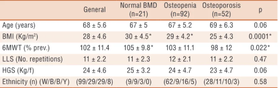
he sample consisted of 165 physically independent elderly women. In relation to bone mineral density, most had osteopenia (n = 92; 56%), 52 participants (31%) had osteoporosis and 21 (13%) had normal BMD. he general sample characteristics are presented in Table 1.
BMD and functional exercise capacity, handgrip strength and
lower limb strength
(Figure 1). Correlation was observed between lumbar spine BMD (g.cm2) and percentage of the predicted value in the 6MWT (r = 0.22; p = 0.02).
Figure 1. Comparison of lumbar spine BMD among elderly women with different performance on the Six-Minute walk test (6MWT). LP: Low Performance; NP: Normal Performance; HP: High Performance. n = 119. * p = 0.003.
Comparing the lumbar spine BMD (g.cm2) among groups with dif-ferent performance in the HGS test, LP group showed tendency to lower BMD when compared to NP group (p = 0.08) (Figure 2). In addition, no correlation was found between lumbar spine BMD (g.cm2) and HGS (r = 0.09; p = 0.32). Regarding LLS, there was no signiicant diference in BMD in the comparison among groups (p = 0.24) (Figure 3) and there was no correlation between BMD and LLS (r = 0.12; p = 0.22). Correlations between BMD of the lumbar spine (g.cm2) and age (r = -0.24; p = 0.01), body mass (r = 0.47; p = 0.0001) and BMI (r = 0.47; p = 0.0001) were found.
Comparison of BMD among VDR gene polymorphisms
In relation to bone mineral density (BMD) in the diferent genotypes (dominant, recessive and heterozygous) of VDR gene polymorphisms (TaqI, BsmI, ApaI and FokI), there was no signiicant diference in the speciic site comparisons to the bone site of the lumbar spine and femoral neck, as shown in Table 2.
Table 1. General sample characteristics
General Normal BMD (n=21)
Osteopenia (n=92)
Osteoporosis (n=52) p Age (years) 68 ± 5.6 67 ± 5 67 ± 5.2 69 ± 6.3 0.06 BMI (Kg/m2) 28 ± 4.6 30 ± 4.5* 29 ± 4.2* 25 ± 4.3 0.0001*
Effect of the interaction among genetic, physical and
functional factors on BMD
here was no efect of the interaction between genetic polymorphisms (TaqI, BsmI, ApaI and FokI) of the VDR gene and functional exercise capacity (6MWT) on BMD (p = 0.58), as well as with other physical and
Figure 2. Comparison of lumbar spine BMD among elderly women with different performance in
handgrip strength (HGS). LP: Low Performance; NP: Normal Performance. n = 119. p = 0.08.
Figure 3. Comparison of lumbar spine BMD among elderly women with different performance
on lower limb strength (LLS). LP: Low Performance; NP: Normal Performance; HP: High Performance. N = 119 p = 0.24.
Table 2. Comparison of bone mineral density (BMD) in different vitamin D receptor gene (VDR) polymorphisms
VDR gene polymorphisms
TaqI BsmI ApaI FokI
TT tt Tt p BB bb Bb p AA aa Aa p FF ff Ff p Sample(n) 63 23 79 - 60 2 103 - 71 18 76 - 24 73 68
-B M D (T-sc o re ) n =1 6 5 S it
e L ± 1.49-1.32 ± 1.19-1.40 -1.74 ± 1.40 0.31 ± 1.41-1.43 ± 0.21-1.35 ± 1.43-1.60 0.94 ± 1.29-1.45 ± 1.20-1.06 ± 1.55-1.72 0.16 ± 1.79-1.98 ± 1.49-1.54 ± 1.15-1.37 0,16
S
it
e F
(n
eck
) -1.70 ± 1.16
-1.47 ± 0.73
-1.89 ± 0.99
0.16 -1.76 ± 1.10 -2.05 ± 0.35 -1.76 ± 1.01 0.57 -1.60 ± 0.91 -1.76 ± 1.14 -1.91 ± 1.11 0.07 -1.90 ± 1.01 -1.77 ± 1.17 -1.69 ± 0.88 0,46
Sample (n) 44 17 58 - 41 2 76 - 51 12 56 - 19 52 48 -BMD Site L
(g.cm2)
functional tests (handgrip strength and sitting and standing test) (p = 0.77; p = 0.17, respectively).
DISCUSSION
his study showed that physically independent elderly women with low functional exercise capacity had lower lumbar spine BMD compared to elderly women with normal performance and high performance. In addi-tion, BMD correlated with functional exercise capacity and anthropometric factors in this population. However, there was no diference in BMD in relation to VDR gene polymorphisms (TaqI, BsmI, ApaI and FokI) and there was no efect of the interaction between VDR gene polymorphisms and physical and functional tests on BMD.
With regard to the association between BMD and exercise capacity, Taafe et al.8 showed that white men with poor aerobic capacity assessed by 400-meter walking test were 2.78 times more likely to have osteoporosis. Although the methodology used by Taafe et al.8 is diferent from that of the present study, results were similar, that is, poor physical performance is associated with lower BMD. It is noteworthy that the present study showed for the irst time association between 6MWT (test widely used in the evaluation of functional exercise capacity in healthy subjects and patients with chronic diseases) and BMD.
sitting and standing test. However, it is noteworthy that studies related to BMD and aging involve mostly large samples; thus, simpler methodologies end up by being used. Since this study involves a subsample of another population study, a test for the evaluation of muscle strength with simple methodology was used so that it could be applied to a large number of individuals without the need for expensive equipment of diicult mobility.
Other studies have shown no association between BMD and peripheral muscle strength 9,10, as in the present study. In the study of Zimmernan et al.9, this may have occurred due to sample size (56 subjects), which may have been insuicient to identify the association, a factor that should also be considered in this research. Foley et al.10 have reported that the lack of association between peripheral muscle strength and BMD is due to that fact that the muscles evaluated were not adjacent to the site where BMD was measured. However, literature shows that the simple handgrip strength measure may represent the peripheral muscle strength in a generalized way24. In addition, the great advantage of assessing handgrip strength is the evaluation method because it is simple, easy, noninvasive, and is important predictor of functionality, nutritional status and mortality in the elderly, which use is highly relevant in clinical practice24. Other stud-ies used peripheral muscle strength tests such as the sitting and standing test also found no correlation with BMD. his is the case of the results of Lindsey et al.22 and those of the present study. he lack of association between these variables may be related to the nature of this functional test18, with dynamic characteristics that involve other skills such as bal-ance, coordination, agility, and may not relect speciically the peripheral muscle strength, as previously mentioned.
Modest and positive correlation between anthropometric variables (body mass and body mass index) and BMD was observed. Kang et al.25 and Salamat et al.26 also found positive correlation between body mass and BMD in their studies. Regarding BMI, Salamat et al.26 found an as-sociation between BMD and body mass index, showing that lower BMI is associated with lower BMD. Higher body mass, whether in relation to lean or fat body mass, leads to greater mechanical stress due to the muscle work to support body or locomotion, which consequently results in stimulation for bone formation 27, explaining the relationships between body mass, BMI and BMD.
It was identiied that age is negatively correlated with BMD. his inding corroborates the indings of Kang et al.25 that advancing age is associated with lower BMD. It is known that age is a risk factor for low BMD, because after the age of thirty years, BMD has a declining rate of one percent per year1. However, the correlation between age and BMD found in this study was weak (r = -0.24; p = 0.01). his inding can be explained by the sample size, the small age variability and the proile of individuals included, i.e., all were physically independent, showing no aspect of fragility that could directly inluence BMD.
However, literature has demonstrated relationships between polymorphic variations of the VDR gene and BMD3,4. Other studies5,6 found no associa-tion between VDR gene polymorphisms (TaqI, ApaI, BsmI) and BMD, which corroborates our indings. It is important to highlight how TaqI, BsmI and ApaI polymorphisms afect the vitamin D receptor action, as they have no efect on the end product of the protein encoded by the VDR gene, but modulate gene expression5. In the present study, it could be inferred that the efects of VDR polymorphisms were discrete in relation to all factors afecting BMD, in addition to the sample size.
Regarding the association of VDR gene polymorphisms and physi-cal and functional tests and bone mineral density, this study found no interaction among such variables. Similarly, Jarvinen et al.12 also found no interaction between VDR gene polymorphism and physical activity in pre menopausal women. he authors of that study showed that bone beneited from physical activity regardless of genotype. Moreover, Blanchet et al.28 and Gentil et al.7 showed that the dominant genotype is associated with increased BMD in physically active women. Tsuritani et al.11 found that the recessive genotype is more responsive to exercise. hese contradictions show that more studies are needed to conirm the interaction between VDR gene polymorphisms, physical and functional tests and BMD. In addition, in relation to polymorphisms, they should be evaluated not only in isolation, but using their combination through the haplotype study.
It should be also mentioned that in addition to physical and functional factors, other environmental components can also inluence BMD. Among them, smoking, alcohol, low calcium intake, vitamin D insuiciency, estrogen deiciency, late menarche and low weight. hese aspects, associ-ated with genetics, make the study of the interaction between physical and functional performance and VDR gene polymorphisms on BMD quite complex, given the numerous existing environmental factors29.
According to the authors, this is the irst study investigating the inter-action between VDR gene polymorphisms, muscle strength and functional exercise capacity measured by objective methods. Until then, interaction studies described in literature were limited to the investigation of physical aspects through questionnaires. Furthermore, the main VDR gene poly-morphism (TaqI, ApaI, BsmI and FokI) were investigated, highlighting the methodological relevance of this study.
transcript of this gene (mRNA) should be performed in order to conirm the bioactivity hypothesis of this protein: how VDR gene polymorphisms could modulate the gene expression and the production of this receptor, which could explain why carriers of certain alleles would show lower BMD. he lack of assessment of physical activity in the daily life can be considered as another limitation, since it could add important information about the physical condition of these individuals.
With regard to the clinical application of the results of this study, the presence of association between functional exercise capacity testing and BMD shows that based on the physical performance assessment of older women, it is possible to identify individuals more likely to have osteopenia and osteoporosis. hus, for subjects with lower BMD and hence increased risk of fracture, a more speciic and early therapeutic approach should be recommended.
CONCLUSIONS
In short, physically independent elderly women with low functional ex-ercise capacity have lower bone mineral density than those with normal and high performance. However, there is no apparent interaction between VDR gene polymorphism and physical and functional performance on bone mineral density.
REFERENCES
1. Riggs BL, Wahner HW, Melton LJ 3rd, Richelson LS, Judd HL, Oford KP.
Rates of bone loss in the appendicular and axial skeletons of women. Evidence of substantial vertebral bone loss before menopause. J Clin Invest 1986;77(5):1487-91.
2. Liu YZ, Liu YJ, Recker RR, Deng HW. Molecular studies of identiication of
genes for osteoporosis: the 2002 update. J Endocrinol 2003;177(2):147-96.
3. Morrison NA, Qi JC, Tokita A, Kelly PJ, Crofts L, Nguyen TV, et al. Prediction
of bone density from vitamin D receptor alleles. Nature 1994;367(6460):284-7.
4. Houston LA, Grant SF, Reid DM, Ralston SH. Vitamin D receptor
polymor-phism, bone mineral density, and osteoporotic vertebral fracture: studies in a UK population. Bone 1996;18(3):249-52.
5. Horst-Sikorska W, Dytfeld J, Wawrzyniak A, Marcinkowska M, Michalak M,
Franek E, et al. Vitamin D receptor gene polymorphisms, bone mineral den-sity and fractures in postmenopausal women with osteoporosis. Mol Biol Rep 2013;40(1):383-90.
6. Zajickova K, Zofkova I, Bahbouh R, Krepelova A. Vitamin D receptor gene
poly-morphisms, bone mineral density and bone turnover: FokI genotype is related to postmenopausal bone mass. Physiol Res 2002;51(5):501-9.
7. Gentil P, de Lima Lins TC, Lima RM, de Abreu BS, Grattapaglia D, Bottaro M,
et al. Vitamin-d-receptor genotypes and bone-mineral density in postmenopausal women: interaction with physical activity. J Aging Phys Act 2009;17(1):31-45.
8. Taafe DR, Simonsick EM, Visser M, Volpato S, Nevitt MC, Cauley JA, et al.
Lower extremity physical performance and hip bone mineral density in elderly black and white men and women: cross-sectional associations in the Health ABC Study. J Gerontol A Biol Sci Med Sci 2003;58(10):M934-42.
9. Zimmermann CL, Smidt GL, Brooks JS, Kinsey WJ, Eekhof TL. Relationship
CORRESPONDING AUTHOR
Vanessa Suziane Probst Laboratory of Research in Respiratory Physiotherapy (LFIP) Physiotherapy Department State University of Londrina (UEL) Av. Robert Koch, 60 - Vila Operária CEP 86038-350 - Londrina, PR, Brasil
Email: [email protected]
10. Foley KT, Owings TM, Pavol MJ, Grabiner MD. Maximum grip strength is not
related to bone mineral density of the proximal femur in older adults. Calcif Tissue Int 1999;64(4):291-4.
11. Tsuritani I, Brooke-Wavell KS, Mastana SS, Jones PR, Hardman AE, Yamada
Y. Does vitamin D receptor polymorphism inluence the response of bone to brisk walking in postmenopausal women? Horm Res 1998;50(6):315-9.
12. Jarvinen TL, Jarvinen TA, Sievanen H, Heinonen A, Tanner M, Huang XH,
et al. Vitamin D receptor alleles and bone’s response to physical activity. Calcif Tissue Int 1998;62(5):413-7.
13. ATS Committee on Proiciency Standards for Clinical Pulmonary Function
Laboratories. ATS statement: guidelines for the six-minute walk test. Am J Respir Crit Care Med 2002;166(1):111-7.
14. Lutz W, K CS. Dimensions of global population projections: what do we know
about future population trends and structures? Philos Trans R Soc Lond B Biol Sci 2010;365(1554):2779-91.
15. Spirduso WW,dimensões físicas do envelhecimento. Barueri: Manole; 2005.
16. Guedes DP, Guedes JERP, manual prático para avaliação em educação física.
Barueri - SP: Manole; 2006.
17. Vianna LC, Oliveira RB, Araujo CG. Age-related decline in handgrip strength
difers according to gender. J Strength Cond Res 2007;21(4):1310-4.
18. Jones CJ, Rikli RE, Beam WC. A 30-s chair-stand test as a measure of lower body
strength in community-residing older adults. Res Q Exerc Sport 1999;70(2):113-9.
19. Britto RR, Probst VS, de Andrade AF, Samora GA, Hernandes NA, Marinho PE,
et al. Reference equations for the six-minute walk distance based on a Brazilian multicenter study. Braz J Phys her 2013;17(6):556-63.
20. Rikli RE, Jones CJ. Functional Fitness Normative Score for Community-Residing
Older Adults, Ages 60-94. J Aging Phys Act 1999(7):162-81.
21. Kim SW, Lee HA, Cho EH. Low handgrip strength is associated with low bone
mineral density and fragility fractures in postmenopausal healthy Korean women. J Korean Med Sci 2012;27(7):744-7.
22. Lindsey C, Brownbill RA, Bohannon RA, Ilich JZ. Association of physical
per-formance measures with bone mineral density in postmenopausal women. Arch Phys Med Rehabil 2005;86(6):1102-7.
23. Frost HM. Bone “mass” and the “mechanostat”: a proposal. Anat Rec 1987;219(1):1-9.
24. Bohannon RW. Dynamometer measurements of hand-grip strength predict
mul-tiple outcomes. Percept Mot Skills 2001;93(2):323-8.
25. Kang D, Liu Z, Wang Y, Zhang H, Feng X, Cao W, et al. Relationship of body
composition with bone mineral density in northern Chinese men by body mass index levels. J Endocrinol Invest 2014;37(4):359-67.
26. Salamat MR, Salamat AH, Abedi I, Janghorbani M. Relationship between Weight,
Body Mass Index, and Bone Mineral Density in Men Referred for Dual-Energy X-Ray Absorptiometry Scan in Isfahan, Iran. J Osteoporos 2013;2013:205963.
27. Reid IR, Ames R, Evans MC, Sharpe S, Gamble G, France JT, et al. Determinants
of total body and regional bone mineral density in normal postmenopausal women--a key role for fat mass. J Clin Endocrinol Metab 1992;75(1):45-51.
28. Blanchet C, Giguere Y, Prud’homme D, Dumont M, Rousseau F, Dodin S.
As-sociation of physical activity and bone: inluence of vitamin D receptor genotype. Med Sci Sports Exerc 2002;34(1):24-31.
29. Kung AWC, Huang Q-Y. Genetic and environmental determinants of osteoporosis.