Association of Severity of Coronary Lesions with Bone Mineral
Density in Postmenopausal Women
Rui Xu,
*1Xin-Chun Cheng,
*1,2Yuan Zhang,
1Hong-Mei Lai,
3Hong-Ni Yang
1Gerontology Center - People’s Hospital of Xinjiang Uygur Autonomous Region, Urumqi, Xinjiang – China1
Department of Pacing and Electrophysiological - The First Affiliated Hospital of Xinjiang Medical University, Urumqi, Xinjiang – China2 Department of Cardiology - People’s Hospital of Xinjiang Uygur Autonomous Region, Urumqi, Xinjiang - China3
*Equally contributed to the work
Mailing Address: Hong-Ni Yang •
N°. 91 Tianchi Road, Tianshan Disztrict, Urumqi, Xinjiang, China. 830001, Tianshan Disztrict, Urumqi, Xinjiang – China
E-mail: [email protected]
Manuscript received April 02, 2017, revised manuscript June 13, 2017, accepted August 23, 2017
Abstract
Background: Coronary artery disease (CAD) and osteoporosis (OP) are common diseases in postmenopausal women. In both cross-sectional and longitudinal epidemiologic studies, low bone mass has been related to increased frequency of CAD. However, available data on the relationship between bone mineral density (BMD) and severity of coronary lesions is limited.
Objective: To investigate association between the BMD and severity of coronary lesions assessed by Gensini score in postmenopausal women.
Methods: This study included 122 postmenopausal women who were diagnosed with CAD. These patients were divided into two groups according to the severity of coronary lesions assessed by the Gensini score – patients with mild coronary lesions (Gensini score < 25) and patients with severe coronary lesions (Gensini score ≥ 25). Femoral neck mineral density was measured with dual energy X-ray absorptiometry (DXA).
Results: The study included postmenopausal women aged 64.31 ± 4.71 years, 85 of whom (69.7%) exhibited severe coronary lesions. Participants with severe coronary lesions had a significantly higher T score than did those with mild coronary lesions at the femoral neck (p < 0.05). The mean T-score was −0.84 ± 1.01 in mild coronary lesions group, −1.42 ± 1.39 in severe coronary lesions group (p < 0.05). Multivariable logistic regression analysis showed that osteopenia-osteoporosis at the Femoral neck (odds ratio 2.73; 95% confidence interval 1.06 to 6.13) was associated with an increased risk of developing severe coronary lesions. The multiple regression model showed that T-scores (β = −0.407, SE = 0.151, p=0.007) were the independent predictors of Gensini score.
Conclusion:The relationship between severity of coronary lesions and BMD was significant in postmenopausal women. BMD, a low-cost technique involving minimal radiation exposure, widely used for osteoporosis screening, is a promising marker of severity of coronary lesions. (Arq Bras Cardiol. 2018; 110(3):211-216)
Keywords: Coronary Artery Disease; Osteoporosis, Postmenopausal; Bone Density; Stroke; Morbidity; Bone Diseases, Metabolic.
Introduction
Atherosclerosis (AS) is one of the most common diseases in elderly people, especially in postmenopausal women. The complications of AS, like coronary artery disease (CAD) and cerebrovascular diseases reduce quality of life and lead to excess morbidity.1 Epidemiology studies found that the CAD morbidity
and mortality rates were significantly higher in postmenopausal women compared with premenopausal women.2 Unlike younger
women, the risk of CAD in older women is higher when there is a decrease in estrogen production, marking the end of the protective effect of endogenous estrogens against CAD.3-5
Therefore, identifying the risk factors associated with CAD in postmenopausal women is critical for improving patients' survival rate and life quality.
Recently, increasing evidence has accumulated to support the correlation between low bone mineral density (BMD) and AS.6-8 AS and osteopenia-osteoporosis syndrome share
some risk factors, among which are parathyroid hormone, lack of estrogen, homocysteine, inflammatory process, vitamins D and K, lipid oxidation products, molecular pathways involved in bone and vascular mineralization, and calcification mechanisms that seem to be similar in vascular structure and bone.9,10
We have previously reported that coronary artery calcium scores, an earlier sign of coronary artery AS, were significantly higher in the osteopenia/osteoporosis groups compared to normal BMD groups, and that these values were negatively associated with T-scores. These findings indicate that decreased BMD may increase the risk of CAD.11 However, little is known
about the association between decreased BMD and severity of coronary lesions in postmenopausal woman.
Methods
Study population
A total of 122 female patients who were admitted to the cardiology clinic with chest pain between January 2014 and August 2016 were included in the study. Inclusion criteria were postmenopausal women aged ≥ 50 years, who were diagnosed with acute coronary syndrome or chronic CAD. This diagnosis was made by history of angina pectoris or myocardial infarction, electrocardiographic findings, cardiac enzymes, and coronary angiography results. These patients underwent a bone densitometry on a routine basis within the previous 12 months, and were not taking any medication with known effect on bone turnover. Exclusion criteria were patients with normal coronary angiography; patients who had moderate-to-severe heart valve disease and decompensated heart failure; patients with severe kidney or liver failure, malignancy, hematological diseases, or autoimmune disorder.
Clinical features and laboratory examination
The weight and height were measured at each of eligible patient. Body mass index (BMI) was calculated as body weight/height2 (kg/m2). Information concerning the history
of diseases (diabetes, hypertension, and hyperlipidemia) was collected using a standard questionnaire.
Hypertension was defined as history of hypertension and/or an average systolic blood pressure (SBP) ≥ 140 mmHg and/or an average diastolic blood pressure (DBP) ≥ 90 mmHg on two separate occasions. Diabetes was defined as history or presence of diabetes and/or a fasting plasma glucose level > 126 mg/dL on 2 separate occasions, or a random glucose value of > 200 mg/dL on one or more occasion. Hypercholesterolemia was defined as a total serum cholesterol level of > 240 mg/dL; high triglyceride (TG) and high LDL-cholesterol (LDL-c) were defined as total serum TG > 200 mg/dL and LDL-C > 160 mg/dL, respectively.
BMD measurement
Participants had undergone a BMD test of the left femoral neck bone by dual-energy X-ray absorptiometry using a QDR 4500A fan beam bone densitometer (Bedford, MA, USA) according to the manufacturer’s instructions within the previous 12 months. BMD results were reported as T-scores, which were also categorized into three groups according to the World Health Organization (WHO) criteria for diagnosing osteoporosis: normal BMD (T-score≥-1 SD); osteopenia (T < -1 SD and > -2.5 SD); and osteoporosis (T-score ≤ -2.5 SD).11
Gensini risk scoring
Coronary angiography was performed in all subjects. Gensini score: angiographic stenosis of a culprit artery in the range of 0% to 25% was scored as 1 point, stenosis in the range of 25% to 50% was scored as 2 points, 50% to 75% was scored as 4 points, 75% to 90% was scored as 8 points, 90% to 99% was scored as 16 points, and total occlusion
was scored as 32 points. A multiplier was assigned to each vascular segment based on the functional significance of the myocardial area supplied by that segment: 5 for the left main coronary artery, 2.5 for the proximal segment of the left anterior descending (LAD) coronary artery and the proximal segment of the circumflex artery, 1.5 for the mid-segment of the LAD, 1.0 for the right coronary artery, the distal segment of the LAD, the mid-distal region of the circumflex artery, the posterolateral artery, and the obtuse marginal artery, and 0.5 for other segments.12 Angiographic evaluations were reviewed
by the consensus of two observers with more than two years of experience. Based on the Gensini score, patients were divided into two groups – 37 patients in the group of mild coronary lesions (Gensini score < 25 points) and 85 patients in the group of severe coronary lesions (Gensini score ≥ 25 points); this grouping was compatible with the literature.13
Statistical analyses
Analyses were carried out using SPSS version 17.0 (SPSS Inc., Chicago, IL). Continuous variables with a Gaussian distribution are presented as mean ± standard deviation (SD), and those with a non-Gaussian distribution are presented as median values with corresponding 25th and 75th percentiles. The normal distribution of different parameters was verified with the Kolmogorov-Smirnov test. Differences between the groups were evaluated using unpaired t-test or the Mann-Whitney U-test. Categorical variables were compared with the chi-square test or Fisher’s exact test (Fisher’s exact test was used for frequencies of osteoporosis in Table 2). The association between BMD and risk for severe coronary lesions was evaluated by multiple logistic regression analysis. Multiple linear regression analysis was performed to assess whether BMD was the independent explanatory factor for the severity of coronary lesions (assessed by the Gensini score) in postmenopausal women. Statistical significance was set at p < 0.05 (2-tailed).
Results
A total of 122 postmenopausal women (mean age 64.31 ± 4.71) were included in the present study, 69.7% of whom exhibited severe coronary lesions. Clinical characteristics of all participants at baseline are summarized in Table 1. In all, 19.6% of the patients were found to have osteoporosis in the femoral neck and 41.8% osteopenia; 39.3% of the women suffered from high blood pressure, 38.5% have diabetes, and 31.1% have hyperlipidemia.
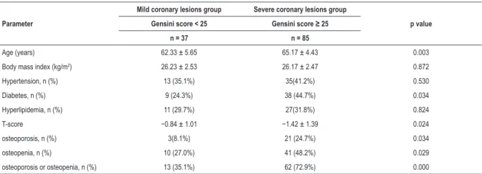
Table 2 shows the comparison between the groups with mild coronary lesions and severe coronary lesions in terms of some clinical parameters. Patients with severe coronary lesions patients were older, and had higher prevalence of diabetes and osteoporosis/osteopenia compared with those with mild coronary lesions (p < 0.05). There were no differences between the groups with respect to BMI, proportions of patients with hypertension and hyperlipidemia.
Table 1 – Characteristics of the study population (n = 122)
Age (years) 64.31 ± 4.71 Body mass index (kg/m2) 26.19 ± 2.49
Hypertension, n (%) 48 (39.3%) Diabetes, n (%) 47 (38.5%) Hyperlipidemia, n (%) 38 (31.1%) T-score –1.24 ± 1.27 Gensini score 43.46 (17.5, 73) osteoporosis, n (%) 24 (19.6%) osteopenia, n (%) 51 (41.8%) osteoporosis or osteopenia, n (%) 75 (61.5%)
Continuous variables with a Gaussian distribution are presented as mean ± SD, and those with a non-Gaussian distribution are presented as median values with corresponding 25th and 75th percentiles. Categorical data are expressed as absolute numbers with (percentages).
Table 2 – Comparison of clinical parameters between the groups with mild coronary lesions and severe coronary lesions
Parameter
Mild coronary lesions group Severe coronary lesions group
p value Gensini score < 25 Gensini score ≥ 25
n = 37 n = 85
Age (years) 62.33 ± 5.65 65.17 ± 4.43 0.003 Body mass index (kg/m2) 26.23 ± 2.53 26.17 ± 2.47 0.872
Hypertension, n (%) 13 (35.1%) 35(41.2%) 0.530 Diabetes, n (%) 9 (24.3%) 38 (44.7%) 0.034 Hyperlipidemia, n (%) 11 (29.7%) 27(31.8%) 0.824
T-score −0.84 ± 1.01 −1.42 ± 1.39 0.024
osteoporosis, n (%) 3(8.1%) 21 (24.7%) 0.034 osteopenia, n (%) 10 (27.0%) 41 (48.2%) 0.029 osteoporosis or osteopenia, n (%) 13 (35.1%) 62 (72.9%) 0.000
Continuous variables with non-Gaussian distribution (except for those expressed as median) were compared using t-tests. For values expressed as median (25th and 75th percentiles), P values were determined by Mann-Whitney U test. Categorical variables were compared by chi-square test, except for osteoporosis, which were
compared by Fisher’s exact test (expected frequencies of ≤ 5).
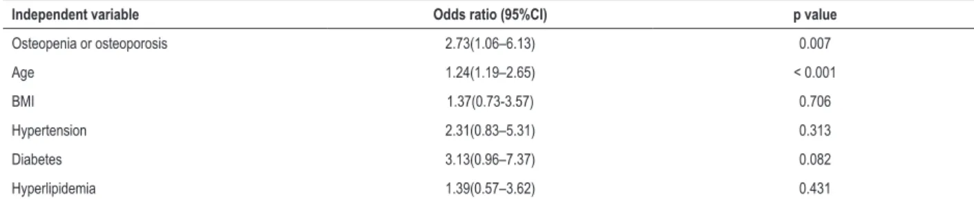
Corresponding to these findings, multivariate logistic regression analysis was used to detect the association between osteoporosis/osteopenia and risk of severe coronary lesions. After adjusting for confounding factors such as age, hypertension, diabetes, and hyperlipidemia, the osteoporosis /osteopenia remained the risk factors for severe coronary lesions (OR = 2.73, 95% CI, 1.06–6.13, p = 0.007, Table 3).
When Gensini score was considered as the dependent variable in a linear regression model, T-score (β = −0.407,
SE = 0.151, p = 0.007) and age (β = 0.295, SE = 0.132,
P = 0.023), but not diabetes, hypertension, BMI, and
hyperlipidemia, were the independent predictors of Gensini score.
In a linear regression analysis with Gensini score as a dependent variable and age, T-score, diabetes, hypertension, BMI, and hyperlipidemia as independent variables (Table 4), only T-score
(β = −0.407, SE = 0.151, p = 0.007) and age (β = 0.295,
SE = 0.132, p = 0.023) correlated with Gensini score.
Discussion
In our study, postmenopausal women with severe coronary lesions are more likely to have osteopenia/osteoporosis compared with mild coronary lesions group, independent of other risk factors. This suggests that postmenopausal women with osteopenia/osteoporosis may have a higher risk of developing severe coronary lesions. Our findings are in accordance with previous studies demonstrating the relationship between BMD and CAD that concluded that BMD is a promising marker of severity of CAD.
Both osteopenia and AS are serious public health problems that can threaten people's health and quality of life.14,15
Previous studies have proved a clear link between AS and BMD. In a retrospective study including 1,335 elderly patients, the incidence of CAD increased in low BMD patients, compared with patients with normal BMD. Multiple logistic regression analysis confirmed that low BMD is associated with CAD, after adjustment for diabetes mellitus, hypertension, smoking, and age.16 Another study with 252 postmenopausal women
showed that osteopenia/osteoporosis at the lumbar spine or femoral neck was associated with coronary AS assessed by 64-row multidetector computed tomography.17 Our previous
study showed that another measure of AS, coronary artery calcification, was associated with BMD of the lumbar spine in healthy postmenopausal women. The odds for coronary artery calcification in osteoporotic women were over three-fold higher compared with those in women with a normal BMD.11
Table 3 – Adjusted odds ratio of risk factors for severe coronary lesions
Independent variable Odds ratio (95%CI) p value
Osteopenia or osteoporosis 2.73(1.06–6.13) 0.007
Age 1.24(1.19–2.65) < 0.001
BMI 1.37(0.73-3.57) 0.706
Hypertension 2.31(0.83–5.31) 0.313
Diabetes 3.13(0.96–7.37) 0.082
Hyperlipidemia 1.39(0.57–3.62) 0.431
BMI: body mass index; 95% CI: 95% confidence interval
Table 4 – Multiple regression analysis of Gensini score (dependent variable) versus age, diabetes, hypertension, body mass index, hyperlipidemia, and T-score (independent variables).
Independent variable β SE p value
T-score 0.407 0.151 0.007
Age 0.295 0.132 0.023
Body mass index 0.183 0.203 0.136
Hypertension 0.147 0.134 0.254
Diabetes 0.113 0.179 0.572
Hyperlipidemia 0.053 0.121 0.697
R2 0.31
β: Values are standardized coefficient; SE: values are standard error of β. R2: values are the total explained variance of the model.
about the relationship between BMD and severity of coronary lesions. A retrospective study carried out with 55 male patients with CAD, confirmed by coronary angiography, showed that decreased BMD was associated with severe coronary lesions assessed by Gensini score, independent of other cardiovascular risk factors.13 Similarly, a study involving 74 male CAD patients
revealed that the incidence of osteopenia/osteoporosis in severe coronary artery lesions group determined by SYNTAX score was significantly higher than mild coronary artery lesions group.20
However, most of these studies have been based on male CAD patients while few studies have involved postmenopausal, CAD women patients. In our study, 186 postmenopausal women with CAD patients identified by coronary angiography were divided into two groups by Gensini scoring: mild coronary lesions patients (Gensini score < 25) and severe coronary lesions patients (Gensini score > 25). We found that there was an increase in the osteoporosis/osteopenia rate in the severe coronary lesions group. Multivariable logistic regression analysis showed that osteopenia/osteoporosis at the femoral neck was associated with an increased risk of developing severe coronary lesions. The multiple regression model showed that T-scores were the independent predictors of Gensini score. Most previous research, if not all, including our results indicate that low BMD not only were associated with increased risk of CAD, but also were an independent predictor of severity of coronary lesions in postmenopausal women.
Although many hypotheses have been proposed to explain the correlation between osteoporosis and CAD, it has not been
thoroughly understood.16,21,22 In spite of common risk factors
of bone metabolism and cardiovascular risk (inflammation, dyslipidemia, menopause, hypertension, smoking, and diabetes mellitus), the possible influence of genetics and vascular calcification also exists.23,24 Hydroxyapatite, an important part
of the mineral phase of bone, is also found in the artery calcified plaque. Moreover, it has been reported that bone matrix proteins such as gla protein, bone morphogenetic protein-2, osteocalcin, and collagen were found in calcified plaques. Studies have suggested that some important gene mutations can lead to the early development of AS and osteoporosis, which indicate the evidence of common genetic basis.25,26 It’s worth
noting that current evidence linking osteoporosis and CAD is far from conclusive. So, further study is needed to explore the relationship between the two common diseases.
Limitations
1. Uppoor RB, Rajesh A, Srinivasan MP, Unnikrishnan B, Holla R. Oxidative stress in obese postmenopausal women: an additive burden for atherosclerosis. J Clin Diagn Res. 2015;9(12):OC03-5. doi: 10.7860/ JCDR/2015/16467.6868.
2. Dou KF, Xu B, Yang YJ, Lu R, Qiu H, Yang WX, et al. Clinical and angiographic characteristics of premenopausal women with coronary artery disease. Chin Med J (Engl). 2008;121(23):2392-6. PMID: 19102955.
3. Calvo RY, Araneta MR, Kritz-Silverstein D, Laughlin GA, Barrett-Connor E. Relation of serum uric acid to severity and progression of coronary artery calcium in postmenopausal White and Filipino women (from the Rancho Bernardo study). Am J Cardiol. 2014;113(7):1153-8. doi: 10.1016/j. amjcard.2013.12.022.
4. Kocic R, Spirovski D, Ciric V, Velija-Asimi Z. Impact of diet, physical activity, lipid status and glycoregulation in estimation of score (systematic coronary risk evaluation) for ten years in postmenopausal women. Bosn J Basic Med Sci. 2007;7(3):197-204. PMID: 17848142.
5. Barton M, Meyer MR, Haas E. Hormone replacement therapy and atherosclerosis in postmenopausal women: does aging limit therapeutic benefits? Arterioscler Thromb Vasc Biol. 2007;27(8):1669-72. doi: 10.1161/ ATVBAHA.106.130260.
6. Liang DK, Bai XJ, Wu B, Han LL, Wang XN, Yang J, et al. Associations between bone mineral density and subclinical atherosclerosis: a cross-sectional study of a Chinese population. J Clin Endocrinol Metab. 2014;99(2):469-77. doi: 10.1210/jc.2013-2572.
7. Hajsadeghi S, Khamseh ME, Larijani B, Abedin B, Vakili-Zarch A, Meysamie AP, et al. Bone mineral density and coronary atherosclerosis. Saudi Heart Assoc. 2011;23(3):143-6. doi: 10.1016/j.jsha.2011.03.001.
8. Hmamouchi I, Allali F, Khazzani H, Bennani L, El Mansouri L, Ichchou L, et al. Low bone mineral density is related to atherosclerosis in postmenopausal Moroccan women. BMC Public Health. 2009 Oct 14;9:388. doi: 10.1186/1471-2458-9-388.
9. Barbarash OL, Lebedeva NB, Kokov AN, Novitskaya AA, Hryachkova ON, Voronkina AV, et al. Decreased cathepsin K plasma level may reflect an association of osteopoenia/osteoporosis with coronary atherosclerosis and coronary artery calcification in male patients with stable angina. Heart Lung Circ. 2016;25(7):691-7. doi: 10.1016/j.hlc.2016.02.002.
10. van Ballegooijen AJ, Robinson-Cohen C, Katz R, Criqui M, Budoff M, Li D, et al. Vitamin D metabolites and bone mineral density: the multi-ethnic study of atherosclerosis. Bone. 2015 Sep;78:186-93. doi: 10.1016/j. bone.2015.05.008.
11. Xu R, Yang HN, Li YQ, Wang QF, Guo AH, Ayiti A, et al. Association of coronary artery calcium with bone mineral density in postmenopausal women. Coron Artery Dis. 2016;27(7):586-91. doi: 10.1097/ MCA.0000000000000402.
12. Luo JY, Xu R, Li XM, Zhou Y, Zhao Q, Liu F, et al. MIF Gene polymorphism rs755622 is associated with coronary artery disease and severity of coronary lesions in a Chinese Kazakh Population: a case-control study. Medicine (Baltimore). 2016;95(4):e2617. doi: 10.1097/ MD.0000000000002617.
13. Alan B, Akpolat V, Aktan A, Alan S. Relationship between osteopenic syndrome and severity of coronary artery disease detected with coronary angiography and Gensini score in men. Clin Interv Aging. 2016 Mar 24;11:377-82. doi: 10.2147/CIA.S104036.
14. Rodrigues IG, Barros MB. Osteoporosis self-reported in the elderly: a population-based survey in the city of Campinas, Sao Paulo, Brazil. Rev Bras Epidemiol. 2016;19(2):294-306. doi: 10.1590/1980-5497201600020007.
15. Whayne TF Jr. Atherosclerosis: current status of prevention and treatment. Int J Angiol. 2011;20(4):213-22. doi: 10.1055/s-0031-1295520.
16. Yesil Y, Ulger Z, Halil M, Halacli B, Yavuz BB, Yesil NK, et al. Coexistence of osteoporosis (OP) and coronary artery disease (CAD) in the elderly: it is not just a by chance event. Arch Gerontol Geriatr. 2012;54(3):473-6. doi: 10.1016/j.archger.2011.06.007.
17. Seo SK, Yun BH, Noe EB, Suh JW, Choi YS, Lee BS. Decreased bone mineral density is associated with coronary atherosclerosis in healthy postmenopausal women. Obstet Gynecol Sci. 2015;58(2):144-9. doi: 10.5468/ogs.2015.58.2.144.
18. Sinning C, Lillpopp L, Appelbaum S, Ojeda F, Zeller T, Schnabel R, et al. Angiographic score assessment improves cardiovascular risk prediction: the clinical value of SYNTAX and Gensini application. Clin Res Cardiol. 2013;102(7):495-503. doi: 10.1007/s00392-013-0555-4.
References
Conclusion
In this study, we investigated the association between BMD and severity of coronary lesions in postmenopausal women. Our results suggested postmenopausal women with low BMD are at high risk for severe coronary lesions. Future research should investigate common pathophysiological pathways between osteoporosis and severity of coronary lesions.
Author contributions
Conception and design of the research: Xu R, Xin-Chun C, Hong-Ni Y; Acquisition of data and Analysis and interpretation of the data: Xu R, Zhang Y, Hong-Mei L; Statistical analysis: Xu R; Writing of the manuscript: Xin-Chun C; Critical revision of the manuscript for intellectual content: Xu R, Hong-Ni Y.
Potential Conflict of Interest
No potential conflict of interest relevant to this article was reported.
Sources of Funding
There were no external funding sources for this study.
Study Association
This study is not associated with any thesis or dissertation work.
Ethics approval and consent to participate
19. Pan HC, Sheu WH, Lee WJ, Lee WL, Liao YC, Wang KY, et al. Coronary severity score and C-reactive protein predict major adverse cardiovascular events in patients with stable coronary artery disease (from the Taichung CAD study). Clin Chim Acta. 2015;445:93-100. doi: 10.1016/j.cca.2015.03.029.
20. Kokov AN, Maliuta EB, Masenko VL, Sigareva AA, Fanaskov VB, Tarasov RS, et al. [Evaluation of coronary artery lesion in men with osteopenic syndrome and coronary artery disease]. Ter Arkh. 2014;86(3):65-70. PMID: 24779073.
21. Lee SN, Cho JY, Eun YM, Song SW, Moon KW. Associations between osteoporosis and coronary artery disease in postmenopausal women. Climacteric. 2016;19(5):458-62. doi: 10.1080/13697137.2016.1200550.
22. Celczynska Bajew L, Horst Sikorska W, Bychowiec B, Wykretowicz A, Wesoly J, Michalak M. The effects of osteoprotegerin (OPG) gene polymorphism in patients with ischaemic heart disease on the morphology of coronary arteries and bone mineral density. Kardiol Pol. 2011;69(6):573-8. PMID: 21678294.
23. Cecelja M, Jiang B, Bevan L, Frost ML, Spector TD, Chowienczyk PJ. Arterial stiffening relates to arterial calcification but not to noncalcified atheroma in women: a twin study. J Am Coll Cardiol. 2011;57(13):1480-6. doi: 10.1016/j.jacc.2010.09.079.
24. Yahagi K, Kolodgie FD, Lutter C, Mori H, Romero ME, Finn AV, et al. Pathology of Human Coronary and Carotid Artery Atherosclerosis and Vascular Calcification in Diabetes Mellitus. Arterioscler Thromb Vasc Biol. 2017;37(2):191-204. doi: 10.1161/ATVBAHA.116.306256.
25. Bostrom K, Watson KE, Horn S, Wortham C, Herman IM, Demer LL. Bone morphogenetic protein expression in human atherosclerotic lesions. J Clin Invest. 1993;91(4):1800-9. doi: 10.1172/JCI116391.
26. Dhore CR, Cleutjens JP, Lutgens E, Cleutjens KB, Geusens PP, Kitslaar PJ, et al. Differential expression of bone matrix regulatory proteins in human atherosclerotic plaques. Arterioscler Thromb Vasc Biol. 2001;21(12):1998-2003. doi: https://doi.org/10.1161/ hq1201.100229.