UNIVERSIDADE DE LISBOA
Faculdade de Medicina Veterinária
EFFECTS OF THE ADMINISTRATION OF A BOLUS OF 7,2% HYPERTONIC SALINE SOLUTION IN HORSES WITH COLIC, AS A PART OF MEDICAL TREATMENT: A
PRELIMINARY STUDY.
CATARINA CAMPOS DE CARVALHO STILWELL
ORIENTADOR Dr. Benjamin Rice Buchanan
CO-ORIENTADOR Doutora Paula Alexandra Botelho Garcia de Andrade Pimenta Tilley
2017 LISBOA
__________________________________________________________ CONSTITUIÇÃO DO JÚRI Doutor José Paulo Pacheco Sales Luís
Doutor Luís Ressano Garcia Pardon Lamas Doutora Paula Alexandra Botelho Garcia de Andrade Pimenta Tilley
UNIVERSIDADE DE LISBOA
Faculdade de Medicina Veterinária
EFFECTS OF THE ADMINISTRATION OF A BOLUS OF 7,2% HYPERTONIC SALINE SOLUTION IN HORSES WITH COLIC, AS A PART OF MEDICAL TREATMENT: A
PRELIMINARY STUDY.
CATARINA CAMPOS DE CARVALHO STILWELL
DISSERTAÇÃO DE MESTRADO INTEGRADO EM MEDICINA VETERINÁRIA
ORIENTADOR Dr. Benjamin Rice Buchanan CO-ORIENTADOR Doutora Paula Alexandra Botelho Garcia de Andrade Pimenta Tilley
2017 LISBOA
CONSTITUIÇÃO DO JÚRI Doutor José Paulo Pacheco Sales Luís
Doutor Luís Ressano Garcia Pardon Lamas Doutora Paula Alexandra Botelho Garcia de Andrade Pimenta Tilley
i
Acknowledgments
Thank you to:
My parents, for showing me the beauty and honor in being a vet. For all their support and precious advices along my life.
My siblings, Diogo, Maria, Tomaz e Sebastião and my grandmother Teresa as they make everything easier and more enjoyable.
Rui, for always bringing out the best in me and for the endless patience.
My friends, in particular Carolina, Mariana, Mariana, Daniela, Marina and Carmo for showing me that the best way to face a hard time is through a good laugh.
Ben, Rolf, Olivia, Valentina and the rest of the staff at the BVEH, for the learning opportunities. Professora Paula and Professor Telmo, for their help with this dissertation.
ii
Effects of the administration of a bolus of 7,2% hypertonic saline solution in horses with colic, as a part of medical treatment: a preliminary study.
Catarina Stilwell Abstract
The present study is a preliminary study that aims to determine the existence of a beneficial effect in the administration of a bolus of 7,2% hypertonic saline solution, as a part of medical treatment, to horses suffering from gastrointestinal colic and in need of IV fluid therapy.
Eight horses were enrolled in this study and divided into two groups: LRS group that received a 2L bolus of lactated Ringer’s solution and HSS group that received a 2L bolus of 7,2% hypertonic saline solution. Blood pressures and several blood parameters were collected for all horses before (T0), immediately after (T1) and 90 minutes after (T2) the administration of the fluid bolus. Between T1 and T2 all horses received approximately 10 litters of plasma - Lyte A, intravenously. No horse received oral fluids for the duration of this study.
Blood pressures increased at T1 for the HSS group while all blood parameters decreased, except for blood urea nitrogen which remained similar to T0. At T2, the blood pressures for the HSS group were lower than at T1 but higher than at T0. Lactate concentrations decreased continuously for the HSS group and increased at T1 for the LRS group.
Despite the small sample of horses used in this study, there seems to exist strong evidence of the beneficial effects of the administration of hypertonic saline solution to horses with medical colics.
iii
Efeitos da administração de um bólus de soro salino hipertónico a 7,2% em cavalos com colica, como parte do tratamento médico: um estudo preliminar.
Catarina Stilwell Resumo
O presente estudo tem como objectivo determinar a existência de um efeito benéfico na administração de um bólus de soro salino hipertónico a 7,2%, como parte do tratamento médico, a cavalos com cólica gastrointestinal e com necessidade de receber fluidoterapia IV. Foram admitidos oito cavalos neste estudo e divididos em dois grupos: grupo LRS que recebeu um bólus de 2L de lactato de Ringer e o grupo HSS que recebeu um bólus de 2L de soro salino hipertónico a 7,2%. Pressões sanguíneas e vários parâmetros sanguíneos foram avaliados em todos os cavalos antes (T0), imendiatamente depois (T1) e 90 minutos depois (T2) da administração do bólus de fluido. Entre T1 e T2 todos os cavalos receberam cerca de 10 litros de plasma - Lyte A, intravenosamente. Nenhum cavalo recebeu fluidos orais durante a duração deste estudo.
As pressões sanguíneas aumentaram em T1 no grupo HSS enquanto todos os parâmetros sanguíneos diminuíram com execepção da ureia, que permaneceu semelhante a T0. Em T2, as pressões sanguíneas para o grupo HSS eram inferiores a T1 mas superiores a T0. As concentrações de lactato diminuíram continuamente para o grupo HSS mas aumentaram em T1 para o grupo LRS.
Apesar da reduzida amostra usada neste estudo, parecem existir fortes evidências dos efeitos beneficos da administração de soro salino hipertónico a cavalos com cólicas médicas.
iv Table of contents Acknowledgments ... i Catarina Stilwell ... ii Abstract ... ii Resumo ... iii Table of contents ... iv Table of figures ... vi
List of tables ... vii
List of graphics ... vii
Acronyms... viii
Internship report ... 1
Introduction ... 5
CHAPTER I – Bibliographic Review ... 6
1.1. Equine Colic ... 6
1.1.1. Hypovolemia and dehydration ... 8
1.1.2. Hemodynamic changes in colic ... 9
1.1.3. Fluid therapy according to type of colic ... 10
1.2. Fluid therapy ... 12
1.2.1. 0,9% Sodium chloride and Isotonic polyionic solutions ... 12
1.2.2. Risks of large volume resuscitation with crystalloids ... 14
1.2.3. Hypertonic Saline Solutions ... 15
1.2.4. Crystalloids Vs Colloids ... 21
1.3. Central Venous Pressure ... 22
1.4. Noninvasive arterial blood pressure ... 26
1.5. Oxygen delivery and uptake ... 30
1.6. Lactate ... 31
1.7. Creatinine and Urea ... 38
1.8. Total plasma proteins, total solids and albumin ... 41
1.9. Packet cell volume ... 43
CHAPTER II ... 46
2.1. Goals ... 46
2.2. Materials and methods ... 46
2.2.1. Case enrolment criteria ... 46
2.2.2. Blinding method ... 47
v
2.2.4. Administered drugs ... 47
2.2.5. Parameter measurements ... 47
2.3. Statistical analysis ... 50
2.4. Results and discussion ... 51
2.4.1 Enrolled animals ... 51
2.4.2. Central venous pressure ... 51
2.4.3. Noninvasive blood pressure ... 53
2.4.4. Packed cell volume ... 55
2.4.5. Total solids, total protein and albumin ... 57
2.4.6. Creatinine and BUN ... 59
2.4.7. Plasma lactate and blood lactate ... 61
2.4.8. Blood pH ... 62
2.5. Study limitations and future considerations ... 63
Conclusion ... 64
References ... 65
Appendix 1 – Results table ... 72
Appendix 2 – Data sheet ... 74
vi
Table of figures
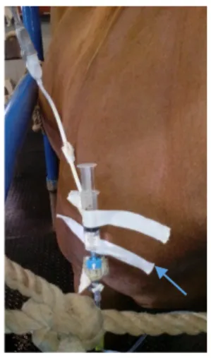
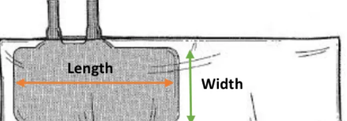
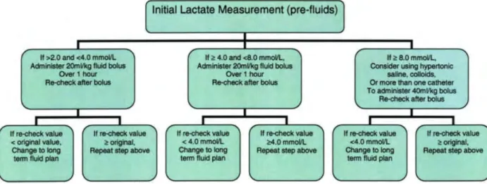
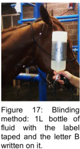
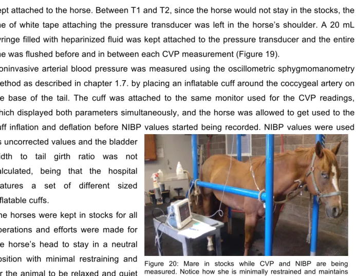
Figure 1: Permanent tracheostomy performed after a temporary one had been performed in a more rostral position. ... 1 Figure 2: Calcified rope removed from the small colon of a 5yo mare with acute colic, very painful and with low response to analgesia and sedation. Nail brush for scale. ... 2 Figure 3: Two large lacerations. The horse on the left had a big sequestrum on the dorsal aspect of the cannon bone that was later removed. ... 2 Figure 4: Horse with Equine Asthma using an aerosol mask. ... 2 Figure 5: One year old zebra with severe diarrhea receiving IV fluids. ... 2 Figure 6: 48h old dummy foal with oxygen line and feeding tube after surgery for correction of three ribs fractured during labor. ... 2 Figure 7: The first hoof to fall from a septic two month old filly. ... 2 Figure 8: Use of acupuncture to manage pain on a 12yo mare with laminitis after colic surgery. 3 Figure 9: First time foaling mare and Dr. Modesto assisting the foal to stand and nurse. ... 3 Figure 10: Laser cauterization of uterine cysts. ... 3 Figure 11: To avoid clipping a line in the horse's shoulder a strip of white tape (arrow) marks the "zero" for the measuring devise. This technique only works if the skin is dry and for a short period of time. ... 24 Figure 12: Neutral position of the head. Notice how the mare is relaxed and no restraining is being used. If repositioning of the head was necessary, holding the lead rope in the right position would be enough for most of the cases. ... 24 Figure 13: Different waveforms and pressure values shown on the monitor of the pressure transducer according to the location of the CVP catheter tip (Tennent-Brown, 2015). ... 25 Figure 14: Inflatable cuff placement for NIBP measurements on the coccygeal artery of a mare. ... 27 Figure 15: Inflatable cuff, bladder and bladder width and length. Adapted form: http://drrajivdesaimd.com/?paged=20 ... 28 Figure 16: Algorithm for using blood lactate as a guide to fluid therapy (Fielding & Magdesian, 2005). ... 37 Figure 17: Blinding method: 1L bottle of fluid with the label taped and the letter B written on it. 47 Figure 18: Mindray™ PM-9000Vet monitor used for measuring CVP and NIBP. ... 48 Figure 19: Pressure transducer taped to the horse’s shoulder tip and a 20 mL syringe filled with heparinized fluid attached to the transducer. The bottom piece of tape was kept in the same place throughout all operations to avoid variations of the “zero” level. ... 48 Figure 20: Mare in stocks while CVP and NIBP are being measured. Notice how she is minimally restrained and maintains her head in a nearly neutral position. ... 49
vii
List of tables
Table 1: History findings, predisposing factors and potential mechanisms for the occurrence of
colics in horses. Adapted from Blikslager and Jones (2010) and Cohen (2002). ... 7
Table 2: Composition of different types of crystalloid solutions used in equine medicine compared to horse plasma. Adapted from Magdesian (2015b) and Cook and Bain (2003). ... 13
Table 3: Types of lactic acidosis in veterinary medicine. Adapted from Magdesian (2004), Allen and Holm (2008) and Radcliffe (2015). ... 34
Table 4: PCV values according to type of horse. Adapted from (Sellon, 2010) ... 44
Table 5: Baseline characteristics of horses enrolled in this study receiving a 2L bolus of ... 51
Table 6: CVP (cmH₂O) values for each horse at each time point. ... 52
Table 7: MAP values (mmHg) for each horse at each time points. ... 53
Table 8: PCV values (%) for each horse at each time points. ... 55
List of graphics Graphic 1: CVP values for HSS and LRS groups at T0, T1 and T2 (mean ± SE) ... 51
Graphic 2: Noninvasive MAP mean ± SE for HSS and LRS groups at T0, T1 and T2. ... 53
Graphic 3 and Graphic 4: noninvasive systolic (SYS.NIBP) and diastolic (DIS.NIBP) arterial pressures mean ± SE for HSS and LRS groups at T0, T1 and T2 ... 54
Graphic 5: PCV mean ± SE for HSS and LRS groups at T0, T1 and T2. ... 55
Graphic 6: Total plasma solids mean ± SE for HSS and LRS groups at T0, T1 and T2. ... 57
Graphic 7: Total plasma protein mean ± SE for both HSS and LRS groups at T0, T1 and T2. .. 57
Graphic 8: Albumin mean ± SE for the HSS and LRS groups at T0, T1 and T2. ... 58
Graphic 9: Creatinine mean ± SE for the HSS and LRS groups at T0, T1 and T2. ... 59
Graphic 10: BUN mean ± SE for the HSS and LRS groups at T0, T1 and T2. ... 59
Graphic 11: Plasma lactate mean ± SE for the HSS and LRS groups at T0, T1 and T2. ... 61
Graphic 12: Whole blood lactate mean ± SE for the HSS and LRS groups at T0, T1 and T2. ... 61
viii
Acronyms
AG Anion gap
ARDS Acute respiratory distress syndrome
BUN Blood urea nitrogen
BW/TG Bladder width/tail girth
CaO₂ Arterial oxygen content
CI Cardia index
CO Cardiac output
CVP Central venous pressure
CvO2 Venous oxygen content
DO₂ Oxygen delivery
Hb Hemoglobin
HSS Hypertonic saline solution
IM Intramuscular
IV Intravenous
LDH Lactate dehydrogenase
LRS Lactated Ringer’s solution
MAP Mean arterial pressure
MCFP Mean circulatory filling pressure NADH Nicotinamide adenine dinucleotide
NIBP Noninvasive blood pressure
OER Oxygen extraction ratio
PCV Packed cell volume
SaO₂ Oxygen saturation
SIRS Systemic inflammatory response syndrome
TP Total protein
TPR Total peripheral resistance
TS Total solids
VO₂ Oxygen uptake
VR Venous return VRst Venous resistance
1
Internship report
This work was performed during the 6th year of the Integrated Masters in Veterinary Medicine of the Faculty of Veterinary Medicine, University of Lisbon, under the supervision of Dr. Benjamin Buchanan and Professor Paula Tilley.
I did my internship at the Brazos Valley Equine Hospital (BVEH) in Navasota, Texas, USA, where I spent about 5 months split in two periods. For the first period I lived at the hospital from September 28 to December 18 and worked with the senior practitioners during the day and with the interns during the night as an intensive training period. For the second time, from April 4 to June 14 I did not live at the hospital but spent my days with the practitioners and interns, only driving to the hospital at night if one of the cases for my study was coming in as an emergency. The BVEH Navasota consists of a fully equipped laboratory, a surgery room with two induction/recovery rooms, two appointment rooms with two sets of stocks each and facilities to hospitalize more than 50 animals including an ICU unit with four stalls and an isolation unit with two stalls. At the time of my internship three board-certified specialists in Surgery, Internal Medicine and Emergency/Critical Care, and Reproduction as well as two more practitioners worked at the hospital. The hospital’s caseload is quite large and consists mostly of horses and ponies (95%) but also donkeys, miniature donkeys, llamas and alpacas. In 2015 the hospital attended 13,903 animals split approximately in 40% sports medicine, 20% internal medicine, 10% dentistry/wellness, 10% reproduction, 5% complimentary medicine, and 5% pre-purchase examinations.
Through my training period I perfected skills like administering medication IM, IV and orally, passing nasogastric tubes and checking for gastric reflux, placing IV catheters, performing a complete physical exam in adults and neonates, flexing limbs for lameness exams and evaluating lameness, applying bandages, suturing and caring for wounds, among others. I also got the chance of learning numerous new skills such as performing an upper airway, tracheal and gastric endoscopy, doing a bronchoalveolar lavage, flushing guttural pouches, performing a temporary tracheostomy on cadavers and
assisting surgery for permanent tracheostomy (Figure 1), learning how to ultrasound lungs, abdomen, umbilicus and limbs, doing nerve and skin blocks and regional limb perfusions, placing subpalpebral lavage systems, performing abdominocentesis, measuring central venous pressure, assisting surgery and anesthesia and using several laboratory tests and machines.
Figure 1: Permanent tracheostomy performed after a temporary one had been performed in a more rostral position.
2
I had the opportunity of witnessing several surgeries including for colics by intestinal strangulation, impaction,
torsion or obstruction by a foreign body (Figure 2), various orthopedic cases of fractures, arthrodesis, joint or tendon sheets lavages, correction of angular and flexural
defects and
osteochondrosis, one case
of an ethmoid hematoma, one resolution of an esophageal fibrotic scar, one paranasal sinus cyst and several sinusitis from rotten teeth roots, many permanent tracheostomies due to nasopharyngeal cicatrix syndrome, various lacerations (Figure 3), several castrations, umbilical hernia resolutions and two enucleations. I also had the chance to watch Dr. Dennis Brook, from the University of Florida, performing three cataract surgeries and one iris abscess removal. In the internal medicine, neonatology and emergency/critical care area I saw several interesting cases including several infections by Streptococcus equi equi (Strangles), two resolutions of large colon right displacement by rolling the horse and trocharising the large colon through the abdominal wall, numerous medical resolutions of gastrointestinal colics, several cases of Equine Asthma (Figure 4), several adult horses, foals and one zebra with diarrhea (Figure 5), two cases of very severe suppurative pneumonia that ended up in one of the horses dying and the other being euthanized, two cases of dummy foals one of which suffered three fractured ribs during labor having to be submitted to surgery at 48h of life and surviving to recover completely (Figure 6), several Rhodococcus equi infections in foals with systemic complications on a miniature Figure 2: Calcified rope removed from the
small colon of a 5yo mare with acute colic, very painful and with low response to analgesia and sedation. Nail brush for scale.
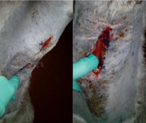
Figure 3: Two large lacerations. The horse on the left had a big sequestrum on the dorsal aspect of the cannon bone that was later removed.
Figure 4: Horse with Equine Asthma using an aerosol mask.
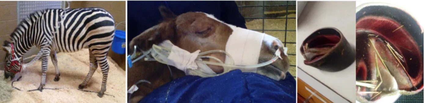
Figure 5: One year old zebra with severe diarrhea receiving IV fluids.
Figure 6: 48h old dummy foal with oxygen line and feeding tube after surgery for correction of three ribs fractured during labor.
Figure 7: The first hoof to fall from a septic two month old filly.
3
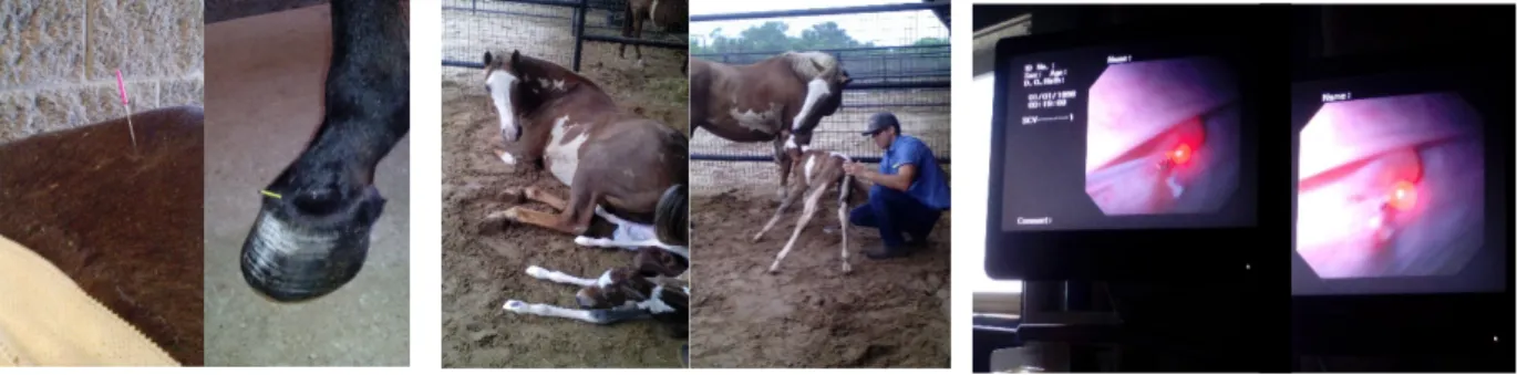
Figure 8: Use of acupuncture to manage pain on a 12yo mare with laminitis after colic surgery.
foal that ended up dying, one hyper-acute laminitis secondary to septicemia in a two month old filly that lost three hooves and had to be euthanized (Figure 7) and one diaphragmatic hernia from unknown cause with entrapment of the liver and spleen and splenic rupture on a 20 year old horse that was euthanized 24h after being admitted to the hospital.
I also observed the use of acupuncture to manage pain in several post-operative cases (Figure 8) and as treatment or therapeutic aid for cases of Equine Asthma, sweeney shoulder, laminitis, back and neck pain and navicular syndrome. On the reproduction area, I assisted with semen collection, natural breeding and artificial insemination, helped synchronizing oestrus and foaling mares (Figure 9) and witnessed laser cauterization of uterine cysts on four mares (Figure 10).
hfhf
I thought that one surgical case was particularly interesting. A nine year old Arabian mare, seven months pregnant, was observed at the farm with acute signs of abdominal colic. The mare had had surgery for colic about a year before that and showed to be very painful. As the response to analgesia and sedation was weak and brief she was immediately refered to the hospital with indication for abdominal surgery. Surgery was conducted normally being that the cause for colic was an entrapment of the small intestine through a net in the mesentery, probably originated on the previous surgery. A resection and anastomosis was performed and the mare was up in the stall shortly after. The in utero foal showed good signs of vitality after surgery but progesterone was still administered orally to the mare for the time of recovery. The post-surgery antibiotic therapy was done following the hospital’s protocol with penicillin and gentamicin IV as well as analgesic and anti-inflammatory therapy with flunixin meglumine IV. For four days following surgery the mare kept refluxing large amounts of fluid and feed material. By the fourth day she received a one liter bolus of 7,2% hypertonic saline solution IV on the grounds that it would decrease the swelling of the anastomosis site, improving motility and reducing the amount of reflux produced. On the fifth day the mare stopped producing reflux and was fed a liquid meal made with senior equine feed dissolved in water. For the next week the same meal was provided every six hours and alfalfa hay was introduced gradually to the mare’s diet. By the end of the second week post-op the mare started showing signs of abdominal discomfort and spiked
Figure 9: First time foaling mare and Dr. Modesto assisting the foal to stand and nurse.
Figure 10: Laser cauterization of uterine cysts.
4
a fever. On abdominal ultrasound the foal showed no signs of vitality and it was spontaneously aborted later that same night. Even though there were no signs of retained placenta or any further complications related to the abortion, the mare’s condition kept worsening and she started refluxing again having to be taken back to surgery by the end of the third week after being admitted to the hospital. Although the anastomosis site showed no signs of complications, there were some small intestine adhesions that were considered the cause of colic and the surgery went on without complications. For the following weeks the mare recovered well from surgery but developed severe laminitis on both front feet. Besides special shoeing and medical treatment, acupuncture was also used to manage pain. At the end this mare stayed at the hospital for about six months and went home almost fully recovered.
5
Introduction
Colics are among the most important medical problems in equine medicine and, despite their relevance, there is still a lot to be understood about this disease (Cohen, 2002).
There is a constant search for better ways to improve recovery and survival of horses with colic and the use of hypertonic saline solution (HSS) has been a target of this quest.
It’s been almost a century since the first benefits of hypertonic saline solutions were discovered but it wasn’t until after 1960 that this solution’s effects on hypovolemic animals started being investigated. The effects of HSS on equine colics only became a focus of investigation in the 80’s when there was an increase of interest in this solution’s properties.
Before the benefits of HSS in hypovolemic animals were discovered, intravascular fluid deficits had to be corrected by administrating large volumes of isotonic crystalloid fluids. The process of administering large volumes of fluids implies a longer resuscitation time and an increased risk of fluid overload, besides the fact that, in field situations, it may reveal hard to have access to such amounts of fluids. Although there are some situations where the use of hypertonic solutions is contraindicated, in hypovolemic animals this type of fluids will cause a considerable expansion of plasma volume, alongside with other effects, which will secure organ perfusion and vital functions for enough time to allow the necessary volume of replacement fluids to be administered.
Other benefits of hypertonic saline solutions, besides plasma expansion, have been studied and include the improvement of cardiac function, venodilation, immunomodulatory and anti-inflammatory properties and reduction of reperfusion injuries, among others ( Schmall, 1989; Reitan, & Moore, 1998; Kien, Kreimeier & Messmer, 2002; Oliveira, Velasco, Soriano, & Friedman, 2002; Pantaleon, 2006; Strandvik, 2009; Fielding & Magdesian, 2011; Urbano et al., 2012 Chimabucuro et al., 2014;).
Although most of the existent literature about the use of hypertonic solutions in horses with colic seems to focus on surgical cases, we believe that medical cases can also benefit from the effects of this solution in a way that allows for faster recovery and lower mortality rates.
6
CHAPTER I – Bibliographic Review 1.1. Equine Colic
The term colic is used to describe the presence of abdominal pain, being, in reality, a generic term for a much more complex setting of problems with a variety of causes (Bentz, 2004a). There are nearly 100 recognized causes for colics in horses and although many go by undiagnosed, especially the ones treated in the field, there seems to be a certain correlation between age, sex and breed and specific types of colic (Cohen, 2002). For example, young animals seem to be more prone to intussusceptions while strangulating lipomas are more common in older horses. Although there isn’t a consistent relation between colic occurrence and gender there are some forms of colic that happen exclusively in one of the genders, such as uterine torsions in mares and scrotal herniation in stallions and geldings, and others that seem to be more frequent in some sexes despite the lack of epidemiologic studies like colonic torsions in mares after parturition (Cohen, 2002). Older horses also appear to be in higher risk of colic and of needing surgical treatment but also have better prognosis for survival (Cohen, 2002). The Arabian breed has been reported to have increased risk of colic, younger miniature horses have a bigger prevalence of fecaliths and impactions of the small colon, and Standardbreds have a higher risk of scrotal herniation (Cohen, 2002).
Some of the most common causes for equine colic not related to the GI tract, also known as “false colics”, include the reproductive tract: uterine torsion, dystocia, ovulation, retained placenta, uterine hematoma or perforation and orchitis; the urinary tract: ruptured bladder, cystitis, pyelonephritis and calculi of different etiology; hepatic or splenic causes: acute hepatitis and splenic abscess; peritonitis and neoplasia (Bentz, 2004b; Mair, 2002b) .
It is important to remember that, when looking for the cause for a colic, one must not forget about other conditions that, even though are not related with abdominal pain, may mimic some clinical signs and lead to confusion about the diagnose. These disorders can be related, among other causes, with the musculoskeletal system: laminitis, rhabdomyolysis; the cardiorespiratory system: pneumonia, pleuritis; the cardiovascular system: acute hemorrhage, myocardial infarction or pericarditis; the nervous system: tetanus, botulism, hypocalcemic tetany or equine motor neuron disease; or unspecific like intoxications by Cantharidin (Bentz, 2004b; Mair, 2002b).
7
Predisposing factors, besides the signalment referred above, for the occurrence of gastrointestinal colics are described on Table 1.
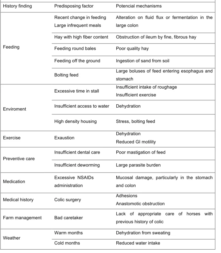
Table 1: History findings, predisposing factors and potential mechanisms for the occurrence of colics in horses. Adapted from Blikslager and Jones (2010) and Cohen (2002).
History findings, predisposing factors and potential mechanisms for the occurrence of gastrointestinal colics.
History finding Predisposing factor Potencial mechanisms
Feeding
Recent change in feeding Large infrequent meals
Alteration on fluid flux or fermentation in the large colon
Hay with high fiber content Obstruction of ileum by fine, fibrous hay
Feeding round bales Poor quality hay
Feeding off the ground Ingestion of sand from soil
Bolting feed Large boluses of feed entering esophagus and
stomach
Enviroment
Excessive time in stall Insufficient intake of roughage
Insufficient exercise
Insufficient access to water Dehydration
High density housing Stress, bolting feed
Exercise Exaustion Dehydration
Reduced GI motility
Preventive care
Insufficient dental care Poor mastigation of feed
Insufficient deworming Large parasite burden
Medication Excessive NSAIDs
administration
Mucosal damage, particularly in the stomach and colon
Medical history Colic surgery Adhesions
Anastomotic obstruction
Farm management Bad caretaker Lack of appropriate care of horses with
previous history of colic
Weather
Warm months Dehydration from sweating
8
Given the presented study was based solely on adult horses suffering from hypovolemia due to gastrointestinal colic, I will focus on a deeper review about gastrointestinal colics in horses and their repercussions in the body’s fluid balance.
1.1.1. Hypovolemia and dehydration
One must be aware of the differences between hypovolemia and dehydration since both conditions require distinct approaches (Corley, 2008a). Even though both conditions may be present in the same horse, hypovolemia is the term used to describe the reduction of circulating blood volume whereas dehydration is the loss of interstitial fluid without decline of volemia (Corley, 2008a).
Hypovolemia can be considered absolute, where there is actual loss of intravascular volume due to diarrhea, hemorrhage or increased capillary permeability, among others; or relative, when the plasma volume remains the same but the intravenous compartment expands like in situations of systemic venodilation following SIRS or the administration of vasodilatory drugs (Magdesian, 2015b). Hypovolemia compromises organ perfusion and might result in permanent organ damage and death if not addressed correctly and in time (Magdesian, 2015b).
Hypovolemic horses that have lost 8 -10L of plasma volume, which represents about one quarter of total blood volume for a 500Kg horse, show clinical signs like slow jugular fill, tachycardia and tachypnoea, reduced urine output, cold extremities and weak pulse (Corley, 2008a; Hassel, 2015; Magdesian, 2015b). The main therapeutic goal for hypovolemia is to quickly replace blood volume, increase blood pressure and cardiac output and secure organ perfusion, which can be achieved by administering large volumes of isotonic crystalloid solutions, smaller volumes of hypertonic solutions or an association of both (Corley, 2008a; Magdesian, 2015b).
Horses that have fluid losses of more than 5% of their body weight may show dehydration signs such as tacky or dry mucous membranes, decreased skin turgor, sunken eyes and tachycardia (Corley, 2008a; Hassel, 2015; Magdesian, 2015a). Due to the body’s physiologic mechanisms to maintain an appropriate circulating volume by drawing extravascular fluid into the vessels, hypovolemia due to dehydration only happens in severely dehydrated animals (Corley, 2008a). Fluid therapy in dehydrated horses is aimed at replacing interstitial fluid losses over a period of 12 to 24 hours, which should be achieved through the administration of isotonic crystalloid solutions (Corley, 2008a; Magdesian, 2015a).
9
1.1.2. Hemodynamic changes in colic
Total body water content of an adult horse is estimated to be about 60% to 70% of the body weight and is divided into three major spaces or compartments: intracellular (ICF), intravascular and interstitial, the last two are commonly referred as extracellular fluid compartment (ECF) (Fielding, 2015a; Seahorn & Seahorn, 2003).
Water present in the adult horse’s gastrointestinal tract is thought to be approximately 6 to 10% of total body weight (Seahorn & Seahorn, 2003). The degree of fluid deficit of a horse with colic may vary from mild dehydration to hypovolemic shock (Seahorn & Seahorn, 2003).
Factors such as type of colic, duration of colic and ongoing losses should be taken into account when assessing the circulatory status of a horse with colic (Divers, 2002; Hassel, 2015). Fluid losses through sweating, increased gastric and intestinal secretions, decreased water intake and decreased intestinal absorption all contribute more or less for the onset and aggravation of fluid derangements (Divers, 2002; Hassel, 2015).
Initial losses will cause a reduction in extracellular fluid but as fluid depletion progresses, intracellular fluid may be lost as well (Divers, 2002; Hassel, 2015). Most horses suffering from colic for more than a day will show clinical signs of dehydration (Hassel, 2015). However, acute processes, such as strangulating lesions and proximal obstructions, will likely be accompanied mainly by hypovolemia secondary to a number of events such as endotoxemia, hypersecretory states, and increases in endothelial permeability (Hassel, 2015).
Loss of integrity of the gastrointestinal barrier will promote the passage of endotoxin producing Gram negative bacteria leading to endotoxemia and, more severely, to endotoxic shock (Pantaleon, 2005). Endotoxemia may originate a capillary leak syndrome which is described as the loss of fluid, with or without protein, into the interstitium due to increased endothelial permeability (Pantaleon, 2005). Endotoxemia also leads to arterial and venous dilation and, eventually, decreased cardiac output, which aggravate even further the patient’s hypovolemic status (Pantaleon, 2005).
Situations of intestinal obstruction can also result in increased capillary filtration or leakage (Seahorn & Seahorn, 2003; Hassel, 2015). Intestinal obstructions result in distension of the proximal portion of intestine due to accumulation of digesta, fluid, gas and secretions. The increased intraluminal pressure compresses the veins in the intestinal wall and increases the capillary filtration rate into the interstitium. If the rate of fluid secretion is greater than the one removed by lymph flow, then edema and accumulation of fluid within the intestinal and/or gastric lumen increase (Freeman, 2002). Distension of the intestinal lumen can be such that causes necrosis of the surrounding tissues (Blikslager & Jones, 2010b).
Plasma protein losses, through diarrhea, into the intestinal lumen, in abdominal effusion, and due to increased permeability of the endothelium contribute for the loss of oncotic pressure
10
within the vessels and to the onset or worsening of intravascular fluid loss and tissue edema (T. Divers, 2002; Seahorn & Seahorn, 2003).
1.1.3. Fluid therapy according to type of colic
Even though there are innumerous causes of colic in horses, it is possible to divide the most common into non-strangulating mechanical obstructions, strangulating obstructions and functional obstructions (Hassel, 2015).
1.1.3.1. Non-strangulating mechanical obstructions
Non-strangulating obstructions can be partial or complete and are caused by an intraluminal obstacle to the progression of digesta without, however, causing obstruction of the blood flow (Bentz, 2004b; Blikslager & Jones, 2010b). Nonetheless, non-strangulating obstructions may result in vascular obstruction as explained on chapter 1.1.1. Some of the most common non-strangulating mechanical obstructions are originated by feed, sand or ascarids’ impactions, colonic displacements, foreign body obstructions, sand impactions, enteroliths, enlargement or thickening of the intestinal wall, masses, and intestinal scarring (Bentz, 2004b; Blikslager & Jones, 2010b; Hassel, 2015).
Horses with non-strangulating obstructions normally show signs of moderate dehydration but not hypovolemia, therefore, fluid therapy for these cases aims mostly at restoring hydration and electrolytes balance using mostly isotonic crystalloid solutions (Hassel, 2015). In situations of impaction, fluid therapy may also have the goal of hydrating and dissolving the impaction, even if the horse’s hemodynamic status is normal (Blikslager & Jones, 2010b; Hassel, 2015).
Colonic impactions have traditionally been treated by administering large volumes of IV fluids to achieve systemic overhydration, associated to oral administration of a laxative like mineral oil, sodium sulfate or magnesium sulfate (Bentz, 2004b; Hassel, 2015; Lopes, White, Donaldson, Crisman, & Ward, 2004), however, the amount of IV fluids administered in these situations involve some risks and haven’t proved to be more effective than the oral administration of similar volumes (Hassel, 2015; Lopes, Walker, White, & Ward, 2002). Aggressive IV fluid therapy is justifiable in horses that do not tolerate enteral fluids, either due to the existence of gastric reflux previous to oral administration of fluids or caused by it, and in severe cases where enteral fluids alone are not enough (Hassel, 2015; Lopes et al., 2004).
Some colonic displacements can also be managed through IV fluid therapy (Hassel, 2015). 1.1.3.2. Strangulating obstructions
A strangulating obstruction results from the complete or partial obstruction of the intestinal lumen and corresponding blood supply and are commonly caused by intestinal volvulus, strangulating
11
lipomas, intussusceptions, inguinal and diaphragmatic hernias and entrapments in the epiploic foramen, gastrosplenic ligament or mesenteric rents (Bentz, 2004b; Blikslager & Jones, 2010a; Hassel, 2015).
Strangulating obstructions can be classified as hemorrhagic or ischemic. Hemorrhagic strangulating lesions happen when the intestine is twisted or compressed only to the point where veins are occluded but arteries are not, due to their more rigid wall. The affected intestine is dark and enlarged as blood flow keeps being pumped into the vessels but not out of them (Blikslager & Jones, 2010a). Ischemic strangulating lesions result from a tightly twisted intestine capable of occluding both veins and arteries and is rarer than the previous. The affected bowel is pale and of normal or thinner thickness (Blikslager & Jones, 2010a).
Both types of strangulating obstructions cause quick deterioration of the patient’s condition and should be handled as surgical emergencies (Hassel, 2015).
Horses suffering from a strangulating obstruction are almost always in need for IV fluid therapy as hypovolemia, endotoxemia and systemic inflammatory response syndrome (SIRS) are frequently present (Blikslager & Jones, 2010a; Hassel, 2015). The first approach to these cases should be to restore volemia and secure organ perfusion. The administration of hypertonic saline solution associated or not to a colloid solution is often advised to quickly promote plasma expansion and improve microcirculation, organ perfusion and cardiac function besides other benefits that will be further explained on chapter 1.4. (Blikslager & Jones, 2010a; Hassel, 2015). To assure the maintenance of these effects one must provide the appropriate amount of isotonic polyionic solutions following the administration of the hypertonic solution (Hassel, 2015).
1.1.3.3 Functional obstructions
Functional obstructions are the consequence of an inflammatory condition known as duodenitis-proximal jejunitis, duodenitis-proximal enteritis or anterior enteritis, characterized by inflammation of the proximal small intestine and resulting in ileus (Bentz, 2004b; Hassel, 2015). Although the etiology is not yet fully understood, it is believed that Clostridium spp., Salmonella spp. and some mycotoxins play an important role in the development of anterior enteritis (McConnico, 2010; Hassel, 2015). The inflammatory process and reduction in motility lead to a large accumulation of fluid in the small intestine and stomach (Bentz, 2004b; McConnico, 2010; Hassel, 2015). As fluid accumulates, the stomach and small intestine become distended and painful and the horse becomes dehydrated and hypovolemic (Bentz, 2004b; Hassel, 2015). Many cases of anterior enteritis are accompanied by SIRS, which contributes even further for the depletion of the horse’s hemodynamic status (Hassel, 2015). Horses with functional obstructions produce large amounts of gastric reflux that can sometimes be spontaneous (Hassel, 2015; McConnico, 2010). These horses almost always have prerenal azotemia and
12
electrolyte derangements (Johnston & Morris, 1987; Bentz, 2004b; McConnico, 2010; Hassel, 2015).
Aggressive IV fluid therapy should be started as quickly as possible. Hypertonic saline solution or colloids can prove useful in restoring plasma and should be followed by large volumes of isotonic polyionic crystalloid solution (McConnico, 2010). When planning fluid therapy for horses with functional obstructions it’s important to have in account that these horses are in great need for fluid replacement and of expansion of the plasma volume, and that the ongoing losses from gastric reflux are very significant. However, because of the high secretion of electrolytes and protein into the intestinal lumen, horses with anterior enteritis tend to have low plasma oncotic pressure which means that, with fluid resuscitation there is also an increased risk of edema formation and further fluid secretion into the intestinal lumen. Therefore, the use of synthetic or natural colloids should be considered for horses with low plasma protein concentrations (Hassel, 2015; McConnico, 2010).
1.2. Fluid therapy
1.2.1. 0,9% Sodium chloride and Isotonic polyionic solutions
Although 0,9% Sodium chloride is usually known as isotonic or normal saline, it is actually slightly hypertonic compared to horse plasma (Magdesian, 2015a). Its composition is mildly hypernatremic and significantly hyperchloremic compared to horse plasma being, therefore, recommended for the treatment of hyponatremic and hypochloremic patients. Also, because of its high concentration in chloride and absence of a buffer component, this solution’s acidifying properties can lead to a hyperchloremic metabolic acidosis (Cazzolli & Prittie, 2015) but, at the same time, make it a suitable treatment for hyperkalemic patients such as postpartum mares with a ruptured bladder (Cook & Bain, 2003). Animals being treated with 0,9% sodium chloride should be closely monitored for electrolyte disturbances since this is a solution lacking all other electrolytes in its composition.
Isotonic polyionic solutions (IPS) are considered balanced solutions and are more physiologic than isotonic saline solution since they have an electrolyte composition similar to plasma as well as a buffer component, making them a better fit for most fluid resuscitation cases (Cazzolli & Prittie, 2015). This type of fluids can be classified as maintenance or replacement solutions according to their composition (Cook & Bain, 2003; Magdesian, 2015a).
Maintenance IPS have a higher concentration of potassium and a lower concentration of sodium which allows for a long-term administration without the risk of hypernatremia or hypokalemia occurring (Cook & Bain, 2003). Maintenance fluid rate is considered to be 2,5 mL/kg/h or 60 mL/kg/d for adult horses (Cook & Bain, 2003; Magdesian, 2015a).
13
Replacement IPS have a relatively low potassium concentration (Table 2) making them similar to the extracellular fluid and ideal for situations where fluid resuscitation is required. The low potassium concentration of these fluids prevents the risk of iatrogenic hyperkalemia due to the high rate of which these fluids need to be administered (Cook & Bain, 2003; Magdesian, 2015b).
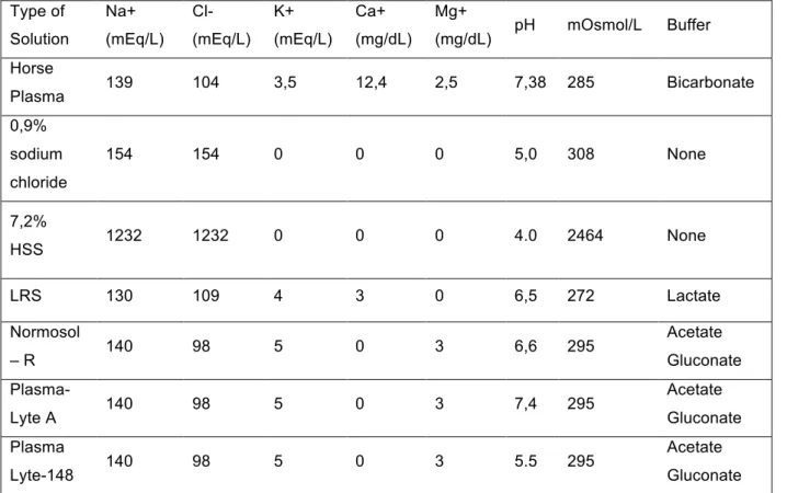
Table 2: Composition of different types of crystalloid solutions used in equine medicine compared to horse
plasma. Adapted from Magdesian (2015b) and Cook and Bain (2003).
Normosol-R, Plasma Lyte-148 and Plasma Lyte- A are the most common replacement solutions for horses. These fluids differ only in their pH, making Plasma Lyte-A the most physiological for horses (Magdesian, 2015b). These fluids contain acetate and gluconate as buffers and therefore are advised in patients with liver dysfunctions since acetate is metabolized by muscle, liver and kidney cells and gluconate is metabolized by the big majority of body cells (Cook & Bain, 2003; Magdesian, 2015b).
Because lactate is metabolized by the liver and kidney, the presence of hepatic abnormalities may be a contraindication for the administration of Lactated Ringer’s Solution or Hartman’s solution (Cook & Bain, 2003; Magdesian, 2015b).
Type of Solution Na+ (mEq/L) Cl- (mEq/L) K+ (mEq/L) Ca+ (mg/dL) Mg+ (mg/dL) pH mOsmol/L Buffer Horse Plasma 139 104 3,5 12,4 2,5 7,38 285 Bicarbonate 0,9% sodium chloride 154 154 0 0 0 5,0 308 None 7,2% HSS 1232 1232 0 0 0 4.0 2464 None LRS 130 109 4 3 0 6,5 272 Lactate Normosol – R 140 98 5 0 3 6,6 295 Acetate Gluconate Plasma-Lyte A 140 98 5 0 3 7,4 295 Acetate Gluconate Plasma Lyte-148 140 98 5 0 3 5.5 295 Acetate Gluconate
14
1.2.2. Risks of large volume resuscitation with crystalloids
Crystalloid fluids bear negligible oncotic pressure and they’re main electrolyte is sodium. Sodium is also the main electrolyte present in the extracellular space being that about 75% of it is located in the interstitium (Cook & Bain, 2003; Cazzolli & Prittie, 2015). This fact explains why about 60 to 80% of the infused volume disseminates into the interstitial space within 30-60 minutes, making it so that considerably large amounts of these fluids must be administered before normovolemia is achieved (Cook & Bain, 2003; Magdesian, 2003; Cazzolli & Prittie, 2015).
The administration of large volumes of isotonic crystalloids may be necessary to revert hypovolemic shock and restore appropriate organ perfusion making fluid overload a common complication in human and small animal medicine (Cazzolli & Prittie, 2015). Fluid infusion should be closely monitored as a cascade of events following fluid overload will worsen the patient’s prognosis (Cazzolli & Prittie, 2015). Although a significant amount of the administered fluids is believed to be excreted in urine, a high infusion rate may still result in interstitial edema formation (Cook & Bain, 2003; Fielding, 2015). As fluid load increases, the extracellular space expands, compressing and damaging the structure of the endothelial surface layer leading to capillary leak and tissue edema formation (Cazzolli & Prittie, 2015; Fielding, 2015). These types of fluids will also increase the production of inflammatory cytokines and endothelial cell activation due to their proinflammatory properties, aggravating the formation of tissue edema and hypoxia (Cazzolli & Prittie, 2015; Fielding, 2015). Other complications resulting from aggressive fluid resuscitation include dilutional coagulopathy, compartment syndromes, cellular swelling, reduced cell activity and impaired tissue healing (Cazzolli & Prittie, 2015).
In equine medicine clinical signs of fluid overload are rare in adult horses with normal renal and cardiac function. However, horses with renal failure (Corley, 2008a), increased capillary permeability and reduced oncotic pressure due to SIRS or horses with acute respiratory distress syndrome (ADRS) are more prone to getting overhydrated and developing life threatening pulmonary edema (Fielding, 2015). In these cases fluids should be administered as conservative boluses instead of continuously infused (Corley, 2008; Fielding, 2015).
Although foals barely tolerate dehydration, fluid overload in equine neonates can represent an equally threatening condition, easily leading to lung and cerebral edema and seriously compromising the cardiorespiratory, renal and hepatic systems. A close monitoring of these patients is of the utmost importance and several authors claim that, as long as renal function is secured, a small restriction of the infused volume is preferable to the risk of overhydrating and that administering fluid as boluses should only be allowed for emergency resuscitation (Corley, 2008a; Knottenbelt, Holdstock, & Madigan, 2004; Palmer, 2006; Wilkins, 2010).
15
Volume overload can be avoided or early detected by monitoring parameters such as central venous pressure (CVP), arterial pressure, jugular fill (Corley & Stephen, 2008; Hardy, 2010; Palmer, 2006), urine output, particularly in the neonate, urinary specific gravity, blood hematocrit and total solids (Knottenbelt et al., 2004; Palmer, 2006; Hardy, 2010). The presence of white-pink foamy nasal discharge accompanied by dyspnea is an indication of pulmonary edema, the most important sign of fluid overload, and should be assessed with furosemide given as an IV bolus and reduction or interruption of fluid administration. Some cases may require intranasal oxygen supplementation (Corley, 2008a). Changes in mental status, especially in the neonate, can also work as an indicator of the hydration status (Palmer, 2006).
1.2.3. Hypertonic Saline Solutions
A hypertonic solution is any solution with a concentration higher than its respective isotonic form. This can be achieved by using high concentrations of sodium, glucose, mannitol and other components (Kreimeier & Messmer, 2002).
Hypertonic saline solutions (HSS) have higher concentrations of sodium and chloride than isotonic saline solutions, meaning, than 0,9%. HSS are normally sold in variations of 3%, 5% or 7%, being that the ones most commonly used in equine practice are 7,2% to 7,5% (Duval, 1995; Magdesian, 2015b). HSS more concentrated than these are usually primary solutions to be used in association with other types of fluids such as dextran or hetastarch (Duval, 1995). Even though HSS are contraindicated in some situations and may involve some risks, they are generally safe and effective as well as much more affordable than colloids.
It’s been 90 years since the use of hypertonic saline solutions was first described by Silbert et al (1926) as treatment for Buerger’s disease or thromboangiitis obliterans (Duval, 1995). But it wasn’t until 1963 that Brook’s et al described the use of a hypertonic saline solution in dogs suffering from hemorrhagic shock as a way to prevent tissue damage and improve recovery. In 1980, Velasco et al increased the popularity of HSS by showing that the administration of a 4 mL/Kg bolus of 7,5% HSS to severely hemorrhagic dogs restored the blood pressure and acid-base equilibrium quickly. From the 80’s forward, the number of studies concerning the efficacy of different hypertonic solutions, concentrations, doses, infusion rates and administration routes increased greatly both in human and in veterinary medicine.
In 1984, Nakayama et al used the concept of small-volume resuscitation (SVR) to describe the fast administration of a small dose of hypertonic saline solution (approx. 4 ml/kg of 2,400 mosm/L HSS) to manage hypovolemic shock as an alternative to large volumes of isotonic crystalloids (Kreimeier & Messmer, 2002).
Several mechanisms of action for the effects of hypertonic saline solutions have been suggested, studied and proven or contested. The most commonly accepted and better explained is the expansion of plasma volume. Several authors have tried to define the real increase in
16
plasma volume that is due to the administration of HSS, being that an increase two to five times the administered volume of HSS seems to be the most consensual values (Kien, Reitan, & Moore, 1998; Cook & Bain, 2003; Magdesian, 2015b). Fielding and Magdesian (2011) concluded that endurance horses requiring IV fluid therapy and treated with a bolus of 7,2% hypertonic saline solution had an increase in plasma volume of 29,1 ± 4% while horses treated with 0,9% isotonic saline solution had significantly smaller increase of 12 ± 14,6%, which roughly corresponds to the amount of infused fluids.
The first explanation for this expansion, to which most authors seem to agree, is a simple rise in the plasma’s osmotic pressure that will cause fluid from the interstitial and/or intracellular spaces to be drawn to the intravascular compartment (Duval, 1995; Kien et al., 1998; Fielding & Magdesian, 2011). Since 96% of plasma osmolality is determined by electrolytes like sodium, chloride, bicarbonate, potassium, calcium, magnesium and phosphate, it’s clear how hypertonic NaCl solutions are capable of such increases is plasma osmolality (Kreimeier & Messmer, 2002).
A study where hypertonic saline solution was administered to anesthetized rats reported not only an increase in plasma volume of four to five times the infused volume of HSS but also a reduction of 10% of the intracellular fluid volume of muscle and liver cells (Kien et al., 1998). Kien et al (1998) suggest skeletal muscle as the major mobilized fluid source for plasma expansion after HSS administration and Fielding and Magdesian (2011) state that the initial shift of fluid is from the interstitial space and only later from the intracellular space. Garrido et al (2006) consider the intracellular fluid drawn from erythrocytes and endothelial cells to be the main responsible for the immediate increase in plasma volume. Constable (2003) also includes water present in the gastrointestinal tract as a fluid source for plasma expansion.
Fettman (1985) references two authors who concluded that, following a physiological response to hemorrhagic shock that creates hyperosmolar plasma conditions, the skin and skeletal muscle represented the main sources of replacement fluid for the intravascular space. In fact, in situations of hypovolemia there is a physiological neurohumoral response that leads to changes in peripheral capillary resistance and increases in glycaemia and plasma osmolality. This mechanism promotes a fluid shift from the intracellular and interstitial compartments to the intravascular space and, at the same time, reduces the intravascular space relatively to blood volume (Fettman, 1985; Schmall, 1989). However, this physiologic response is not enough to endure a severe hypovolemic state (Schmall, 1989), so hypertonic solutions are considered to synergize with this intrinsic mechanism in order to obtain a more effective result (Fettman, 1985).
17
Another possible explanation is that there is a considerable thirst response triggered by the increase in plasma’s sodium concentration meaning that, in cases where the patient is allowed to drink, the water intake will contribute for the plasma expansion (Fielding & Magdesian, 2011). Hypertonic saline solutions also appear to reduce swelling of erythrocytes (Oliveira, Velasco, Soriano, & Friedman, 2002; Magdesian, 2015b) and capillary endothelial cells, often seen during hypovolemic and hemorrhagic shock and systemic inflammatory response syndrome ( Kien et al., 1998; Oliveira et al., 2002; Corley, 2008a). Reducing erythrocyte swelling associated to the plasma expansion will reduce blood viscosity and improve microhemodynamics (Pantaleon, 2005). Endothelial swelling causes the capillary lumen to reduce, increasing capillary resistance and affecting the involved microcirculation, ultimately leading to tissue ischemia. Endothelial swelling also restricts the passage of large white blood cells across the vessel’s walls (Kien et al., 1998). A study where rabbits were subjected to hemorrhagic shock observed that, within one minute after resuscitation with HSS, the blood volume was similar to values previous to hemorrhage and that endothelial swelling had reduced by 20% (Kien et al., 1998).
Adding to the plasma expansion effects, hypertonic saline solutions also carry vasodilator properties, mainly on peripheral vasculature. This effect is considered to be the cause for some cases of short-lasting hypotension immediately after infusion (Kien et al., 1998; Kreimeier & Messmer, 2002; Oliveira et al., 2002). HSS infusion causes the release of vasodilating substances such as prostacyclin and increases the 6-keto-prostaglandin F1α : thromboxane B2
ratio (Oliveira et al., 2002). Prostacycline is a vasodilator and inhibitor of platelet adhesion and aggregation, released by endothelial cells ( Frandson, Wike, & Fails, 2009). 6-keto-prostaglandin F1α and thromboxane B2 are stable hydrolysis products of prostacyclin and thromboxane A2,
respectively. As said before, prostacyclin is a vasodilator and inhibitor of platelet aggregation while thromboxane A2 is a vasoconstrictor and an inducer of platelet aggregation, therefore an
increase in their ration will favor the prostacyclin’s effects (Saldeen & Saldeen, 1983).
Kien et al (1998) reported a severe decrease in mean arterial pressure 45 seconds after the beginning of the administration of hypertonic saline solution at a rate of 2 mL/Kg/min and 3 mL/Kg/min but not with rates of and under 1 mL/Kg/min. At five minutes post-infusion the mean arterial pressure had risen to values similar to baseline for all groups (0,5, 1, 2 and 3 mL/Kg/min), suggesting that the initial hypotension was due to peripheral vasodilation leading to a reduction in peripheral resistance with distribution of the total plasma volume. The authors do not state the solution’s concentration or the species of the animals used. Kien et al (1998) also showed that the hypotensive effect of HSS in dogs treated with endothelium-derive relaxing factor was significantly smaller when compared to dogs treated with autonomic nervous system blocking drugs and to not treated dogs, indicating the involvement of mechanisms directly related to the endothelium. In a study using a model for hemorrhagic shock in foals, the
18
administration of 32 mL/Kg isotonic saline solution or 4mL/Kg of 7% NaCl in 6% Dextran 70 produced the same significant increases in mean arterial pressure and cardiac output but the reduction in total peripheral resistance was not observed with the administration of 0,9% NaCl (Schmall, 1989). The vasodilator properties of HSS have also been proven to exist in salt-free hypertonic solutions suggesting this effect may be due to the hyperosmolar effect rather than to the presence of a salt (Kien et al., 1998).
Most of the existing literature agrees that hypertonic saline solutions increase cardiac contractility, cardiac output, stroke volume, and improve arterial blood pressures (Schmall, 1989; Schmall, Muir, & Robertson, 1990; Pantaleon, 2005; Taylor & Clark, 2007; Robertson, 2010; Theobaldo, Barbeiro, Barbeiro, Petroni, & Soriano, 2012; Magdesian, 2015b). Some authors discuss if the increase in cardiac contractility caused by HSS administration should be considered a direct inotropic effect (Kien et al., 1998; Magdesian, 2015b) as the hyperosmolality of the extracellular compartment and subsequent reduction in intracellular water may promote the release of calcium ions from the sarcoplasmic reticulum and t-tubules, resulting in higher concentrations of intracellular calcium and favoring the cardiac muscle fibers contractility (Kien et al., 1998; Pantaleon, 2005). On the other hand, Constable (2003) considers HSS to actually have a negative inotropic effect caused by the exchange of extracellular sodium for intracellular calcium promoted by the high extracellular sodium concentration, resulting, therefore, in lower intramyocyte calcium availability. This contradiction can be explained by some findings reporting that moderate degrees of hyperosmolarity will increase cardiac contractile forces whereas the opposite happens under severe hyperosmolarity conditions (Oliveira et al., 2002). Some authors seem to agree that HSS will have a vasodilator effect on the coronary arteries, improving blood supply to the myocardium (Fettman, 1985; Kien et al., 1998) while others consider the improvements in cardiac performance to be independent of the increased coronary blood flow (Oliveira et al., 2002).
There is still some discussion about the role of lung innervation on the action mechanisms of hypertonic saline solutions. It has been suggested that the high concentration of sodium stimulates pulmonary receptors, sending an impulse to the brain through the vagal nerve and resulting in a shift of blood flow from the skin and muscle to the vital organs. This was called the pulmonary arc reflex (Duval, 1995). Initial studies showed that HSS infusions to the aorta or pulmonary vein of hemorrhagic dogs, meaning post-pulmonary circulation, did not produce the same resuscitation effects as the intravenous or pre-pulmonary infusions (Schmall, 1989; Oliveira et al., 2002). On the other hand, a study conducted with sheep showed no differences in the results obtained with peripheral or central administration of HSS (Oliveira et al., 2002). In the same way some studies showed the need for the innervation to the lung to be intact for HSS to produce a systemic result (Schmall, 1989). However, other authors showed that similar effects of
19
resuscitation with hypertonic saline are obtained in hypovolemic and hemorrhagic dogs with innervated and denervated lungs (Duval, 1995; Oliveira et al., 2002). The vagal nerve is known to have an important function on the restoration of hemodynamics in hypovolemia situations and several authors showed that animals with vagal blockade and vagotomy produced a reduced response or even no response to the administrations of HSS ( Fettman, 1985; Schmall, 1989; Duval, 1995; Oliveira et al., 2002;). The number of contradicting literature leaves several unanswered questions and creates doubt around the real involvement of this reflex on hypertonic saline solution therapy.
Hypertonic saline solutions have also been showed to have immunomodulatory and anti-inflammatory effects (Theobaldo et al., 2012; Chimabucuro et al., 2014; Magdesian, 2015b). Chimabucuro et al. (2014) studied the effects of 7,5% HSS (4 mL/Kg) and 0,9% normal saline (32 mL/Kg) in gut perfusion after ischemia by occluding the superior mesenteric artery (SMA) in rats and concluded that the immunologic effects of the administration of a small dose of HSS are similar to the ones obtained with a large dose of normal saline without the increased risk of edema production. The animals used were distributed in four groups: subjected to SMA occlusion treated with HSS, subjected to SMA occlusion treated with normal saline, subjected to SMA occlusion but not treated and animals not subjected to SMA occlusion and not treated with any type of fluid. Inflammatory and oxidative stress markers were collected for a six hour times after gut reperfusion. It is important to notice that the animals not subjected to SMA occlusion were still exposed to general anesthesia, abdominal wall incision, gut manipulation and SMA handling, having, therefore, relatively increased levels of inflammatory and oxidative stress markers. The authors noticed that both groups receiving crystalloids had significant decreases in oxidative stress markers and an improved inflammatory response when compared to the not treated group. Furthermore, the hypertonic saline solution group had higher plasma levels of interleukin 6 and 10, which are pro-inflammatory and anti-inflammatory respectively, but the levels of these interleukins in these animal’s tissues were very similar to the group that was not subjected to gut ischemia. Even though the levels of pro-inflammatory cytokines are not completely annulled by HSS, authors agree that the production of IL-6 will regulate the synthesis of IL-10 and an adequate ratio between anti and pro-inflammatory cytokines is crucial for a positive outcome (Pantaleon, 2005; Chimabucuro et al., 2014). Several other studies have shown the increases in anti-inflammatory cytokines caused by hypertonic saline solution ( Pantaleon, 2005; Theobaldo et al., 2012; Magdesian, 2015b).
The mechanisms for neutrophil activation, migration and infiltration are well known to play a major role in the inflammatory response to sepsis or ischemia and have been associated with damage to the endothelium, lung, intestines and other tissues, capillary leakage and tissue
20
edema, aggravating even further the tissue hypoxia (Garrido, Cruz, Poli de Figueiredo, & Rocha e Silva, 2006; Kreimeier & Messmer, 2002; Pantaleon, 2005; Magdesian, 2015b).
Neutrophil infiltration was reduced on the HSS and normal saline treatment groups to levels similar to the group free of gut ischemia in the study by Chimabucuru et al. (2014). The administration of hypertonic saline solution in experimental hemorrhagic shock showed to reduce neutrophil activation resulting in reduced lung injury (Corley, 2008a). Elevated levels of sodium reduce the neutrophil exaggerated response (Pantaleon, 2005). Moreover HSS reduces neutrophil accumulation in the lung, neutrophil - endothelial adhesion and neutrophil degranulation decreasing end organ injury (Pantaleon, 2005).
Other benefits of hypertonic saline solutions may include the indirect release of vasopressin and activation of aquaporin channels (Fielding & Magdesian, 2011). Vasopressin is released in both plasma hyperosmolality and hypovolemia situations being that the stimulation of volume receptors predominates over the osmoreceptors. In this way hypertonic solutions will not inhibit this hormone’s secretion through their hyperosmolarity (Fettman, 1985). In hypovolemic animals, the increases in vasopressin will prevent water diuresis in order to maintain an adequate plasma volume while the activated aquaporin channels will facilitate the water to flow out of the cell (Magdesian, 2015b).
There are many conditions where the use of HSS may be indicated, either alone or combined with colloid agents. Severe burns, traumatic brain injury, lung injury, before general anesthesia and as resuscitation during circulatory and endotoxic shock are some of the most common situations that justify the use of this type of fluids (Duval, 1995; Kien et al., 1998; Garrido et al., 2006; Magdesian, 2015b).
Hypertonic saline has been used for some time to help balance fluid and electrolyte’s losses in severely burnt patients and to reduce intracranial pressure in traumatic brain injury, many times as pre-hospital treatments (Fettman, 1985; Kien et al., 1998; Magdesian, 2015b).
The expansion in plasma volume and improved cardiac output promote the redistribution of blood flow from peripheral beds to vital organs and the reestablishment of microhemodynamics, reducing tissue lesions due to ischemia or inflammatory processes. There seems to be a bigger increase in blood flow to organs like heart, kidney, liver and intestines after treatment with hypertonic solutions when compared to isotonic solutions (Schmall, 1989; Kien et al., 1998). The improved kidney perfusion and higher plasma sodium concentration after HSS infusion lead to a shorter time to urination, increased urinary frequency and more diluted urine, compared to treatment with isotonic crystalloids, which is believed to act as a protective effect against renal failure associated with hypoperfusion (Fielding & Magdesian, 2011). With the normalization of renal function, HSS is also believed to restore the renal interstitium osmolality and the countercurrent mechanisms by providing the needed electrolytes (Fettman, 1985). This effect
21
will help avoid medullary washout (Fettman, 1985). Medullary washout describes the loss of the medullar interstitium osmolality gradient, consequent inability to concentrate urine after restoration of renal blood flow and increased diuresis following high-volume fluid administration (Schott II, 2010; Magdesian, 2015b).
Treatment with hypertonic saline is contraindicated in the presence of hypernatremia, hyperchloremia or hyperosmolarity as these are already intrinsic risks to the administration of HSS (Fettman, 1985; Kien et al., 1998; Magdesian, 2015b).
Due to its low pH and absence of a buffer component, some clinicians worry about a possible iatrogenic acidosis caused by the administration of hypertonic saline. But according to Constable (2003) the decrease in pH caused by HSS in large animals is less than 0.08 units and it lasts little time making this a safe solution in terms of acid-base balance.
While treatment with hypertonic saline solution is indicated in some cases of hemorrhagic shock, the presence of uncontrolled hemorrhage excludes the possibility of using HSS as these solutions are reported to increase bleeding and reduce clot stabilization (Kien et al., 1998; Kreimeier & Messmer, 2002; Magdesian, 2015b).
Even though the benefits of hypertonic solutions are numerous one should have in mind that the effects of hypertonic saline solutions are of short duration and work at the expenses of the body’s fluid reserves. There’s some concern about giving hypertonic solutions to dehydrated patients since these may have intracellular fluid deficits and may not be able to compensate the hyperosmolar conditions. In such situations Magdesian (2015b) advises the administration of isotonic crystalloids prior or simultaneously to the HSS infusion. In patients with mild or no signs of dehydration, the administration of hypertonic solutions should be followed by adequate amounts of isotonic or hypotonic IV fluids or the patient should be allowed to drink ad libitum (Magdesian, 2015b).
1.2.4. Crystalloids Vs Colloids
Colloids are solutions containing large molecular weight molecules and can be divided in natural: plasma (the most used in equine medicine), whole blood and albumin; or synthetic: hydroxyethyl starch solutions (the most used in equine medicine), dextran, gelatin and polymerized hemoglobin (Magdesian, 2015b). There is an ongoing debate in the medical field about the fluid choice for resuscitation purposes in both human and veterinary medicine (Pantaleon, 2005; Chan, 2008; Magdesian, 2015b; Cazzolli & Prittie, 2015). Both crystalloids and colloids present advantages and disadvantages in their use and numerous studies have been conducted to clarify which one overcomes the other with no obvious conclusions in either human or veterinary medicine (Chan, 2008; Magdesian, 2015b). Due to the elevated price of colloids and lack of obvious benefits over crystalloids, the last ones are usually preferred over the first (Magdesian, 2015b). Although colloid solutions carry increased risks of anaphylactic