Universidade de Lisboa
Faculdade de Ciências
Departamento de Biologia Animal
Effects of Stress on CA3 Pyramidal !eurons in
the Pregnant Female Rat
Andreia Barbosa Valença
Mestrado em Biologia Humana e Ambiente
2010
Universidade de Lisboa
Faculdade de Ciências
Departamento de Biologia Animal
Effects of Stress on CA3 Pyramidal !eurons in
the Pregnant Female Rat
Andreia Barbosa Valença
Dissertação orientada por:
Orientador interno: Doutora Tatyana Strekalova, Centro de Biologia
Ambiental, Faculdade de Ciências da Universidade de Lisboa
Orientador externo: Doutora Jodi Pawluski, Department of
!euroscience, Maastricht University
Mestrado em Biologia Humana e Ambiente
2010
i
Acknowledgements
I would like to thank everyone that in one way or another took part in this big journey:
Dr. Jodi Pawluski, for her guidance, knowledge, encouragement, patience, time and wise advices throughout the elaboration of this thesis. Besides being my supervisor, her friendship along the way was very important.
Dr. Tatyana Strekalova, for believing in me, making this project possible and all her support and time.
Professor Harry Steinbusch, for the great opportunity that was working in the Department of Neuroscience and for his support throughout the time in Maastricht.
Marisela, for her endless patience, support, friendly smile and the awesome mexican dinner.
My mom, with all my heart, for her unconditional love, never ending support and patience and for always believing in me and in my future endeavours and giving me strength to go ahead and follow my dreams. She is the best mom anybody could wish for! And my sister Cristina for supporting me in her own way. Thank you both for being by my side, always.
Margarida, for being the best rommie I could ever wish for! In health and sickness, in joy and tears, we made it through and survived to this big journey and kind of adventure, with distinction. Together we laughed, learned, fell and raised again, and shared really good and not so good moments but together we kept always the best side (which really brought on my psychologist skills and made me see how big my zen side is). Together we grew and we were stronger and made the best of it! And Margarida’s parents, for their endless support and good mood. And Bruno “pops”, for taking part in a bit of everything, listening, laughing, cooking, biking, flying.
Bianca, for always being there for me along the way for so many years. I know I can count on her for everything. Thank you.
ii Rodrigo “Zukinha”, for being one in a million. It was just impossible not to laugh around him! Countless moments and healthy discussions to remember by a lucky chance that made us cross paths, through Maastricht flying all the way to Amsterdam. Friends have the gift to turn everything richer, fuller, better. Tot volgende keer!
Julie and João “Xangue”, for sharing worries and making everything easier with their help. Julie, our favorite Belgian girl, even with all the work, there was always time for fun and Pecherese! Xangue, in the middle of Dutch, we rocked it in Portuguese! Together we made it, thank you guys!
Marta, Edgar, Joana and Rocha, for endless afternoons in Starbucks and dinners, and really special funny bike rides, just having fun and not driving crazy with the thesis work. Their support and friendship mean the world to me.
Paulo for being a great friend and such an informatics genius!
All my friends, family, lab mates and peers. Thank you.
“DSU room”, for being our “office” throughout our time in Maastricht and kind of the Portuguese speakers’ place. There we were happy and sometimes less happy. There we laughed a lot, made friends and spent great moments, and even celebrated 7 goals!
All the awesome people I met along the way that sure made the best of Maastricht, with great discovery weekends, protocol nights, crazy Amsterdam feelings, lunches and dinners to laugh and remember.
The organizations and people that made my work possible: International Stichting Alzheimer Onderzoek (ISAO), The Netherlands, grant N 09501 to Dr. Tatyana Strekalova; Buddha Biopharm, Finland; Fundação para a Ciência e a Tecnologia (FCT) and Centro de Biologia Ambiental (CBA), Portugal; Professor Ana Santos, Dr. Martti Vallila, Professor Deodália Dias, Professor Margarida Reis, Dr. Cláudia Oliveira, Luís Marques and Raquel Vaz.
iii
Abstract
Stress is one of the primary factors leading to many disorders, including depression, one of the most prevalent psychiatric disorders. Additionally, it has been well documented that hippocampal plasticity is vulnerable to the effects of stress and these effects are often sexually differentiated. Women are twice as likely as men to experience stress-related disorders during the lifespan. In fact, a growing number of women experience psychological stress, such as depression and anxiety, during pregnancy and the postpartum period. This maternal stress may have detrimental effects on maternal mood and maternal care of offspring. In turn, recent research has documented a significant impact of pregnancy and motherhood on hippocampus plasticity in the mother. However, very little research has focused the impact of stress during gestation on the neurobiology of mother. Therefore, the present study investigated how stress affects dendritic morphology of CA3 pyramidal neurons in the hippocampus of pregnant females, and whether these effects differ from those in virgin females. Age-matched pregnant and virgin female Wistar rats were divided into two conditions: 1) Stress and 2) Control. Females in the stress condition were restrained for 1 hour/day for 2 weeks, beginning on gestation day 8 and at matched time-points in virgin females. Females were sacrificed the day after the last restraint session, prior to giving birth, and the brains were processed using Golgi impregnation technique. The results obtained show that repeated restraint stress results in dendritic atrophy in the apical region of CA3 pyramidal neurons in both pregnant and virgin females. Moreover, pregnant females resulted in less complex CA3 pyramidal neurons compared to virgin females. Stress had no effect on weight gain in virgin and pregnant, or litter characteristics and sex of fetuses in pregnant females. These factors were also not associated with CA3 dendritic morphology. Further work is needed to determine how restraint stress affects dendritic morphology in other regions of the hippocampus.
Key words: Stress, hippocampus, CA3 pyramidal neurons, dendritic morphology,
iv
Resumo
O quotidiano é preenchido por diversos episódios stressantes que podem representar uma grande ameaça ao bem-estar físico e emocional. De facto, o stress é um dos principais factores que leva a diversos transtornos, incluindo depressão, um dos transtornos psiquiátricos mais prevalentes. Assim, para lidar adequadamente com situações de stress, ajustes fisiológicos ou estratégias comportamentais são de extrema importância e são normalmente acompanhadas pela activação da resposta ao stress, com a intenção de manter ou alcançar a homeostase interna. Uma activação e desactivação da resposta ao stress bem sucedidas são, então, vitais para a sobrevivência. A resposta ao stress é coordenada pelo cérebro, que interpreta as experiências como ameaçadoras ou não e, de acordo com a situação, determina as respostas comportamentais e psicológicas. Portanto, quando uma ameaça real ou percebida ocorre, a resposta ao
stress é activada no cérebro e envolve a libertação de hormonas pelo sistema nervoso
simpático e pelo eixo hipotálamo-pituitária-adrenal (HPA). Os glucocorticóides (GC), cortisol nos humanos e corticosterona em roedores, desempenham um papel central na mediação de aspectos essenciais à resposta ao stress e retorno à homeostase. A duração do stress também está implicada nesta resposta neuronal, sendo que uma duração prolongada por mais de uma semana acarreta efeitos mais profundos ao nível dos neurónios. O hipocampo, constituído principalmente pelas regiões do cornu ammonis (CA) e pelo giro denteado (DG), para além de desempenhar um papel essencial na aprendizagem e memória, tem também a função de regulação de “feedback” negativo da resposta ao stress através do eixo HPA. A grande concentração de receptores de GC na formação hipocampal sugere que os efeitos desta hormona no hipocampo sejam directos, tornando esta área do cérebro particularmente sensível ao stress e aos GC. De facto, tem sido bem documentado que a plasticidade do hipocampo é vulnerável aos efeitos do stress, através de níveis elevados de GC, causando alterações estruturais e funcionais no hipocampo. Os neurónios piramidais da região CA3 do hipocampo são particularmente sensíveis ao efeito do stress crónico, apresentando remodelação dendrítica. Sendo que esta região está envolvida na formação de memórias e processamento espacial, é interessante que eventos stressantes repetitivos resultem em atrofia dos neurónios piramidais CA3, caracterizada pela redução da complexidade dendrítica e do comprimento dendrítico total em machos, o que igualmente afecta a função do hipocampo, incluindo perda de memória espacial. Esta remodelação
v dendrítica pode ter duas interpretações: uma resposta mal adaptada, com a retracção dendrítica a contribuir para uma maior vulnerabilidade do hipocampo a outros eventos, como doenças, e factores stressantes crónicos, ou uma resposta compensatória para protecção contra efeitos neurotóxicos. É também importante ter em consideração que estes efeitos do stress são muitas vezes sexualmente diferenciados. A propensão para desenvolver transtornos relacionados com stress é estimada em duas vezes mais para mulheres em relação aos homens, durante a vida. Esta tendência é marcada pelo envolvimento das hormonas gonadais femininas, progesterona e estradiol, e a sua acção no eixo HPA. Tendo em consideração que, para além de desempenharem um papel chave no desenvolvimento diferencial do cérebro, estas hormonas estão também envolvidas na formação da plasticidade cerebral nos principais centros emocionais e podem exercer um papel importante na modulação da resposta ao stress, é cada vez mais reconhecida uma ligação entre género e transtornos relacionados com stress, com as discrepâncias entre géneros atribuídas ao efeito das hormonas gonadais. O ciclo reprodutivo da mulher está intimamente relacionado com os níveis de GC, com elevada libertação desta hormona e elevada sensibilidade ao stress durante a fase folicular do ciclo menstrual bem como da fase proestro do ciclo estral em roedores, quando os níveis de estrogénio estão elevados. Assim, uma potencial combinação de GC e hormonas gonadais pode levar a uma maior incidência de transtornos relacionados com stress em fêmeas. De facto, um número crescente de mulheres sofre stress psicológico, como depressão e ansiedade, durante a gravidez e o período pós-parto. Por outro lado, pesquisas recentes têm documentado um impacto significativo da gravidez e maternidade na plasticidade do hipocampo da mãe. Este impacto pode estar relacionado com o envolvimento do hipocampo nas importantes adaptações hormonais, neurológicas e comportamentais necessárias na mãe para assegurar a sobrevivência da prole, na transição para a maternidade. A placenta, os ovários e o feto contribuem para as flutuações dramáticas de hormonas esteróides e peptídicas que ocorrem durante a gravidez e o período pós-parto e são importantes para a indução do circuito maternal e o inicio dos comportamentos maternos. Além disso, visto os efeitos que as hormonas esteróides têm nas propriedades estruturais do hipocampo, estas flutuações hormonais no período reprodutivo podem ter também um impacto na plasticidade desta área do cérebro. O stress e os níveis de GC têm também um impacto na mãe. Apesar das alterações normais nos níveis de GC serem importantes para diversos aspectos da maternidade, o stress durante a gestação leva ao aumento da concentração basal de GC
vi e pode ter efeitos prejudiciais sobre o humor materno e os cuidados maternos da prole. No entanto, pouca pesquisa tem focado o impacto do stress durante a gestação sobre a neurobiologia da mãe. Assim, o presente estudo investigou o efeito do stress sobre a morfologia dendrítica dos neurónios piramidais da região CA3 do hipocampo de fêmeas grávidas e se, estes efeitos, diferem em fêmeas virgens.
Ratos Wistar fêmeas, grávidas e virgens de idades correspondentes, foram divididos em duas condições: Stress e Controlo. As fêmeas na condição de stress foram contidas em caixas de contenção uma hora/dia durante duas semanas, começando no oitavo dia de gestação e em tempos correspondentes em fêmeas virgens. As fêmeas foram sacrificadas no dia a seguir à última sessão de contenção, antes do parto. O útero das fêmeas grávidas foi dissecado para permitir a contagem dos fetos, tendo também em conta o seu sexo. Os cérebros foram processados usando a técnica de impregnação de Golgi, que consiste numa impregnação metálica e permite detectar as árvores dendríticas e as espinhas dendríticas. Para a análise da morfologia dendrítica, seis células piramidais CA3 por cada cérebro foram escolhidas e o número de pontos de ramificação, bem como o comprimento total da árvore dendrítica, foram avaliados separadamente para a região apical e basal. A distribuição e complexidade das dendrites foram analisadas recorrendo à contagem das intersecções das dendrites com círculos concêntricos equidistantes (análise de Sholl).
Os resultados obtidos mostraram que as fêmeas grávidas e virgens, na condição de
stress, tiveram atrofia dendrítica significativa na região apical dos neurónios piramidais
CA3, em comparação com as fêmeas controlo. Para além disso, as fêmeas grávidas apresentaram neurónios piramidais CA3 significativamente menos complexos, em comparação com as fêmeas virgens. O stress não teve efeito sobre o peso em virgens e grávidas, nem afectou as características das ninhadas. Este estudo forneceu novas evidências de que o stress e a gravidez têm um impacto na morfologia dendrítica dos neurónios piramidais CA3. Pesquisa futura irá avaliar a morfologia dendrítica e a densidade das espinhas dentríticas na região CA1 e DG bem como o possível papel do
stress e da maternidade no desempenho de tarefas dependentes do hipocampo na fêmea
adulta.
Palavras-chave: Stress, hipocampo, neurónios piramidais CA3, morfologia dendrítica,
vii
Table of Contents
Acknowledgements ...i
Abstract ... iii
Resumo ...iv
Table of Contents ... vii
Abbreviations Index ...ix
Figure Index ... x
Table Index ...xi
CHAPTER I. INTRODUCTION ... 1
Stress in the Brain ... 1
The Structure of Hippocampus ... 3
The Role of Hippocampus in the Stress Response ... 4
Stress and the CA3 Region of the Hippocampus ... 5
Sex Differences and Stress Effects in the CA3 Region of the Hippocampus ... 8
Visualization of Dendritic Morphology via Golgi Impregnation ... 10
Impact of Pregnancy and Motherhood in the Hippocampus ... 11
Stress Effects in the Mother ... 13
Thesis Objectives ... 15
CHAPTER II. MATERIAL AND METHODS ... 16
Animals and Housing ... 16
Breeding ... 16
Group Formation ... 16
Restraint Stress Procedure ... 17
Sacrificing and Dissection ... 18
Brain Removal ... 18
Uterine Horns Dissection in Pregnant Females ... 18
Histological Procedures ... 19
Golgi Impregnation Technique... 19
Dendritic Morphology ... 20
viii CHAPTER III. RESULTS ... 24
Pregnant Females Gained Significantly More Weight than Virgin Females ... 24 Regardless of Reproductive State, Stressed Females Showed Dendritic Atrophy in the Apical Tree of CA3 Pyramidal Neurons ... 25 CA3 Pyramidal Neurons Are Less Complex in Pregnant Female Rats ... 29 There Was No Significant Effect of Stress on Litter Characteristics ... 30 Size of Litter and Sex of Fetuses Was Not Associated with CA3 Dendritic
Morphology in Pregnant Female Rats ... 30
CHAPTER IV. DISCUSSION ... 32 Stress Decreased Dendritic Morphology in the Apical Region of CA3
Pyramidal Neurons in Pregnant and Virgin Female Rats ... 32 Litter Size or Sex of the Fetuses Was Not Affect by Stress and Not Associated
with CA3 Dendritic Morphology in Pregnant Female Rats ... 35 Stress Had No Significant Effect on Body Weight in Pregnant and Virgin
Female Rats ... 37 Possible Consequences of Hippocampal CA3 Dendritic Remodeling in Response to Repeated Stress for Maternal Behavior ... 37
CHAPTER V. CONCLUSION AND FUTURE DIRECTIONS ... 40
ix
Abbreviation Index
ACTH Adrenocorticotropin hormone
AIDS Acquired immune deficiency syndrome
ANOVA Analysis of variance
BDNF Brain-derived neurotrophic factor
CA Cornu ammonis
CBG Corticosterone binding globulin
CRH Corticotropin-releasing hormone
CUMS Chronic ultramild stress
DG Dentate gyrus
DSU Disk spinning unit
EC Entorhinal cortex
e.g. exempli gratia
FCM Faculdade de Ciências Médicas
GABA Gamma-aminobutyric acid
GC Glucocorticoids
Glu Glutamate
GR Glucocorticoid receptors
HPA axis Hypothalamic-pituitary-adrenal axis
LSD Least significant difference
LTP Long-term potentiation
MR Mineralocorticoid receptors
NMDA N-methyl-D-aspartate
SEM Standard error of the mean
SNS Sympathetic nervous system
Sub Subiculum
x
Figure Index
Figure Description Page
1 Human hippocampus ... 3
2 Basic circuitry of the hippocampus... 4
3 Representation of a CA3 pyramidal cell ... 6
4 Mechanism of CA3 dendritic retraction following chronic stress ... 7
5 Golgi impregnation reaction ... 11
6 Representation of dendrites and spines with Golgi technique ... 11
7 Relative hormone profile across rat pregnancy and parturition ... 12
8 Timeline of the experiment for the stress condition ... 17
9 Close-up of a pregnant rat dissection ... 19
10 Final slides of the Golgi impregnation technique ... 20
11 Photomicrograph of Golgi impregnated dorsal hippocampus ... 21
12 Photomicrograph and drawing of a CA3 pyramidal neuron ... 22
13 Sholl analysis representation... 22
14 Mean (± SEM) percentage of weight change ... 24
15 Neurolucida drawings of representative CA3 pyramidal neurons ... 27
16 Mean (± SEM) total CA3 dendritic length ... 28
17 Mean (± SEM) total number of CA3 branch points ... 28
xi
Table Index
Table Description Page
1 Group information... 17 2 Mean (± SEM) total litter size and number of male and female fetuses ... 30 3 Correlations between dendritic morphology and litter characteristics in
stressed and control pregnant females ... 31 4 Correlations between dendritic morphology and litter characteristics in
stressed pregnant rats ... 31 5 Correlations between dendritic morphology and litter characteristics in
1
Chapter I Introduction
A growing number of women experience stress-related diseases, such as depression and anxiety, during pregnancy and the postpartum period. In fact, more than 20% of women suffer from mood disorders during this time (Bennett et al., 2004a, b; Oberlander et al., 2006). These stress-related mood disorders can have detrimental effects on the mother and offspring, resulting in difficulties in mother-offspring bonding and increased susceptibility to mood disorders and cognitive problems in adult offspring (Lindgren, 2001; Smith et al., 2004; Maccari & Morley-Fletcher, 2007; Darnaudéry & Maccari, 2008).
Unfortunately our understanding of how stress affects the neurobiology of the mother is limited as very little research in humans and rodent model has aimed to determine the effect of stress during gestation on neurobiology of the maternal brain.
Stress in the Brain
The body's ability to physiologically regulate its inner environment, ensuring its stability in response to changes in the outside environment, can be defined as homeostasis. One of the major threats to homeostasis is stress (Bartolomucci & Leopardi, 2009). Life is manifested by single or recurring stressful episodes that can be a major threat to one’s physical or emotional health. Events, such as the loss of a spouse or the onset of disease, may set in motion fear, helplessness and emotional distress that can develop into stress-related disorders, such as depression and anxiety (Kessler, 1997; Kim et al., 2007). To adequately cope with major stressful events, adjustments in physiology or behavioral strategies are of major importance and are usually accompanied by activation of the stress response (Joëls & Baram, 2009), intending to maintain the initial homeostasis or achieve a new homeostasis (Bartolomucci & Leopardi, 2009). In fact, as stress has an impact in emotional states and cognitive abilities, leading to variety of mental disorders and diseases (McLaughlin et al., 2009; Strekalova & Steinbusch, 2010), a successful activation and termination of a stress response is vital for survival (de Kloet et al., 2005; Conrad, 2008).
The brain is the organ that interprets experiences as threatening or nonthreatening and, according to the situation, determines the behavioral and psychological responses (McEwen, 2007). Thus, when a real or perceived threat (stressor) occurs, the stress
2 response is activated in the brain and includes the sympathetic nervous system (SNS) and the hypothalamic-pituitary-adrenal (HPA) axis (Fuchs & Flügge, 2002; McEwen, 2002; Conrad, 2008). A rapid stress response occurs through the SNS, involving the release of catecholamines within seconds of the onset of the stressor, providing the regulation of blood pressure, heart rate and cardiovascular tone. On the other hand, the slower onset stress response by the HPA axis involves the release of glucocorticoids (GC: the primary glucocorticoid is cortisol in humans and corticosterone in rodents) within minutes of stressor onset (Conrad, 2008). The release of GC occurs via the release of the corticotropin-releasing hormone (CRH) in the hypothalamus that stimulates synthesis and release of the adrenocorticotropin hormone (ACTH) from the pituitary into the blood circulation (Cannon, 1914; Selye, 1936a, b). ACTH activates the adrenal glands where subsequently a variety of hormones, such as GC, are released into the blood to mediate essential aspects of the stress response and help the body return to homeostasis (de Kloet et al., 1998; Miller & O’Callaghan, 2002). For example, the GC hormones, derived from the cortex of the adrenal glands, convert proteins and lipids into carbohydrates, which can be directly used as energy resources (Sapolsky et al., 2000).
Despite the fact that a stress response is crucial for a successful adaptation to a threat, detrimental consequences can result from a persistent stress response or from inefficient management of allostasis, the active process by which the body responds to daily events and maintains homeostasis (McEwen 2001, 2007; McEwen & Gianaros, 2010). In the brain, the limbic system is particularly vulnerable to the effects of stress (Kim et al., 2007; Conrad 2008). For example, the nature of the neuronal responses is closely linked to the duration of the stressor. When acute stressors are present, a rapid surge of neurotransmission, neuronal activation and hormone release is established, followed by rapid return to baseline levels (McEwen, 2007). On the other hand, when stress is prolonged for a week or more (chronic stress), more profound changes will be induced, such as expression of particular genes, structural alterations in neurons and changes in neuronal firing patterns throughout the brain (Joëls et al., 2007; Krugers et
al., 2010). In addition, the diversity of the stressors and their impact on an individual’s
brain also relies on many factors, such as interaction of sex and genetic factors with life events (Magariños & McEwen, 1995; Joëls & Baram, 2009).
3
The Structure of Hippocampus
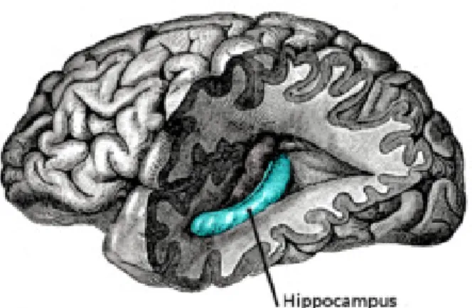
Multiple brain regions are likely involved in the organization of responses to stressful stimuli. In the limbic system, the hippocampus (Figure 1) has long been studied for its potential involvement in mental illness in general. The hippocampus is a neural structure comprised of three main areas: the cornu ammonis (CA) regions, CA1 and CA3, and the dentate gyrus (DG). The hippocampal formation also includes the entorhinal cortex, subiculum, parasubiculum, and the presubiculum (Amaral & Lavenex, 2007; Scharfman, 2007). The CA regions are primarily constituted of pyramidal neurons, while the DG, where neurogenesis occurs throughout adulthood, consists mainly of granule neurons (Amaral & Witter, 1989; Leuner & Gould, 2010). The hippocampus is comprised of a trisynaptic circuitry (Figure 2) where fibres from the entorhinal cortex project to the distal granule cell dendrites of the DG, via the perforant path (synapse 1), and projections from the DG granule cells project, via the mossy fibers, to the proximal dendrites of CA3 pyramidal cells (synapse 2). The pyramidal cells of the CA3 region send their major input via the Schaffer collaterals to the CA1 (synapse 3) (Amaral & Witter, 1989; Amaral & Lavenex, 2007; McEwen, 2007; Scharfman, 2007).
Figure 1. The human hippocampus.
4
The Role of Hippocampus in the Stress Response
This hippocampus has been primarily established to play a critical role in learning and memory, but it also has an important role in general cognition, mood regulation, and even in encoding predictions for future events (Dranovsky & Hen, 2006; Conrad, 2008; DeCarolis et al., 2010). However, a less well known function of the hippocampus is its role as a negative feedback regulator of the stress response via the HPA axis (Vyas
et al., 2002; Dranovsky & Hen, 2006; Leuner & Gould, 2010). The high concentration
of GC receptors in the hippocampal formation suggests that the effect of GC on the hippocampus may be direct, therefore making this brain area particularly sensitive to stress and GC (Lucassen et al., 2001; McEwen, 2001; Kim et al., 2007; Conrad, 2008; Leuner & Gould, 2010). In the rat, binding sites for the high-affinity mineralocorticoid receptors (MR) and low-affinity glucocorticoid receptors (GR) are located in the nuclei of granule cells in the DG and pyramidal neurons in the CA1 and CA3 subfields of the hippocampus, where corticosterone is highly taken up and retained (de Kloet et al., 1998; Jöels & Baram, 2009). These two types of receptors are responsible for translation of GC into specific cellular actions (Joëls, 1997; de Kloet et al., 1998). In fact, as a target for adrenal “stress” hormones, the hippocampus provides a crucial
Figure 2. Basic circuitry of the hippocampus, shown using a modified drawing by Ramon y
Cajal. CA3: cornu ammonis region 3; CA1: cornu ammonis region 1; DG: dentate gyrus; Sub: subiculum; EC: entorhinal cortex.
5 model for studying neurobiological consequences of stress (Magariños et al., 1997). An increasing body of research suggests that stress, via elevated levels of GC, can cause changes in the hippocampus, affecting both hippocampal structure and function (McEwen, 2006; Jöels et al., 2007). These changes include decreasing neurogenesis, alterating spine density, hindering synaptic output, impeding long-term potentiation (LTP), changing inhibitory and excitatory tone, altering pyramidal cell morphology, and reducing dendritic complexity (McEwen, 2007; Conrad, 2008).
Stress and the CA3 Region of the Hippocampus
Stress greatly influences CA3 pyramidal neurons in the hippocampus. In particular, the neurons of the CA3 region are very sensitive to chronic stress effects and show dendritic remodeling after chronic stress has been experienced (Magariños & McEwen, 1995; Sousa et al., 2000). The CA3 region is crucial to memory formation and spatial processing (Cerasti & Treves, 2010) and presents remarkable anatomical features: CA3 neurons are the largest pyramidal neurons within the hippocampus (Fitch et al., 1989) and are homogenously placed in cell bands that are easily identifiable, without interneurons and glial cells (Newrzella et al., 2007). Despite the fact that the main projections are from the DG, via a few, but very strong, tens of inputs from the granule neurons, the CA3 pyramidal neurons also receive many thousands of weak inputs from other sources, such as perforant path connections from the entorhinal cortex. This scarcity in the connectivity is unclear, however it is purposed to help the DG drive CA3 activity during the storage of new memories (McLaughlin et al., 2009; Cerasti & Treves, 2010).
The pyramidal neurons are comprised of two regions, apical and basal (see Figure 3). The apical and basal regions differ in functionality (Spratling, 2002), but the extent of this difference in the hippocampal pyramidal neurons is not well documented. The CA3 apical dendrites receive input from all parts of the DG while the basal dendrites receive input mainly from the infrapyramidal blade of the DG (Witter, 1989).
Considering these significant roles and inputs of CA3 cells, it is interesting that repeated stressful experiences result in atrophy of hippocampal CA3 pyramidal neurons, characterized by reduced dendritic complexity and reduced total dendritic length in males, shown in several studies (Watanabe et al., 1992; Magariños & McEwen, 1995; Conrad, 2008). Therefore, stress-induced changes in hippocampal CA3 neurons are
6 consistent with deficits in hippocampal function, including spatial memory impairment in male rats (McLaughlin et al., 2007).
CA3 atrophy found in rats is a relatively slow process, taking normally at least three weeks to develop under daily stress, but the atrophy produced by stress is reversible, within a week or so after the termination of stress, and factors, such as physical exercise, can speed up this process (McEwen & Magariños, 1997). Chronic stress-induced CA3 dendritic remodeling may be a maladaptive response and this dendritic retraction may contribute to hippocampal vulnerability to other life events and chronic stressors (Conrad 2006, 2008). For example, cognitive dysfunction can result when chronic stress precedes or coincides with other conditions, such as AIDS (Oberfield et al., 1994), depression (Sheline et al., 1996), and Alzheimer’s disease (de Leon et al., 1993). However, another interpretation is that CA3 dendritic remodeling may be a compensatory response to protect against extended excitatory amino acid stimulation, which can compromise and kill neurons (Conrad 2006, 2008).
An illustration of the mechanism underlying stress-induced CA3 dendritic retraction is in Figure 3. Moreover, as shown in previous studies, dendritic retraction typically occurs on the CA3 apical region while the basal region seems not to be affected (Watanabe et al., 1992; Magariños & McEwen, 1995; Magariños et al., 1996). The mossy fiber input to the CA3 region at the stratum lucidum appears to drive the dendritic remodeling, as it is the apical dendrites above this input that retract (McEwen, 1999). In addition, the middle part of the apical CA3 dendritic tree, corresponding to the
Figure 3. A) A schematic representation of a CA3 pyramdidal cell and its inputs and B)
a photomicrograph of a representative Golgi stained CA3 pyramidal cell. (From http://www.pitt.edu/~german/ (A) and Magariños et al., 2006 (B))
B
7 region expressing chronic stress-induced changes in the N-methyl-D-aspartate (NMDA) glutaminergic receptor sensitivity, suffers drastic remodeling by chronic stress (Kole et
al., 2002, 2004). Electrophysiological investigations in the CA3 region have shown that
after chronic stress, NMDA-receptor mediated responses are enhanced (Kole et al., 2002), whereas LTP, long-lasting enhancement in signal transmission between two neurons that results from stimulating them synchronously, is largely impaired (Pavlides
et al., 2002). Consequently, the commissural-associational collaterals are implicated as
contributing to CA3 dendritic retraction (Conrad, 2006). Aside from the vulnerability of hippocampal morphology in the CA3 region to the effects of stress, it is also important to take into consideration that often these effects are sexually differentiated (McLaughlin et al., 2009).
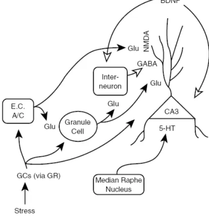
Figure 4. Mechanism of CA3 dendritic retraction following chronic stress. Repeated GC
elevations from chronic stress directly influence the CA3 pyramidal cells and CA3 afferents (dentate gyrus granule cells, commissural/associational fibers [C/A], entorhinal cortex [E.C.]) because all of these cells express receptors for GC. The GR most likely mediates dendritic retraction in rodents, but the MR probably plays a role in primates. Excess glutamate (Glu via N-methyl-D-aspartate [NMDA] receptor) and serotonin (5-HT) as well as altered inhibitory tone from interneurons and gamma-aminobutyric acid (GABA) modulate CA3 dendritic retraction. Reduced levels of brain-derived neurotrophic factor (BDNF), which is retrogradely transported to CA3 neurons, may permit CA3 dendritic remodeling. Solid arrows = enhanced tone permits CA3 dendritic retraction; open arrows = reduced tone permits CA3 dendritic retraction (From Conrad, 2006).
8
Sex Differences and Stress Effects in the CA3 Region of the Hippocampus
There is a clear pattern for the sex-specific prevalence rates of mental and physical disorders (Wang et al., 2007). In general, men are more prone to infectious diseases, cardiovascular disease, aggressive behavior, abuse of drugs or alcohol and schizophrenia, which has been associated with prenatal and early life exposures to stress (Wang et al., 2007). Women are more susceptible to autoimmune diseases and chronic pain, and tend to show heightened stress sensitivity and an increased predisposition to affective disorders, such as depression and anxiety (Wang et al., 2007; Goel & Bale, 2009; Lin et al., 2009). In rodents, the initial response of the HPA axis to a stressor is similar between males and females, however adult females generally have elevated levels of GC compared to males (Romeo, 2003). Prior to puberty, when the activation of gonadal hormones has not occurred, males and females also have a similar predisposition to stress-related disorders (Arnold & Gorski, 1984; Romeo & McEwen, 2006). However, the presence of an increase of testosterone beginning in puberty can affect active coping behaviors and stress physiology by exerting additional modulatory actions on serotonergic and GABAergic systems (Goel & Bale, 2009). In fact, during adolescence, a blunted male responsiveness, as a result of maturation of stress neurocircuitry, is likely associated with an increase in testosterone (Gomez et al., 2004). Following adolescence, there is an increased predisposition to affective disorders in females compared to males (Romeo & McEwen, 2006). This may be due to the effects of female gonadal hormones, estradiol and progesterone, and their action on the HPA system. These gonadal hormones can act in the HPA responsiveness with sluggish cortisol feedback on the brain and less or delayed containment of the stress response (Young & Altemus, 2004). For example, it has been proposed that a compromised cortisol feedback effect on HPA arousal in women plays a role in the neurobiological pathway mediating the greater tendency of women to develop depression (Young & Altemus, 2004). These findings suggest that gonadal hormones besides having a key role in differential brain development (Gomez et al., 2004), are also involved in shaping brain plasticity in key emotional centers, and may play an important role in modulating stress responsivity (Romeo & McEwen, 2006; Goel & Bale, 2009; Lupien et al., 2009). Thus, gender discrepancies may be partly attributed to the effect of gonadal hormones and a link between gender and stress-related disorders is gaining recognition.
9 In animal models, chronic stress or stressful life events often lead to depressive-like symptoms, with females and males coping differently with stressful situations (Luine, 2002; Bowman et al., 2003; Westenbroek et al., 2003). For example, female rodents exhibit a greater physiological stress response than males, as seen by higher release of GC (Handa et al., 1994) and decreased corticosterone binding globulin (CBG) (Galea et
al., 1997), following a variety of stressors through-out the estrous cycle, with greater
peaks in proestrous rats (Viau & Meaney, 1991; Conrad et al., 2004). Fluctuations in estradiol and prolactin can also stimulate corticosterone secretion (Lo & Wang, 2003; McLaughlin et al., 2005). Furthermore, women’s reproductive cycle is intimately linked to GC levels, as increased GC release and stress sensitivity is commonly observed during the follicular phase of the menstrual cycle as well as in the proestrous phase of the estrous cycle in rodents, when estrogen levels are high (Viau & Meaney, 1991; Kajantie & Phillips, 2006).
Importantly, sex differences may also be present in innervations of the CA3 region (Galea et al., 1997). As previously mentioned, the main input to the CA3 region is from the DG, and interestingly male rats have a larger DG than female rats (Madeira et al., 1991). Furthermore, sex differences exist in central NMDA receptor function, with a stronger NMDA receptor activation in the DG after high frequency stimulation of the perforant path in adult male rats compared to adult female rats (Maren et al., 1994). Sex differences are also found in the apical tree of short-shaft pyramidal neurons of the CA3 area, with dendritic trees being more complex in the proximal portion in females, while the distal dendritic tree is more complex in males (Juraska et al., 1989). The pattern of sex differences in the proximal region of the apical dendritic tree may be influenced by the principal afferents to this strata, the mossy fibers from the granule cells, and appears to be more active in females (Juraska et al., 1989). Galea et al. (1997) documented that chronic stress resulted in dendritic atrophy in the apical CA3 pyramidal cells in adult male rats while in females atrophy occurred in the basal region. Thus, stress appears to differentially affect hippocampal morphology in the CA3 pyramidal cells of males and females.
Interestingly, estrogens buffer the SNS and HPA arousal (Kajantie & Phillips, 2006) and the effects of these gonadal hormones on the structure and function of the hippocampus of the female have been well documented (for review see Woolley & McEwen, 1993; McEwen, 2002). Therefore, it is likely that these hormone induced changes contribute significantly to the activation of neural circuits necessary for certain
10 behaviors (Gould et al., 1990; Kinsley et al., 2006). Taken together, a potential combination of GC and gonadal hormones leads to a higher incidence of stress-related disorders in females, contributing to gender discrepancies in developing stress-related disorders (McLaughlin et al., 2009). However, care and treatment of women has been derived predominantly from studies performed on males. Therefore, more research on females is necessary to better understand the effects of stress on the brain and thus, improve women’s health.
Visualization of Dendritic Morphology via Golgi Impregnation
The Golgi technique has been widely used in many studies to examine dendritic structure and dendritic spines in brain sections (for review see Leuner & Gould, 2010). The technique, discovered by Camillo Golgi in the late 1800s, was used to provide the first reports on morphology of neurons throughout the brain (Cajal, 1909). Over the past several decades Golgi impregnation has been used widely to investigate behavioral-morphological relationships (Galea et al., 1997; Gibb & Kolbe, 1998; Pawluski & Galea, 2006) There are several variations of Golgi’s method of impregnating nerve cells (Golgi, 1873) but all with the same metallic impregnation principle. This staining technique is achieved by impregnating fixed nervous tissue with potassium chromate and silver nitrate, resulting in microcrystallization of silver chromate, according to the reaction illustrated in Figure 5. The microcrystalline precipitate either grows directly from the surface of the tissue block into transected neuronal processes or spreads from nucleation centers inside the block into nerve cell processes like in preformed channels - until the neuron has been completely filled.Finally, dendrites, as well as the cell soma and spines, are clearly stained in brown and black (Figure 6) and can be followed in their entire length (Harry et al., 1980; Spacek, 1989, 1992). The popularity of this technique is due to the fact that standard histopathological methods are not able to stain dendrites and/or spines while Golgi impregnation detects the soma along with entire dendritic arbors and dendritic spines of the neurons. Moreover, it is less expensive and less time consuming compared to other techniques, such as cell filling, that also detect dendritic arbors and dendritic spines (Gibb & Kolb, 1998). Furthermore, the ability to detect early and progressive neuronal atrophy and show neuroplasticity and recovery from injury (e.g., re-growth of branching and re-gain of spine density) is also of great importance. This technique is really effective, however it is also capricious
11 and unpredictable, as it only stains a limited number of cells, approximately 5% at random, and the mechanism by which this happens is still unknown (Smit & Colon, 1969; Shimono & Tsuji, 1987).
Impact of Pregnancy and Motherhood in the Hippocampus
Pregnancy and mothering are major biological events that can have dramatic effects on the physiology and psychology of the mother. Recent research has documented a significant impact of pregnancy and motherhood on the hippocampus, an area not traditionally associated with the “maternal circuit” and maternal behavior, in the mother (Kinsley et al., 1999; Pawluski & Galea, 2006). For example, there is a decrease in the hippocampus volumes during pregnancy in both the human and rodent (Galea et al., 2000; Oatridge et al., 2002) and previous motherhood enhances both hippocampus-dependent learning and memory (Kinsley et al., 1999, Pawluski & Galea, 2006; Pawluski et al., 2006a, b) and LTP (Tomizawa et al., 2003). This may be due to an involvement of the hippocampus in the remarkable number of hormonal, neurological and behavioral adaptations required in the mother to ensure offspring survival in the transition to motherhood (Kinsley et al., 2006; Kinsley & Lambert, 2006; Pawluski & Galea, 2006, 2007; Numan, 2007; Pawluski et al., 2009a, 2010).
Figure 5. Golgi impregnation reaction. When aqueous solutions of silver nitrate (AgNO3) and potassium chromate (K2CrO4) are mixed, insoluble silver chromate (Ag2CrO4) forms, leaving potassium nitrate (AgNO3) in solution.
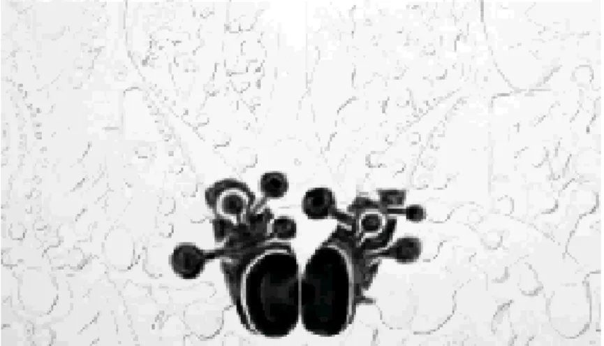
Figure 6. Representation of dendrites and spines during impregnation with Golgi technique.
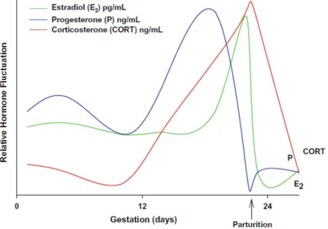
12 Pregnancy and the postpartum period are accompanied by dramatic fluctuations in the levels of steroid (estrogen, progesterone and corticosteroids) and peptide (oxytocin and prolactin) hormones (Numan, 1988). During pregnancy, the ovaries, placenta, and fetus contribute to these fluctuations (Kinsley & Lambert, 2006), which are continued following parturition and throughout lactation (Russell et al., 2001). In rodents, estradiol levels increase from day 11 until the end of pregnancy (Rosenblatt et al., 1979; Nelson, 2000), while progesterone remains elevated throughout pregnancy (Rosenblatt
et al., 1979, 1988). Prior to parturition, progesterone levels fall drastically followed by a
decreased in estradiol levels during the postpartum period (Rosenblatt et al., 1979; Garland et al., 1987). Basal corticosterone levels increase during late pregnancy and remain elevated during the postpartum period, during the first two weeks of lactation (Atkinson & Waddell, 1995; Fisher et al., 1995) (See Figure 5). Prolactin levels increase at the onset of pregnancy, followed by a decrease until parturition and a new increase in response to the suckling stimulation during lactation (Rosenblatt et al., 1979). Similarly, increased levels of oxytocin are present primarily during parturition and lactation (Russel et al., 2001). These fluctuations in circulating hormones during late pregnancy, parturition, and the early postpartum (Numan et al., 2006), as well as the response of receptors in several brain areas to these hormones (Numan, 1988; Numan et al., 2006), are important for the induction of the maternal circuit and onset of maternal behaviors (Numan, 1988; Rosenblatt et al., 1988).
Figure 7. A profile of relative levels of estradiol (pg/mL), progesterone (ng/mL) and
corticosterone (ng/mL) across pregnancy and parturition in the female rat (From Pawluski et al., 2009a).
13 Given that, steroid hormones markedly affect structural properties of the hippocampus (Gould et al., 1990; Galea et al., 1997), it is not surprising that the great hormonal fluctuations that occur during in pregnancy and postpartum period may have an impact in hippocampus plasticity. Recent work has shown that neurogenesis in the DG of the hippocampus is affected by motherhood and reproductive experience, with regards to a decrease in cell proliferation and survival during the early postpartum period. (Pawluski & Galea, 2007; Darnaudéry et al., 2007; Leuner et al., 2007; Pawluski et al., 2009b, 2010). In addition, motherhood significantly impacts dendritic morphology in the hippocampus: primiparous rats (first time pregnancy) showed significant dendritic atrophy in CA3 and CA1 pyramidal neurons compared to multiparous (having been pregnant and mothered at least twice) and nulliparous rats (Pawluski & Galea, 2006). This dendritic remodeling seen in primiparous rats is similar to the one seen following chronic stress, leading to a significant role of corticosterone, as high levels of this hormone are present in both pregnancy and prolonged stress (Woolley et al., 1990a; Magariños & McEwen, 1995; Galea et al., 1997).
Stress Effects in the Mother
Stress and elevated levels of GC have also been shown to impact the mother. However, normal changes in GC are very important for many aspects of motherhood. For example, in human mothers, cortisol is important for a mother’s attraction to her infant, particularly in a first pregnancy (Fleming et al., 1997). Studies from rodents have also shown an important role for the elevation in GC during pregnancy and postpartum in maternal pup-directed behaviors (Graham et al., 2006; Pawluski et al., 2009b). In addition, increased GC levels late in pregnancy are important for mobilization of maternal energy stores to be able to stand fetal demands (Knopp et al., 1973; Metcalfe
et al., 1988) and for milk production (Tucker, 1988; Casey & Plaut, 2007). The
elevation in GC during late pregnancy is also very important for many aspects of fetal growth and development, such as development and maturation of fetal organs before birth (Smith & Shearman, 1974).
Exposure to stress can significantly impact GC and maternal and fetal health. Unfortunately, a growing number of women experience severe and chronic stressors during pregnancy (Bennett et al., 2004a, b). Nowadays, life events, such as problems at work, domestic issues, financial instability, young age, and unplanned pregnancy
14 (Pajulo et al., 2001; Ryan et al., 2005), together with problems with the pregnancy and the responsibilities and challenges that come with a care of a newborn, can be overwhelming for the mother. This can lead to an increased incidence of psychological stress, such as depression and anxiety, during pregnancy and the postpartum period (Bennett et al., 2004a, b). Stress can have detrimental effects on maternal mood and maternal care of offspring (Smith et al., 2004). Moreover, maternal stress during gestation can also have a negative impact on the offspring (Maccari & Morley-Fletcher, 2007; Darnaudéry & Maccari, 2008). For example, gestational stress during critical periods of fetal brain development can result in increased anxiety-like and depressive-like behavior, increased HPA axis reactivity, and memory deficits in adulthood (Welberg & Seckl, 2001; Kofman, 2002; Weinstock, 2008). Taken together, it is of great importance to fully determine and understand how stress affects the maternal brain, and thus improve the health and well being of the mother and child.
Chronic stress models using immobilization, administration of high levels of corticosterone, or chronic ultramild stress (CUMS), have recently been used to investigate the effects of gonadal hormones and stress on the affective-like behavior and physiology of the mother during pregnancy and postpartum (Darnaudéry et al., 2004; Smith et al., 2004; Brummelte et al., 2006). For example, repeated restraint stress of pregnant rodents during gestation can induce a postpartum depressive-like state in female rats (Smith et al., 2004) and dams stressed during gestation show an increase in basal corticosterone concentrations and a decrease in corticosteroid binding globulin during the late pregnancy (Takahashi et al., 1998; Maccari et al., 2003). Gestational and postpartum stress also affects maternal care of offspring (Pardon et al., 2000; Smith et
al., 2004; Brummelte et al., 2006; Brummelte & Galea, in press) and persistently affects
the affective-like behavior of the mother long after the stress has stopped (Darnaudéry
et al., 2004; O’Mahony et al., 2006). For example, dams stressed during pregnancy are
more anxious (Darnaudéry et al., 2004) and can exhibit increased depressive-like behavior (O’Mahony et al., 2006; Brummelte & Galea, in press) one month after the last restraint stress session has occurred (Maccari et al., 2003; Darnaudéry et al., 2004). Unfortunately, very little research has investigated the effect of gestational stress on hippocampal plasticity in the mother. A recent study has shown that administration of elevated levels of corticosterone during late pregnancy and postpartum results in decreased neurogenesis in the hippocampus of the mother (Brummelte & Galea, in
15
press). Clearly, further work is needed to understand how stress during gestation affects
other measures of neural plasticity in the maternal brain.
Thesis Objectives
The present thesis aims to determine the affects of stress on dendritic morphology of CA3 pyramidal neurons in the hippocampus of pregnant female rats, and whether these effects during pregnant females differ from those in virgin female rats. In order to do this, a repeated restraint stress paradigm will be applied and, through Golgi impregnation, dendritic morphology of the CA3 region of the hippocampus will be assessed to evaluate the effects of stress.
This study will increase our understanding of how stress affects the maternal brain, and thus contributes to improve the health and well being of the mother and child.
16
Chapter II Material and Methods
Animals and Housing
Twenty-one adult female Wistar rats (four months old) obtained from Faculdade de Ciências Médicas (FCM) da Universidade Nova de Lisboa, were used in this study. The breeding colony at FCM originated from Charles River Laboratories in Barcelona. Rats in the present study were individually housed in clear polyurethane cages with absorbent bedding throughout the study (from impregnation to decapitation and at matched time points in virgin females). The animals were kept isolated in order to strictly control for enriched social environmental influences on brain morphology. All rats were given pellet food (maintenance chow) and tap water ad libitum. All rats were maintained in a 12h:12h light/dark with lights on at 5 a.m. in a standard laboratory environment (18-24°C, 55% humidity, ventilation: 8-10 changes/hour). Cages and water bottles were changed weekly. All protocols were in accordance with the European Union’s Directive 86/609/EEC and Council Directive 93/119/EC, Portuguese law Law-Decrees DL129/92 (July 6th), DL197/96 (October 16th) and Ordinance Port.131/97 (November 7th) and approved by FCM’s ethical committee board.
Breeding
For breeding, one female and one male were housed together in a wire mesh cage until a vaginal plug was released. Upon release of a vaginal plug, indicating copulation had occurred, females were individually housed in clear polyurethane cages for the duration of the experiment. Virgin females were singly housed throughout the experiment.
Group Formation
This study required twenty-one adult females, divided over two groups (Virgin versus Pregnant females), and two conditions/group (Control versus Stress) (Table 1). For all groups a n=5-6 per group was used as this is a minimum required for histological measures utilizing Golgi impregnation, based on previous investigations (Galea et al., 1997; Pawluski & Galea, 2006).
17
Table 1. Group information. Virgin and Pregnant female were divided into control or stressed
conditions.
Restraint Stress Procedure
For this experiment, pregnant and virgin females assigned to the stress condition were subjected to restraint stress by being placed in transparent plastic cylinders (diameter 6 cm: See Figure 8). Restraint took place between gestation days 8-21 and at matched points in virgin females. This was done to determine how reproductive status may account for changes in neuroplasticity in response to stress. Briefly females were subjected to daily, 1 hour restraint stress that occurred once between 11 am and 2 pm. Control groups were left undisturbed except for regular weight measurements.
Figure 8. Timeline of the experiment for pregnant and virgin females assigned in stress
condition.
Group Condition !umber of animals
Virgin Control 6
Stress 5
Pregnant Control 5
18
Sacrificing and Dissection
On gestation day 21, and at matched time points in virgins, females were deeply anesthetized with pentobarbital (100 mg/kg, intra-peritoneal) and decapitated.
Brain Removal
For the removal of the brain, the dorsal portion of the skull was skinned and the skin-flaps were peeled to the right and left side, from the backof the head forward to betweenthe eyes. After localization of the foramen magnum, a large opening at the back ofthe head where the spinal column enters the skull, the rongeurs were placed into it and used to cracked andpullaway the bone and tissue on either side of the opening. Then, the tips of the rongeurs were placed in the eye sockets, and were used tocrackthe piece of skull that lies between them. Using the rongeurs, the skull was carefully removed, starting atthe top of the foramen magnum and chipping away the bone,up overthe cerebellum and then forward toward the eyes. The skull bone was removed by breaking up the bone piece by piece always holding the rongeurs horizontally until the dorsal brain was exposed on three sides. Holding the head upsidedown, the brain was carefully pried away from the base of the skull witha flat metalspatula. Theoptic and trigeminal nerves attaching the brain to the skull were severed with the edge of the spatula and the brain was removed and longitudinally sectioned with a sharp scalpel (Schneider, 2007).
Uterine Horns Dissection in Pregnant Females
In order to quantify the possible effect of stress on litter size and number of male and female fetus, the uterine horns were dissected after decapitation. To do this, a vertical 2 cm abdominal skin incision was made with a scalpel. The skin was pulled apart toward the head and tail to expose the abdomen. The peritoneum was grasped with forceps and cut to expose the abdominal cavity. The reproductive organs in the dorsal region of the body cavity were located: two uterine horns, the oviduct and the ovaries (Figure 9). The uterine horns were removed by grasping the uterus below the oviduct and cutting it free along the mesoterium. A vertical incision was made in the uterus at the union of the two horns and the pup–placental units were delivered. Each embryo was separated by cutting between implantation sites along uterine horn. The muscular uterine lining was grasped by sliding watchmaker's forceps between the surrounding
19 muscle layer and enveloped decidua tissue. The muscle layer was pulled back, exposing the decidua. A portion of the exposed decidua at the apex was clipped off (approximately 1/5 of the decidua tissue) exposing the midventral or distal tip of the enclosed embryo. The embryos were shelled out using the tips of forceps. The decidua was pierced with forceps surrounding the embryo and open forceps to tear decidua apart. The number of fetuses in a litter was measured, taking in account as well the number of male and female fetuses (Shea & Geijsen, 2007).
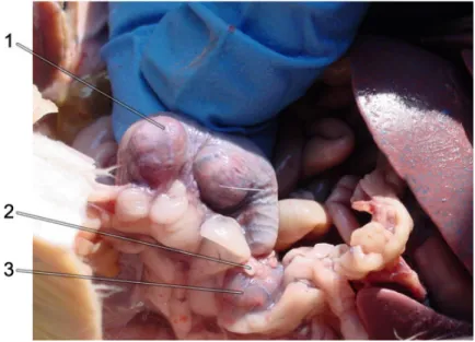
Figure 9. Close-up of the left side of a pregant female rat, preserved and dissected.
1. Embryo in left uterine horn; 2. Oviducts (fallopian tubes, uterine tubes); 3. Ovary (greatly enlarged from normal, non-pregnant, state).
(From http://faculty.orangecoastcollege.edu/mperkins/zoo-review/rat-repro/rat-repro3.html)
Histological Procedures
Golgi Impregnation Technique
After brain removal, the left hemispheres of the each brain were processed for Golgi impregnation using the FD Rapid GolgiStain Kit™ (FD Neurotechnologies Consulting & Services, Elliot City, MD, U.S.A.) adapted for Vibratome (as previously described in Dalla et al., 2009; Gibb & Kolb, 1998). The right hemisphere was used in a separate analysis not discussed here. For the Golgi impregnation, 1 cm blocks of brain tissue including the hippocampus were rinsed with distilled water and immersed in an impregnation solution containing potassium dichromate, mercuric chloride and
20 potassium chromate (provided in the kit). Brains were left undisturbed in the dark for 2,5 weeks. After the 2,5 weeks, brains were immersed in 30% of sucrose at 4°C to protect them from drying. Two to four days later coronal sections (200µm) of the entire hippocampus were cut using a vibratome (Leica VT6000, Leica Microsystems, Germany) in a bath of 15% sucrose and the slices stored in the dark at 4°C in 15% sucrose solution until mounting. Sections were mounted on gelatin coated Superfrost slides (Thermo Fisher Scientific Inc., Waltham, MA, U.S.A.) and firmly pressed using moist filter paper to prevent the slices from falling off the slide during development (Gibb & Kolb, 1998). Slides were placed in a humidity chamber in the dark and were stored overnight at 4°C. For development, slides were rinsed with distilled water twice for 2 minutes and were then placed in developing solutions (provided in the FD GolgiStain Kit). The slides stayed for 10 minutes in the developing solution, then were rinsed in distilled water twice for 2 minutes, taken through a graded alcohol series (50%-96%, 4 minutes each rinse), cleared with xylene for 8 minutes, and coverslipped with Permount (Fisher) (Figure 10).
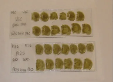
Figure 10. Final slides of the Golgi impregnation technique.
Dendritic Morphology
Dendritic morphology in the CA3 region of the hippocampus was analyzed blind to experimental conditions as previously described (Galea et al., 1997; Pawluski & Galea, 2006). For analysis of dendritic morphology, a pyramidal cell was chosen using the following criteria: 1. the cell body and its dendrites were fully impregnated; 2. the cell was relatively isolated from surrounding impregnated cells to obtain a clear image of
21 the entire cell; 3. the cell was located in the CA3 region of the dorsal hippocampus (Figure 11).
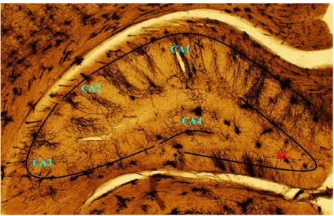
Figure 11. Photomicrograph of Golgi impregnated dorsal hippocampus showing the CA regions
and the dentate gyrus (DG). The main focus of this thesis is the CA3 region. The photomicrograph is taken under 40x magnification. CA = cornu ammonis.
Six CA3 pyramidal cells from each brain were analyzed. For each cell the following variables were measured separately in the apical and basal regions of each cell: the number of branch points - total number of branch points in the dendritic arbor; and the total dendritic length - total length of dendrites connecting to a given cell body. Sholl analysis (Sholl, 1953) was also used to estimate the distribution and complexity of the dendrites by counting numbers of intersections of dendrites with an overlay of concentric rings centered at the cell body (Figure 6). This consecutive-circles (cumulative intersections) analysis is a method for quantifying a specific scaling property of the dendritic tree and specifies dendritic geometry, ramification richness, and dendritic branching patterns. The Sholl analysis consists of: (i) construction of concentric and equidistantly organized spherical shells (in 3 dimensions (3D) case), which are centered in the cell body, (ii) counting the numbers of intersections of dendrites with the circles of increasing radii (10 µm).
To quantify dendritic length, branch points and Sholl analysis, the Neurolucida program (MicroBrightField, Inc., Williston, VT, U.S.A.) was used. When a cell of interest was identified, a 3D morphological image of the cell was manually obtained using the Neurolucida neuronal tracing system (made under 400x) attached to a DSU
22 microscope (Olympus BX51WI, Olympus America Inc., Center Valley, PA, U.S.A.) (Figure 12). For example, Figure 12 depicts a Golgi impregnantion CA3 pyramidal neuron (A) and the corresponding neuronal tracing (B).
Figure 12. A. CA3 pyramidal neuron (made under 400x); B. CA3 pyramidal neuron drawing
obtained using the Neurolucida neuronal tracing system (made under 400x).
Figure 13. Scholl analysis showing the overlay of concentric rings centered at the cell body.
23
Statistical Analysis
The number of CA3 branch points and dendritic length were each analyzed using repeated-measures analysis of variance (ANOVA) with two factors (pregnant vs. virgin, stress vs. control) as the between-subjects factors and region (apical vs. basal) as the within-subjects factor. For the Sholl analysis, the number of dendritic intersections was analyzed using repeated-measures analysis of variance (ANOVA) with two factors (pregnant vs. virgin, stress vs. control) as the between-subjects factors. Post hoc comparisons utilized the Fisher’s LSD procedure. Independent t-tests were conducted on litter size, number of male and female pups in pregnant females. Pearson product moment correlations were performed between apical and basal CA3 morphology and litter size, number of male pups, number of female fetuses. All statistical procedures were set at α = 0.05. All statistical analysis was performed using the software Statistica 9 (StatSoft, Inc., Tulsa, OK, U.S.A.).
24
Chapter III Results
Pregnant Females Gained Significantly More Weight than Virgin Females
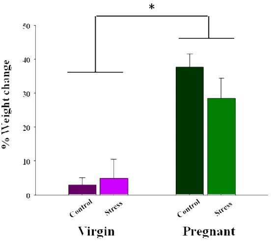
A factorial ANOVA on the weight change between groups revealed a significant main effect of reproductive state (F1,17 = 42.25 , P ≤ 0.0001; Figure 14), with pregnant females gaining significantly more weight than virgin females, as expected. There was no significant main effect of stress or a significant interaction between the effect of stress and reproductive state on weight (0.1 ≤ P ≤ 0.5).
Figure 14. Mean (± SEM) percentage of weight change across the duration of pregnancy and at
matched time points in virgin females. Pregnant females gained significantly more weight than virgin females (P ≤ 0.0001), regardless of stress. *denotes pregnant females significantly different from virgin females (n=5-6/group).
25
Regardless of Reproductive State, Stressed Females Showed Dendritic Atrophy in the Apical Tree of CA3 Pyramidal 1eurons
There were region differences in the effects of repeated restraint stress on dendritic length, with stressed females showing a decrease in the number and length of apical dendrites. Figure 15 represents neurolucida drawings of a representative cell for each of the conditions of female rats. The mean dendritic length of pyramidal cells in the CA3 region of the hippocampus of stressed and control virgin and pregnant female rats is shown in Figure 16. For dendritic length, there was a significant interaction between the effect of stress and region (apical vs. basal) (F1,17 = 6.03, P ≤ 0.025), with stressed pregnant and virgin females having shorter apical dendritic lengths than control pregnant and virgin females. Post hoc tests revealed that pregnant and virgin females had shorter apical dendritic lengths compared to control pregnant and virgin females (P ≤ 0.05) and there was no difference between groups in basal dendritic lengths (P ≤ 0.37). There was also a significant main effect of region (F1,17 = 6.23, P ≤ 0.023), resulting in significantly longer dendrites in the apical region compared to the basal region, but no significant main effect of stress (P ≤ 0.5). There was also no significant main effect of reproductive state (P ≤ 0.096) and no significant interactions between reproductive state and stress (P ≤ 0.14), reproductive state and region (P ≤ 0.46), or reproductive state, region and stress (P ≤ 0.85).
Figure 17 shows the mean number of branch points of pyramidal cells in the CA3 region of the hippocampus of pregnant and virgin female rats. There was a significant interaction between the effect of stress and region (F1,17 = 7.29, P ≤ 0.008), with stressed pregnant and virgin females having fewer apical branch points than control pregnant and virgin females. Post hoc tests revealed that there were fewer apical branch points in stressed females, compared to control females (P ≤ 0.04), regardless of reproductive state. There was also a significant main effect of region on the number of branch points (F1,17 = 10.65, P ≤ 0.005), with a greater number of branch points in the apical region than in the basal region, but no significant main effect of stress (P ≤ 0.32). There was no significant difference between stressed and control females in the total number of basal branch points (P ≤ 0.81). There was a tendency towards a significant interaction between the effect of reproductive state and region (F1,17 = 3.81, P ≤ 0.068). There was also no significant main effect of reproductive state (P ≤ 0.28) and no