Effect of simplified chemical moisture control protocols on the bond durability
of fiber posts cemented to root dentin
Efeito de protocolos simplificados de controle químico de umidade na
durabilidade da união de pinos de fibra cimentados à dentina radicular
DOI:10.34117/bjdv6n8-585
Recebimento dos originais:08/07/2020 Aceitação para publicação:26/08/2020
Ricardo Lopes Rocha
Adjunct Professor, Department of Dentistry; School of Biological and Health Sciences, Federal University of the Jequitinhonha and Mucuri Valleys, Diamantina, MG, Brazil
Adress: Rua da Glória, nº 187, Centro, Diamantina, MG, CEP 39100-000 E-mail: [email protected]
Maria Olívia Alves Dourado
DDS, Department of Dentistry; School of Biological and Health Sciences, Federal University of the Jequitinhonha and Mucuri Valleys, Diamantina, MG, Brazil
Adress: Rua da Glória, nº 187, Centro, Diamantina, MG, CEP 39100-000 E-mail: [email protected]
Andreza Costa Dayrell
Department of Dentistry; School of Biological and Health Sciences, Federal University of the Jequitinhonha and Mucuri Valleys, Diamantina, MG, Brazil
Adress: Rua da Glória, nº 187, Centro, Diamantina, MG, CEP 39100-000 E-mail: [email protected]
Marcos Luciano Pimenta Pinheiro
Adjunct Professor, Department of Basic Sciences, School of Biological and Health Sciences, Federal University of the Jequitinhonha and Mucuri Valleys, Diamantina, MG, Brazil
Adress: Rua da Glória, nº 187, Centro, Diamantina, MG, CEP 39100-000 E-mail: [email protected]
João Victor Frazão Câmara
Department of Biological Sciences, Bauru Dental School, University of São Paulo, Bauru, São Paulo, Brazil
Adress: Alameda Dr. Octávio Pinheiro Brisolla, 9-75 - Jardim Brasil, Bauru - SP, 17012-901 E-mail: [email protected]
Josué Junior Araujo Pierote
PhD, Department of Restorative Dentistry, Piracicaba Dental School, University of Campinas, Piracicaba, Sao Paulo, Brazil
Adress: Av. Limeira, 901 - Areião, Piracicaba - SP, 13414-903 E-mail: [email protected]
Cintia Tereza Pimenta de Araújo
Adjunct Professor, Department of Dentistry; School of Biological and Health Sciences, Federal University of the Jequitinhonha and Mucuri Valleys, Diamantina, MG, Brazil
Adress: Rua da Glória, nº 187, Centro, Diamantina, MG, CEP 39100-000 E-mail: [email protected]
ABSTRACT
Objective: Investigate the influence of simplified chemical moisture control protocols on the bond durability of fiber posts through an evaluation of push-out bond strength. Materials and methods: Forty-eight roots from single-rooted human teeth were treated endodontically and randomly distributed among groups with different dehydration protocols (n = 8): simplified ethanol wet bonding/100 (100G) – irrigation with 100% ethanol; simplified ethanol wet bonding/50 (50G) – irrigation with 50% ethanol; wet bonding (WG) – removal of excess water with paper. Fiber posts were cemented with the Ambar/Allcem (FGM) luting system, following the manufacturer's instructions. The bonded roots were cut cross-sectionally, producing slices 1 mm in thickness. The specimens were tested for push-out bond strength after 24 h (T1) and 12 months (T2) of storage.
Specimens from each group were also processed for the nanoleakage analysis. The statistical analysis was performed considering a 5% significance level. Results: Significant differences were
found among groups at T1 (p = 0.01) and T2 (p = 0.007). Bond stability in 100G was maintained
after one year of aging, demonstrating greater bond strength (9.51 ± 3.28) in comparison to the other groups, with no significant differences in the analysis of the roots by thirds. The nanoleakage results confirmed that the simplified 100% ethanol wet bonding method improved the quality of the interface. Conclusions: Simplified 100% ethanol wet bonding contributes to the bond stability of the fiberglass posts on root dentin.
Keywords: Tooth Root, Post and Core Technique, Humidity.
RESUMO
Objetivo: Investigar a influência de protocolos simplificados de controle químico de umidade na durabilidade da união de pinos de fibra através de uma avaliação da resistência da união push-out. Materiais e Métodos: Quarenta e oito raízes de dentes humanos unirradiculares foram tratadas endodonticamente e distribuídas aleatoriamente entre grupos com diferentes protocolos de desidratação (n = 8): ligação úmida simplificada com etanol / 100 (100G) - irrigação com etanol a 100%; ligação úmida simplificada com etanol / 50 (50G) - irrigação com etanol a 50%; colagem úmida (GT) - remoção do excesso de água com papel. Os pinos de fibra foram cimentados com o sistema de cimentação Ambar / Allcem (FGM), seguindo as instruções do fabricante. As raízes coladas foram cortadas transversalmente, produzindo fatias de 1 mm de espessura. As amostras foram testadas quanto à resistência de união ao empuxo após 24 h (T1) e 12 meses (T2) de armazenamento. Amostras de cada grupo também foram processadas para a análise de nanoinfiltração. A análise estatística foi realizada considerando um nível de significância de 5%. Resultados: Foram encontradas diferenças significativas entre os grupos em T1 (p = 0,01) e T2 (p = 0,007). A estabilidade da união em 100G foi mantida após um ano de envelhecimento, demonstrando maior resistência da união (9,51 ± 3,28) em comparação aos demais grupos, sem diferenças significativas na análise das raízes por terços. Os resultados da nanoinfiltração confirmaram que o método simplificado de ligação a 100% de etanol por via úmida melhorou a qualidade da interface. Conclusões: A união úmida simplificada a 100% de etanol contribui para a estabilidade da união dos pinos de fibra de vidro na dentina radicular.
1 INTRODUCTION
Adhesive restorations are challenging for clinicians, researchers and manufacturers of dental products. To achieve an optimal adaptation with a strong, stable bond,[1,2] it is necessary to overcome the inherent difficulties of the adhesion of resins to dentin, such as the organic content and the natural moisture of the substrate.[3] Due to its anatomical conformation, moisture control is even more difficult when working with root dentin, which affects the diffusion of the adhesive to critical regions,[4] resulting in excess moisture that compromises bond strength.[5]
The moisture in dentin tissue contributes to the degradation of the hybrid layer.6 Excess water generates the phenomenon of phase separation and compromises the diffusion of the adhesive.[7] However, the complete removal of moisture from the conditioned dentin significantly reduces bond strength,8 since water is essential to maintaining the integrity of the collagen network, which, in turn, enables the adequate penetration of resinous monomers.[9] Thus, the control of dentin moisture by chemical means to promote dehydration without drying the substrate (keeping it moist with ethanol) is a promising method that has demonstrated good results in terms of bond strength and durability.[10,11]
Therefore, the purpose of the present study was to determine the effects of different chemical moisture control techniques (ethanol wet bonding) in conditioned dentin on bond strength and durability evaluated using the push-out test. The null hypothesis is that such treatments will not affect bond strength between fiber post and root dentin after 24 hours and one year of storage.
2 MATERIALS AND METHODS
This study received approval from the Human Research Ethics Committee (certificate number: 19231813.8.0000.5108). The sample size was calculated using data from a study by Cechin et al.[12] and considering an 80% test power. A minimum of n=7 was determined, which was increased by 10% to compensate for possible losses (n = 8). Intact straight single-rooted human teeth with similar root diameters and lengths were selected for the study. Prior to use, the teeth were disinfected in 0.1% aqueous thymol solution for seven days.
a) Preparation of roots
The root canals were submitted to the conventional biomechanical preparation technique13 and filled with gutta percha by the same trained operator. Next, a post space measuring 12 mm in
depth was prepared using a low-speed Largo drill (#1 to #4 - Dentsply-Maillefer) until a suitable diameter was obtained for a #1 fiberglass post (Reforpost - Ângelus Dental Products), maintaining 3 mm sealed with gutta percha.
b) Adhesive procedures
The bonding procedures were performed using the Ambar APS/Allcem fixation system (FGM Dental Products Ltd.) following the manufacturer's instructions and light polymerization through the post for 40 s with a minimum irradiance of 600 mW/cm2 (Optilight LD Max Gnatus). The posts were cleaned with 37% phosphoric acid (3M ESPE) for one minute, washed and dried with compressed air.
A single layer of silane coupling agent (FGM Dental Products Ltd.) was then applied to the post surface. The roots were identified and coated with utility wax to mimic the conditions of the oral access tissues from the light to the root. The specimens were randomly divided into three experimental groups (n = 8) according to the dehydration protocol. The proposed protocols for the control of root dentin moisture are detailed in Table 1. After cementation, eight restored roots from each group were stored for 24 hours (T1) in distilled water at 37°C. Eight others remained stored for one year (T2) under the same conditions.
Table 1: Proposed moisture control protocols.
Groups Moisture control (after acid rinse)
Simplified alcoholic technique 1 (100G)
Irrigation with 100% ethanol for 30 sec; aspiration and removal of excess ethanol with paper cone. Simplified alcoholic technique 2
(50G)
Irrigation with 50% ethanol for 30 sec; aspiration and removal of excess ethanol with paper cone.
Wet technique (WG) Removal of excess water with two paper cones.
c) Push-out test
The roots were sectioned perpendicularly to their long axes into a series of 1-mm slices using a low-speed diamond saw (Isomet Buehler) under a water coolant after storage in distilled water at 37° for 24 hours (T1). Two slices from each root third (cervical, middle and apical) were used for the push-out test, one of which was tested immediately and the other was first stored in distilled
water for one year. Push-out bond strength tests were conducted using a universal testing machine (EZ Test- L, Shimadzu Corporation).
Loading was performed with a crosshead speed of 0.5 mm/min until bond failure occurred. Push-out strength (MPa) was calculated by dividing the load at bond failure (N) by area (mm2). The cervical side of each slice was identified with graphite.
d) Fracture pattern
The fracture pattern was evaluated under a stereomicroscope (magnification: 10 x). Fractures were classified as adhesive (at bonding interface between cement and dentin), cohesive (in the cement layer, pin or dentin) or mixed (both adhesive and cohesive).
e) Nanoleakage
For the analysis of nanoleakage, a slice from each group/time was prepared according to the protocol described by Tay et al.,[14] embedded in polystyrene resin and viewed by scanning electron microscopy (SEM) using backscattered electrons.
f) Statistical analysis
After collection and tabulation, the distribution of the data was assessed using the Shapiro- Wilk test. One-way ANOVA and the Kruskal-Wallis test were used to compare means of the push- out test and percentages of nanoleakage among the groups. Tukey's test and the Mann-Whitney test were used for post hoc multiple comparisons. The Student's t-test was used to compare push-out means between the two storage periods. The level of statistical significance was set at 5% (α ≤ 0.05).
3 RESULTS
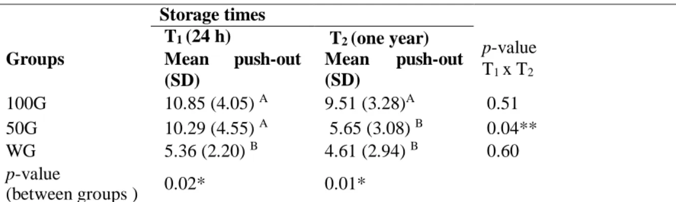
In the push-out analysis after 24 hours of storage (T1), significant differences were found among the groups (p = 0.01), specifically between the 100G and WG (p = 0.02) and between the 50G and WG (p = 0.04). After aging the samples for one year (T2), significant differences were found between the 100G and the other two groups (p = 0.007). In the intra-group analyses (means at T1 and T2), a statistically significant reduction was found in the 50G (p = 0.04) (Table 2).
Braz. J. of Develop., Curitiba, v. 6, n. 8, p.62121-62132 aug. 2020. ISSN 2525-8761 Table 2: Mean push-out values in different groups at different storage times (T1 and T2)
Storage times Groups T1 (24 h) Mean push-out (SD) T2 (one year) Mean push-out (SD) p-value T1 x T2 100G 10.85 (4.05) A 9.51 (3.28)A 0.51 50G 10.29 (4.55) A 5.65 (3.08) B 0.04** WG 5.36 (2.20) B 4.61 (2.94) B 0.60 p-value (between groups ) 0.02* 0.01*
Legend: 100G = simplified alcoholic technique 1; 50G = simplified alcoholic technique 2; WG = wet technique * significant difference (α ≤ 0.05, ANOVA)
** significant differences (α ≤ 0.05, T-test)
A,B: Different letters in column indicate significant differences (α ≤ 0.05, Tukey's post hoc test).
In the intra-group analysis by thirds, cervical slices had significantly higher means than the middle and apical slices in the 50G and WG at T1. In contrast, no significant differences were found between the cervical and middle slices or between the middle and apical slices in the 100G (Table 3).
Table 3: Mean push-out values in root thirds in different groups at T1
Groups Root thirds
Number of slices
(slices lost)
Push-out values MPa (SD)
Post hoc test (p-value) Cervical (C) 15 (1) 12.58 (5.42)A CxM (0.17) Middle (M) 15 (1) 8.8 (5.63)AB MxA (0.85) 100G Apical (A) 16 (0) 7.69 (5.97)B AxC (0.05)
Comparison between groups
(p-value) ANOVA (0.05)
Cervical (C) 16 (0) 13.97 (7.37)A CxM (0.002) Middle (M) 16 (0) 6.36 (4.63)B MxA (0.76)
50G Apical (A) 16 (0) 7.8 (5.2)B AxC (0.012)
Comparison between groups
(p-value) ANOVA (0.001)
Cervical (C) 16 (0) 8.93 (3.85)D CxM (0.03) Middle (M) 16 (0) 5.45 (5.01)E MxA (0.22) WG Apical (A) 14 (2) 2,46 (2.16)E AxC (<0.0001)
Comparison between groups
(p-value) K. Wallis (0.001)
Legend: 100G = simplified alcoholic technique 1; 50G = simplified alcoholic technique 2; WG = wet technique; MPa: Megapascal;
A,B Different letters in columns indicate statistically significant differences (α ≤ 0.05, Tukey's test); D,E: Different letters in columns indicate statistically significant differences (α ≤ 0.05, Mann-Whitney test)
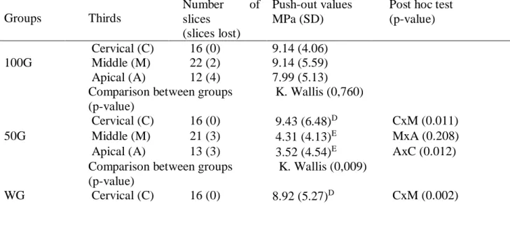
At T2, the cervical slices had significantly higher means than the middle and apical slices in the 50G and WG, whereas no significant differences among thirds were found in the 100G (Table 4).
The distribution of the fracture patterns of the specimens after the push-out test are illustrated in Figure 1. A predominance of adhesive fractures was found in the 50G and WG, whereas a higher frequency of mixed fractures was found in the 100G at both times (Figure 2).
Figure 1: Fracture pattern at T1 and T2.
Table 4: Mean push-out values in root thirds in different groups at T2.
Groups Thirds Number of slices (slices lost) Push-out values MPa (SD)
Post hoc test (p-value)
Cervical (C) 16 (0) 9.14 (4.06) 100G Middle (M) 22 (2) 9.14 (5.59) Apical (A) 12 (4) 7.99 (5.13) Comparison between groups
(p-value)
K. Wallis (0,760)
Cervical (C) 16 (0) 9.43 (6.48)D CxM (0.011)
50G Middle (M) 21 (3) 4.31 (4.13)E MxA (0.208)
Apical (A) 13 (3) 3.52 (4.54)E AxC (0.012) Comparison between groups
(p-value)
K. Wallis (0,009)
Middle (M) 20 (4) 3.39 (3.66)E MxA (0.17) Apical (A) 11 (5) 1.33 (1.65)E AxC (<0.0001) Comparison between groups
(p-value)
K. Wallis
(<0.0001)
Legend: 100G = simplified alcoholic technique 1; 50G = simplified alcoholic technique 2; WG = wet technique; MPa: Megapascal;
D,E,F: Different letters in columns indicate statistically significant differences (α ≤ 0.05, Mann-Whitney test)
Figure 2: Representative stereomicroscopic images of fracture types (magnification: 10 X) (images from 100G). A) Adhesive fracture, B) Cohesive fracture and C) Mixed fracture.
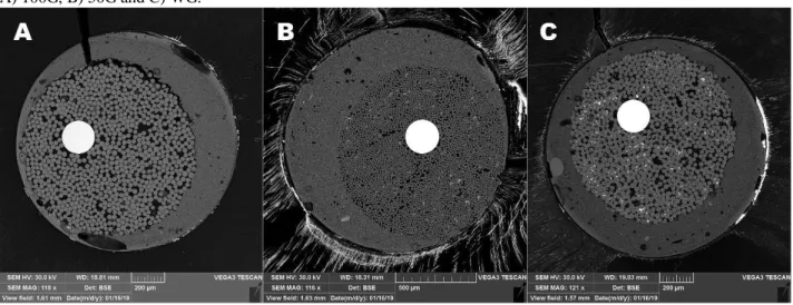
Figure 3: Representative scanning electron microscopy (SEM) images of nanoleakage in slices from each study group. A) 100G, B) 50G and C) WG.
Analysis of the nanoleakage
In the ultramorphological analysis of the bonding interface, the samples from all groups exhibited nanoleakage along the hybrid layer. The WG had the worst nanoleakage pattern, with all specimens exhibiting a considerably greater leakage of silver nitrate at the interface in comparison to the 100G. The 50G exhibited similar leakage to the WG (Figure 3).
4 DISCUSSION
The null hypothesis was rejected at both T1 and T2. In the push-out analysis at T1, the means in the 100G and 50G groups were significantly higher than those in the WG, which is in agreement
with the results of previous studies using ethanol for moisture control.[5,10,15] Thus, maintaining the conditioned dentin wet with ethanol leads to an improvement in bond strength even when used at a concentration of 50%.
In the push-out analysis at T2, the results were better in the 100G compared to the other groups. Indeed, when dentin is maintained wet with 100% ethanol, the adhesion of hydrophobic resins seems to be less susceptible to degradation.[10,15,16] The simplified method using 100% ethanol enables better infiltration of the adhesive into the substrate, creating more homogeneous hydrophobic adhesive interfaces,[16] which may have contributed to the better bond strength and stability as well as less nanoleakage in the 100G (Fig. 3A).
In the comparison of means of the push-out tests between the T1 and T2, stability was not maintained in the 50G group, as demonstrated by the decrease in bond strength (Table 2). When the diffusion of the adhesive is not adequate, the collagen of the fibrils not enveloped by the resin can degrade during over time and the space is replaced with water,[17] resulting in a decrease in bond strength. In the 50G, the water present in the ethanol may have compromised diffusion, leading to the phenomenon described, which is also confirmed by the considerable infiltration of silver nitrate, as shown in Fig. 3B. In contrast, stability was maintained in the 100G, which had higher values compared to the other groups. Moreover, the stability found in the WG does not signify any advantage, as the values were low at both evaluation times, which can also be confirmed by the considerable infiltration of silver nitrate, as shown in Fig. 3C.
In the analysis of push-out means by thirds at time T1, significantly higher values were found for cervical slices in all groups (Table 3). This result is in agreement with data described in previous studies.[18,19] One of the explanations for this finding is the proximity of the cervical third to the light source; thus, a more efficient polymerization is expected in this region.[20] In deeper regions, however, polymerization depends on the light transmission rate. The type of post used in this study (Reforpost) has a low light conduction rate compared to other posts available on the market.[21] This low transluminescence impairs the diffusion of light and leads to less polymerization in the middle and apical thirds of the root,[22,23] which may also have contributed to the results observed in this study.
Significant differences were found in the analysis of push-out values by thirds at time T2, with higher values for the cervical third in the 50G and WG, whereas the differences were non- significant in the 100G (Table 4). The use of 100% ethanol may have provided similar dehydration
In the analysis of the fracture pattern, the 100G had a greater frequency of mixed and cohesive fractures than the other groups at both T1 and T2 (Fig. 1). In contrast, Yesilyurt et al.[24] compared the wet technique to the simplified ethanol technique and found a predominance of adhesive fractures in the aged samples. These authors attributed this fracture pattern to the storage of the slices. Although the push-out test is considered the most adequate for measuring bond strength in cases of adhesion to the dental root,[25] the extruder tip was calibrated to allow the extrusion from the apical portion. Consequently, it is possible that a greater concentration of forces occurred in the center of the slices in the cervical region, where the diameter of the slices is larger. However, we attribute the higher frequency of cohesive fractures in the slices of the cervical third in the 100G to the greater push-out resistance in the cervical slices of this group, resulting in a higher incidence of cohesive and mixed fractures.
The advantage of using ethanol to treat dentin prior to the application of adhesive resides in the chemical affinity with the adhesive and its solubility.[26] Such properties respectively enable a more homogeneous, deep diffusion of the adhesive and the prevention of the formation of hydrogen bonds between collagen peptides in demineralized dentin. Moreover, ethanol is a polar solvent that can be used either alone or with water as a co-solvent.[26] The 50G was proposed to reduce the effect of collagen network stiffness in the presence of ethanol[27] and reduce the technical sensitivity, as it is a less volatile blend than 100% ethanol.[28] However, in the push-out analysis of the deeper thirds in the 50G and 100G, the water in the 50% ethanol mixture exerted a negative influence on the diffusion of the adhesive, demonstrating that the mixed solvent offers no advantage. A dual curing resin cement (Allcem - FGM) was used in the present investigation, which demonstrated a good performance in a recent study.[29] Although this material is not considered the "gold standard", the present results show that, despite limitations in terms of the penetration of light to the deeper regions of the canal, the better results achieved with the simplified technique involving 100% ethanol demonstrates the clinical applicability of this method.
5 CONCLUSIONS
In view of the results obtained, wetting conditioned dentin with ethanol contributes to an increase in bond strength and the preservation of bond stability when cementing fiberglass posts to root dentin. Future studies should combine the simplified ethanol technique with other adhesive systems to confirm these results and possibly reduce nanoinfiltration.
ACKNOWLEDGMENTS
The authors thank the support of LMMA sponsored by FAPEMIG (CEX-112-10), SECTES / MG and RQ-MG (FAPEMIG: CEX-RED-00010-14).
Declaration of interest statement: The authors have declared no conflict of interest. Funding details: None.
Word count: 2096.
REFERENCES
1. Armstrong S, et al. Adhesion to tooth structure: a critical review of “micro” bond strength test methods. Dent Mater 2010;26:e50-e62.
2. Siqueira F, et al. Three-Year Effects of Deproteinization on the In Vitro Durability of Resin/Dentin-Eroded Interfaces. Oper Dent 2018;43:60-70.
3. Pashley DH. The effects of acid etching on the pulpodentin complex. Oper Dent 1992;17:229-42. 4. Bouillaguet S, et al. Microtensile bond strength between adhesive cements and root canal dentin. Dent Mater 2003;19:199-205.
5. Duan SS, et al C. Effects of ethanol-wet bonding technique on root dentine adhesion. Chin J Dent Res 2011;14:105-11.
6. Tjäderhane L. Dentin bonding: can we make it last? Oper Dent 2015;40:4-18.
7. Spencer P, Wang Y. Adhesive phase separation at the dentin interface under wet bonding conditions. J Biomed Mater Res 2002;62:447-56.
8. Cardoso Pde C, et al. Effect of solvent type on microtensile bond strength of a total-etch one- bottle adhesive system to moist or dry dentin. Oper Dent 2005;30:376-81.
9. Carvalho RM, et al. In vitro study on the dimensional changes of human dentine after demineralization. Arch Oral Biol 1996;41:369-77.
10. Nagpal R, et al. Effect of ethanol wet bonding technique on the durability of resin- dentin bond with contemporary adhesive systems. J Clin Pediatr Dent 2015;39:133-42.
11. Sadek FT, et al. One-year stability of resin-dentin bonds created with a hydrophobic ethanol- wet bonding technique. Dent Mater 2010;26:380-6.
12. Cecchin D, et al. Effect of chlorhexidine and ethanol on the durability of the adhesion of the fiber post relined with resin composite to the root canal. J Endod 2011;37:678-83.
13. Siqueira JF, et al. Mechanical reduction of the bacterial population in the root canal by three instrumentation techniques. J Endod 1999;25:332-5.
14. Tay FR, et al. Two modes of nanoleakage expression in single-step adhesives. J Dent Res 2002;81:472-6.
15. Hosaka K, et al. Durability of resin-dentin bonds to water- vs. ethanol-saturated dentin. J Dent Res 2009;88:146-51.
16. Talungchit S, et al. Ethanol-wet bonding and chlorhexidine improve resin-dentin bond durability: quantitative analysis using raman spectroscopy. J Adhes Dent 2014;16:441-50.
17. Tjaderhane L, et al. Strategies to prevent hydrolytic degradation of the hybrid layer-A review. Dent Mater 2013;29:999-1011.
18. Rezende EC, et al. Effects of Dentin Moisture on Cementation of Fiber Posts to Root Canals. J Adhes Dent 2016;18:29-34.
20. Perdigão J, et al. The effect of dowel space on the bond strengths of fiber posts. J Prosthodont 2007;16:154-64.
21. Goracci C, et al. Light-transmitting ability of marketed fiber posts. J Dent Res 2008;87:1122-6. 22. Alves Morgan L, et al. Influence of light transmission through fiber posts: Quantitative analysis, microhardness, and on bond strength of a resin cement. Indian J Dent Res 2018;29:74-80.
23. Stylianou A, et al. Light-transmitting fiber optic posts: An in vitro evaluation. J Prosthet Dent 2017;117:116-123.
24. Yesilyurt C, et al. Effect of simplified ethanol-wet bonding on dentin bonding durability of etchand-rinse adhesives. Dent Mater J 2015;34:441-8.
25. Soares CJ, et al. Finite element analysis and bond strength of a glass post to intraradicular dentin: comparison between microtensile and push-out tests. Dent Mater 2008;24:1405-11.
26. Barton AF. Solubility parameters. Chem Rev 1975;75(6):731-53.
27. Maciel KT, et al. The effects of acetone, ethanol, HEMA, and air on the stiffness of human decalcified dentin matrix. J Dent Res 1996;75:1851-8.
28. Garcia FC, et al. Effects of solvents on the early stage stiffening rate of demineralized dentin matrix. J Dent 2005;33:371-7.
29. Pereira JR, et al. The influence of different cements on the pull-out bond strength of fiber posts. J Prosthet Dent 2014;112:59-63.