Helena Paula Fernandes Pereira
Outubro de 2009
Escola de Ciências
Study of the role of monocarboxylate
transporters in colorectal carcinoma
Tese de Mestrado
Genética Molecular
Trabalho efectuado sob a orientação da
Professora Doutora Maria de Fátima Monginho Baltazar
Helena Paula Fernandes Pereira
Universidade do Minho
Escola de Ciências
Study of the role of monocarboxylate
transporters in colorectal carcinoma
Nome
Helena Paula Fernandes Pereira
Endereço electrónico: [email protected] Telefone: 00351966867670 Número do Bilhete de Identidade: 9877872
Título tese:
Study of the role of monocarboxylate transporters in colorectal carcinoma
Estudo do papel dos transportadores de monocarboxilatos no carcinoma colorectal
Orientador:
Maria de Fátima Monginho Baltazar
Ano de conclusão: 2009 Designação do Mestrado: Mestrado em Genética Molecular
É AUTORIZADA A REPRODUÇÃO INTEGRAL DESTA TESE/TRABALHO APENAS PARA EFEITOS DE INVESTIGAÇÃO, MEDIANTE DECLARAÇÃO ESCRITA DO INTERESSADO, QUE A
TAL SE COMPROMETE;
Universidade do Minho, 20/10/2009
Acknowledgements/Agradecimentos
À Professora Doutora Fátima Baltazar, orientadora desta tese, um agradecimento muito especial por todo o empenho, disponibilidade e acompanhamento durante todas as etapas da realização desta tese. Foi de facto um enorme prazer desenvolver este trabalho sob a sua orientação.
Aos Professores Doutores Rui Reis e Adhemar Longatto, quero agradecer a simpatia, a oportunidade que me concederam e as boas condições de trabalho que me proporcionaram.
A todos os colegas de laboratório (Sandra, Céline, Olga, Bruno, Inês, Marta, Ísis, Sara, Ashley, Vera), obrigada pelo apoio, amizade e pelos momentos que passamos juntos. Um agradecimento especial à Céline, pela paciência e ajuda prestada, sem ti teria sido mais difícil.
A todas as pessoas que tive a oportunidade de conhecer no ICVS/ECS, obrigada por tudo.
A todos os meus amigos, estrelas que iluminaram o meu caminho nas noites da minha vida.
Ao meu irmão, Paulo, obrigada pelos incentivos e por me dizeres aquilo que por vezes eu não queria ouvir. Admiro a tua coragem e força.
À minha mãe e ao meu pai, agradeço todo o amor e apoio que me deram. Este trabalho é dedicado a vocês. Amo-vos.
Por último, ao meu melhor Amigo, minha paz, minha alegria, meu bem-querer. Sem Ti nada seria possível. Obrigada.
SUMMARY
Study of the role of monocarboxylate transporters in colorectal
carcinoma
Colorectal carcinoma (CRC) is the second leading cause of cancer-related death in industrialized countries. Despite 5-fluorouracil (5-FU) being one of the most efficient therapeutics in the treatment of CRC, new strategies of combination of other agents have been developed, aiming to improve its anti-tumour efficacy and decrease side-effects. Similar to most malignant tumours, CRC is highly glycolytic, producing large amounts of lactic acid, which is effluxed to the tumour microenvironment via lactate transporters, namely monocarboxylate transporters (MCTs). MCTs are potential therapeutic targets in solid tumours including CRCs.
The aim of this work was to explore the role of MCTs in CRC, by evaluating the effects of MCT inhibitors on human colon carcinoma cell viability, migration, lactate efflux, and MCTs expression. Furthermore, we intended to evaluate the effect of the combination of the MCT inhibitor alfa-cyano-4-hidroxycinnamic (CHC), with 5-FU in human colon carcinoma cell viability and migration.
Our results showed that CHC has an inhibitory effect on HCT-15 and Co-115 colon carcinoma cell viability being this effect greater in HCT-15 cells than in Co-115 cells. CHC also inhibited HCT-15 cell migration, reinforcing the sensitivity of these cells to CHC. Furthermore, our results suggested that CHC was also able to decrease lactate efflux and probably MCT1 expression in HCT-15 cells. On the other hand, AR-C155858 had no effect on both HCT-15 and Co-115 cell viability, as indicated by MTT assay and morphological analysis. However, it will be necessary to evaluate its effect on cell viability, by trypan blue assay, and cell proliferation.
In this work, it was also demonstrated, for the first time, that CHC potentiated the cytotoxic effect of 5-FU in HCT-15 and Co-115 colon carcinoma cells.
Our findings provide important evidence for the role of MCTs in CRC as well as for the exploitation of MCTs as therapeutic targets in CRC. Additionally, our results show evidences for the benefit of combining MCT inhibitors with conventional anticancer chemotherapy.
RESUMO
Estudo do papel dos transportadores de monocarboxilatos no carcinoma
colorectal
O carcinoma colorectal (CCR) é a segunda causa de morte por cancro nos países industrializados. Apesar do 5-fluorouracilo (5-FU) ser uma das terapêuticas mais eficazes no tratamento do CCR, novas estratégias de combinação de outros agentes têm sido desenvolvidas, no sentido de diminuir as suas reacções adversas. Tal como a maioria dos tumores malignos, o CCR é altamente glicolítico e produz grandes quantidades de ácido láctico, que é transportado para o microambiente tumoral através de transportadores de lactato, nomeadamente os transportadores de monocarboxilatos (MCTs). Os MCTs são potenciais alvos terapêuticos em tumores sólidos, inclusive nos CCRs.
O objectivo deste trabalho foi explorar o papel dos MCTs no CCR, através da avaliação dos efeitos dos inibidores dos MCTs na viabilidade, migração, efluxo de lactato e expressão dos MCTs em células humanas do carcinoma do cólon. Além disso, avaliou-se o efeito da combinação do inibidor dos MCTs, ácido alfa-ciano-4-hidroxicinâmico (CHC), com o 5-FU, na viabilidade e migração em células humanas do carcinoma do cólon.
Os nossos resultados mostraram que o CHC tem um efeito inibitório na viabilidade das células HCT-15 e Co-115, sendo este efeito maior nas células HCT-15. O CHC inibiu também a migração das células HCT-15, reforçando a sua sensibilidade ao CHC. Além disso, os nossos resultados também sugeriram que o CHC foi capaz de diminuir o efluxo de lactato e provavelmente a expressão do MCT1. Por outro lado, o AR-C155858 não teve efeito na viabilidade de ambas as linhas celulares, HCT-15 e Co-115, como indicado pelo ensaio MTT. Contudo, será necessário avaliar o seu efeito na viabilidade celular, pelo ensaio do trypan blue, bem como na proliferação celular.
Neste trabalho, foi também demonstrado, pela primeira vez, que o CHC potenciou o efeito citotóxico do 5-FU nas células HCT-15 e Co-155.
Os nossos resultados fornecem evidências importantes para o papel dos MCTs no CCR, bem como para a exploração dos MCTs como alvos terapêuticos. Além disso, este estudo mostra evidências para o benefício da combinação de inibidores dos MCTs com a quimioterapia convencional.
Index
Summary ... v
Resumo ... vii
Abbreviations ... xi
Figures and Tables ... xiii
1 Introduction ... 1 1.1 Colorectal Cancer ... 1 1.1.1 Risk factors ... 1 1.1.2 Colorectal Carcinogenesis ... 2 1.1.3 Histopathology ... 5 1.1.4 Treatment ... 6 1.1.4.1 Chemotherapeutic agents ... 7
1.1.4.2 The need of new therapeutic strategies ... 9
1.2 Glucose Metabolism and Cancer ... 10
1.2.1 Prevalence of aerobic glycolysis in cancer ... 11
1.2.2 Molecular mechanisms ... 13
1.2.3 Consequences of increased glucose metabolism ... 15
1.3 Monocarboxylate ransporters (MCTs) ... 15 1.3.1 MCT1 ... 17 1.3.2 MCT2 ... 18 1.3.3 MCT3 ... 19 1.3.4 MCT4 ... 19 1.3.5 Regulation of MCTs in cancer ... 19 1.3.6 Inhibitors of MCTs ... 20
1.3.6.1 α-cyano-4-hydroxycinnamic acid (ACCA; CHC) ... 21
1.3.6.2 Other inhibitors ... 22
1.3.6.3 New MCT1 specific inhibitors ... 22
x
1.5 Aims ... 24
2 Materials and methods ... 25
2.1 Cell lines ... 25
2.2 Chemicals ... 25
2.3 Cell culture conditions ... 25
2.4 Cell viability assays ... 26
2.4.1 MTT dye reduction assay... 26
2.4.2 Trypan blue dye exclusion assay ... 26
2.5 Western blotting ... 27 2.6 Immunocytochemistry ... 28 2.7 Lactate determination ... 29 2.8 Wound-healing assay ... 29 2.9 Statistical analysis... 30 3 Results ... 31
3.1 Assessment of MCT1, MCT4 and CD147 expression in colon carcinoma cells ... 31
3.2 Effect of CHC on the viability of colon carcinoma cells ... 33
3.3 Effect of AR-C155858 on the viability of colon carcinoma cells ... 35
3.4 Effect of CHC on HCT-15 cell migration ... 36
3.5 Effect of CHC on HCT-15 cells lactate export ... 38
3.6 Effect of CHC on MCT1, MCT4 and CD147 protein expression in HCT-15 cells .... 39
3.7 Effect of CHC and 5-FU combined treatment on the viability of colon cancer cells .. 40
3.7.1 Effect of 5-FU on the viability of HCT-15 and Co-115 cells ... 41
3.7.2 Effect of 5-FU combined with CHC on the viability of HCT-15 and Co-115 cells... ... 42
3.8 Effect of CHC and 5-FU combined treatment on HCT-15 cell migration ... 43
4 Discussion ... 45
5 Conclusion ... 48
6 Future perspectives ... 49
Abbreviations
CHC Alpha-cyano-4-hydroxycinnamate CRC Colorectal carcinoma
DIDS 4,4’-diisothiocyanostilbene-2,2’-disulphonate DMSO Dimethylsulfoxide
FBS Fetal bovine serum
FDG-PET Fluorodeoxyglucose positron emission tomography 5-FU 5-fluorouracil
GLUTs Glucose transporters HIF Hypoxia-inducible factor IC50 50% inhibition concentration MCTs Monocarboxylate transporters
MTT 3-(4,5-dimethylthiazol-2-yl)-2,5-diphenyl tetrazolium bromide NHE1 Na+/H+ exchangers
OXPHOS Oxidative phosphorylation PBS Phosphate-buffered saline
pCMBS p-chloromercuribenzene sulphonate SDS Sodium dodecyl sulphate
TBS Tris Buffered Saline
Figures and Tables
Fig. 1. Progression model of colorectal cancer ... 3
Fig. 2. Overview of the Wnt signaling pathway ... 4
Fig. 3. Adenocarcinoma progression ... 6
Fig. 4. Mechanisms of action of 5-FU and its metabolites . ... 8
Fig. 5. Transport of glucose and its metabolites in mammalian cells ... 10
Fig. 6. Positron-emission tomography imaging with 18fluorodeoxyglucose . ... 11
Fig. 7. Model for cell–environment interactions during carcinogenesis ... 13
Fig. 8. A summary of the HIF system ... 14
Fig. 9. Proposed membrane topology of the MCT family ... 16
Fig. 10. The proposed topology of CD147 ... 18
Fig. 11. Hypothetical model of the topology of CD147 and MCT1in the plasma membrane . .. 18
Fig. 12. Structure of α-cyano-4-hydroxy cinnamic acid (CHC) ... 22
Fig. 13. Western blot analysis of MCT1, MCT4 and CD147. ... 32
Fig. 14. Immunocytochemical expression of MCT1 and MCT4 ... 32
Fig. 15. Effect of CHC (5-25 mM) on colon carcinoma cell viability . ... 34
Fig. 16. Effect of 10 mM CHC on HCT-15 colon carcinoma cell viability ... 34
Fig. 17. Effect of AR-C155858 on colon carcinoma cell viability ... 35
Fig. 18. Migration of HCT-15 cells in response to 5-15 mM CHC ... 36
Fig. 19. Cell migration in vitro in the presence of CHC (images) . ... 37
Fig. 20. Effect of CHC on lactate export in colon carcinoma cells ... 38
Fig. 21. Effect of treatment with 10 mM CHC on MCT1, MCT4 and CD147 expressions ... 39
Fig. 22. Immunicytochemical expression of MCT1 and MCT4 in the presence of CHC ... 40
Fig. 23. Effect of 5-FU on colon carcinoma cell viability. ... 41
Fig. 24. Effect of 5-FU + CHC on colon carcinoma cell viability ... 42
Fig. 25. Migration of HCT-15 cells in response to 10 mM CHC and/or 1 mM 5-FU ... 43
Fig. 26. Cell migration in vitro in the presence of 5-FU and/or CHC (images) ... 44
Introduction
1 Introduction
1.1 Colorectal Cancer
Colorectal carcinoma (CRC) is the third most common type of cancer in both men and women and the second leading cause of cancer related-death in developed countries [1]. The incidence of CRC in Europe is 58/100 000 per year and the mortality is 30/100 000 per year [2]. Worldwide, the mortality rate is 8.9 per 1000 people for men and 8.1 for women. These rates vary from 4.6 and 3.9, in less developed regions, respectively to 27.5 and 25.1, in more developed regions, respectively [3]. The probability of developing colorectal cancer rises sharply with age. In the younger population, the risk of CRC is very low; between the ages of 45 and 49 years, the incidence rate is approximately 20 per 100,000 for both males and females [4]. Among those over 75 years of age, the incidence rate for CRC is over 300/100 000 per year for males and over 200 for females [4]. The median age of patients at diagnosis is over 70 years [4].
1.1.1 Risk factors
Hereditary, experimental and epidemiological studies [5, 6] suggest that colorectal cancer results from complex interactions between genetic and environmental factors.
Approximately 75 percent of colorectal tumours are sporadic and occur in people with no specific risk factors. The remaining 25 percent of cases develop in people with significant risk factors. Most (15-20%) colorectal cancers occur in people with either a personal history or a positive family history of polyps or colorectal cancer [7]. The remaining cases develop in people with genetic predispositions, such as hereditary non-polyposis colorectal cancer (HNPCC, 4-7%) or familial adenomatous polyposis (FAP, 1%) or in people with inflammatory bowel disease (IBD, 1%) [8]. A diet that is high in red meat and fat and low in vegetables, folate and fibre may increase the risk of CRC [11]. A high intake of animal fat in the diet is linked with an increase in
Introduction
2 Study of the role of monocarboxylate transporters in colorectal carcinoma
faecal bile acids, such as cholic and deoxycholic acid. These bile acids act as potential carcinogens on the colonic mucosa. In contrast to fat, fibre decreases bowel transit time and therefore exposure of the bowel to these carcinogens [9]. Other risk factor associated with colon cancer is lack of physical activity.
1.1.2 Colorectal Carcinogenesis
Colorectal cancer includes cancerous lesions in the colon, rectum and appendix. Cancer cells can invade nearby tissues and organs and subsequently spread to distant parts of the body (metastases). The liver and the lungs are common metastatic sites of CRC [10].
The large majority of colorectal cancers develop from premalignant polyps, commonly referred to as adenomas. These lesions consist in well-demarcated masses of epithelial dysplasia, with uncontrolled cell division; most of them remain benign, but a small fraction may evolve to malignancy. The malignant potential of adenomas of the colon and rectum varies with size, histological type and grade of epithelial atypia. The adenomatous polyp is usually small and has a low malignant potential, whereas tumours with a villous structure are usually larger and have a much higher cancer rate. Severe atypia is more common in villous adenomas than in adenomatous polyps [11].
Colorectal cancer typically progresses from normal epithelium through dysplasia and adenoma stages to carcinoma in situ and finally to invasive cancer. Generally, CRC results from the cumulative effect of multiple sequential genetic alterations (multistep carcinogenesis) (Figure 1). These alterations can either be acquired, as happens in the sporadic forms, or be inherited, as in genetic cancer predisposition syndromes [12, 13]. It is now widely accepted that activation of certain oncogenes and simultaneous inactivation of a variety of tumour suppressor and DNA-repair genes are required for tumour development and progression. In addition, epigenetic alterations by promoter methylation have been found to play a major role in carcinogenesis of a large proportion of sporadic colon cancers. Consequently, it is now apparent that multiple molecular pathways exist in colorectal carcinogenesis in addition to the classic mechanisms [14].
Figure 1. Progression model of colorectal cancer. The changes that occur in the outlined in this figure [Garnis et al, 2004].
Inactivating mutations of both copies (alleles) of the (APC) gene, a tumour-suppressor gene on chromosome 5q, events in colon cancer formation
protein ß-catenin, which, when activated, translocates to the nucleus and stimulates cell proliferation by transcriptional activation of
proliferator-activated receptor delta
proliferation program is activated. In concert with other factors, the APC protein cellular proliferation and promotes apoptosis by phosphorylating ß
its ubiquitination and degradation
the case of an inactivating mutation, APC nuclear concentrations of ß
adenoma. Germline mutation of the
second APC allele cause the inherited familial adenomatous polyposis syndrome (FAP) [17, 18]. This syndrome is characterized by the existence
colonic adenomatous polyps. If these polyps are left untreated, colorectal cancer develops. Somatic (or acquired)
neoplasms, whether these cancers are
Mutation leading to dysregulation of the an early event in colorectal carcinogenesis
12 encodes for a protein which transmits extracellular growth signals to the nucleus.
Introduction
of colorectal cancer. The changes that occur in the progression of colorectal cancer , 2004].
Inactivating mutations of both copies (alleles) of the adenomatous polyposis coli ppressor gene on chromosome 5q, mark one of the earliest colon cancer formation [15]. The APC protein interacts with the intracellular catenin, which, when activated, translocates to the nucleus and stimulates cell proliferation by transcriptional activation of c-myc, cyclin D1 and the
activated receptor delta (PPARδ). Once ß-catenin levels increase proliferation program is activated. In concert with other factors, the APC protein cellular proliferation and promotes apoptosis by phosphorylating ß-cat
its ubiquitination and degradation through the proteosome pathway (Figure
the case of an inactivating mutation, APC-mediated ß-catenin degradation is lost and nuclear concentrations of ß-catenin remain high, which results in the formation of the Germline mutation of the APC gene and subsequent somatic mutation of the allele cause the inherited familial adenomatous polyposis syndrome (FAP)
is characterized by the existence of hundreds to thousands of colonic adenomatous polyps. If these polyps are left untreated, colorectal cancer
Somatic (or acquired) APC alterations are seen in most (85– neoplasms, whether these cancers are of familial or sporadic origin [19].
Mutation leading to dysregulation of the K-RAS oncogene is also thought to be colorectal carcinogenesis [20]. The oncogene K-RAS
12 encodes for a protein which transmits extracellular growth signals to the nucleus.
progression of colorectal cancer are
adenomatous polyposis coli k one of the earliest with the intracellular catenin, which, when activated, translocates to the nucleus and stimulates cell and the peroxisome-catenin levels increase, a cell proliferation program is activated. In concert with other factors, the APC protein stops catenin, leading to Figure 2) [16]. In catenin degradation is lost and e formation of the gene and subsequent somatic mutation of the allele cause the inherited familial adenomatous polyposis syndrome (FAP) of hundreds to thousands of colonic adenomatous polyps. If these polyps are left untreated, colorectal cancer –90%) colorectal
.
oncogene is also thought to be on chromosome 12 encodes for a protein which transmits extracellular growth signals to the nucleus.
Introduction
4 Study of the role of monocarboxylate transporters in colorectal carcinoma
Mutations in K-RAS are always activating missense alterations that lead to continuous growth signals. It has been demonstrated that mutations in K-RAS correlate with the size of lesions and progression to dysplasia [21, 22]. Mutations of K-RAS are found in 50% of large polyps and colorectal cancers [23].
Conversely, loss of heterozygosity (LOH) on the long arm of chromosome 18 (18q) occurs later in the sequence of development from adenoma to carcinoma. Loss of the 18q region is thought to contribute to inactivation of the deleted in colorectal carcinoma gene (DCC) tumour-suppressor gene [20]. More recent evidence suggests that allelic loss on chromosome 18q may also contribute to the inactivation of SMAD-4 and SMAD-2 tumour-suppressor genes [21]. LOH on 18q occurs in approximately 70% of all colon cancers, and this mutation may predict poor prognosis [20].
Introduction
In addition, mutation of the tumour suppressor gene TP53 on chromosome 17p appears to be a late phenomenon in tumorigenesis of colorectal cancer and can be found in 50-60% of sporadic colorectal carcinomas [20, 24]. This mutation may allow the growing tumour with multiple genetic alterations to evade cell cycle arrest and apoptosis.
Subsequently, a third class of genes, DNA repair genes, has been implicated in colorectal carcinogenesis [11]. DNA mismatch repair deficiency, due to germline mutation of the hMSH2, hMLH1, hMSH6, hPMS1, hPMS2, or hMLH3 genes, contributes to development of hereditary nonpolyposis colorectal cancer (HNPCC) [25]. Loss of DNA mismatch repair genes leads to a hypermutable state in which simple repetitive DNA sequences, called microsatellites, are unstable during DNA replication, giving rise to widespread alterations in these repeats [26]. Most of these alterations occur in noncoding region of the genome, but a few in coding or promoter region of genes involved in regulation of cell growth, such as type II TGF-ß receptor [27] and BAX [28]. The majority of tumours in patients with HNPCC and 10% to 15% of sporadic colon cancers exhibit microsatellite instability (MSI) [25].
Moreover, hypomethylation or hypermethylation of DNA sequences may alter gene expression without nucleic acid mutation. The idea that epigenetic changes can be a mechanism for altering gene expression and driving tumorigenesis has been supported by previous work [29, 30]. In 1998, evidence begun to accumulate that hMLH1 was inactivated by epigenetic modification (hypermethylation) of its promoter in most of the sporadic tumours with MSI [31, 32].
1.1.3 Histopathology
The most common colorectal cancer cell type is adenocarcinoma which accounts for 95% of cases. Other, rarer types include lymphoma, leiomyosarcoma and squamous cell carcinoma [33, 34].
Adenocarcinoma is a malignant epithelial tumour, originating from glandular epithelium of the colorectal mucosa. This tumour invades the wall, infiltrating the muscularis mucosae, the submucosa and the muscularis propria (Figure 3) [35]. Adenocarcinoma cells describe irregular tubular structures, presenting multiple lumens,
6 Study of the role of
and reduced stroma ("back to back" aspect). Occasionally which invades the interstitium produci
spaces) - Mucinous adenocarcinoma, poorly differentiated. If the mucus remains inside the tumour cell, it pushes the nucleus at the
Cell adenocarcinoma. Depending on glandular architecture, cellular pleomorphism, and mucosecretion of the predominant pattern, adenocarcinoma may present three degrees of differentiation: well, moderately, and poorly differentiated
Figure 3. Adenocarcinoma progression [portal de oncologia português, www.pop.eu.com]
1.1.4 Treatment
The treatment for CRC depend cancer, treatment may consist of treatments such as chemotherapy or
Curative surgical treatment can be offered if the multiple metastases, palliative
Introduction
Study of the role of monocarboxylate transporters in colorectal carcinoma
back to back" aspect). Occasionally, tumour cells
interstitium producing large pools of mucus (optically "empty" adenocarcinoma, poorly differentiated. If the mucus remains inside cell, it pushes the nucleus at the periphery ("signet-ring cell"
Depending on glandular architecture, cellular pleomorphism, and mucosecretion of the predominant pattern, adenocarcinoma may present three degrees of differentiation: well, moderately, and poorly differentiated [35, 36].
Adenocarcinoma progression [portal de oncologia português, www.pop.eu.com]
for CRC depends, to a large extent, on the cancer
t may consist of surgery alone while for more advanced cancers, other chemotherapy or radiation therapy may also be required [
treatment can be offered if the tumour is localized. palliative (non curative) resection of the primary
monocarboxylate transporters in colorectal carcinoma
cells secrete mucus, (optically "empty" adenocarcinoma, poorly differentiated. If the mucus remains inside ring cell") - Signet Ring Depending on glandular architecture, cellular pleomorphism, and mucosecretion of the predominant pattern, adenocarcinoma may present three degrees
Adenocarcinoma progression [portal de oncologia português, www.pop.eu.com].
stage. For early more advanced cancers, other may also be required [37].
is localized. In case of of the primary tumour is still
Introduction
offered in order to reduce further morbidity caused by tumour bleeding, invasion, and its catabolic effect [38].
Radiation therapy can be delivered preoperatively, intraoperatively, or postoperatively and sometimes chemotherapy agents are used to increase the effectiveness of radiation by sensitizing tumour cells [39].
Chemotherapy is used to reduce the likelihood of metastasis development, decrease tumour size, or slow tumour growth. Chemotherapy is often applied after surgery (adjuvant), before surgery (neo-adjuvant), or as the primary therapy (palliative) [40]. The chemotherapeutic treatment of CRC has undergone revolutionary changes in the last 5-10 years, with a variety of new drugs and regimens being either approved or under investigation [41].
1.1.4.1 Chemotherapeutic agents
5-fluorouracil (5-FU), an antimetabolite fluoropyrimidine analogue, is one of the most effective chemotherapeutic agents for colorectal carcinoma and has been used to treat this type of tumour for about 50 years [42]. 5-FU and its metabolites possess a number of different mechanisms of action (Figure 4). In vivo, fluorouracil is converted to the active metabolite 5-fluoroxyuridine monophosphate (F-UMP); replacing uracil, F-UMP incorporates into RNA and inhibits RNA processing, thereby inhibiting cell growth. Another active metabolite, 5-5-fluoro-2'-deoxyuridine-5'-O-monophosphate (F-dUMP), inhibits the activity of thymidylate synthase, resulting in the depletion of thymidine triphosphate (TTP) and leading to inhibition of DNA synthesis and G1/S cell cycle arrest [43]. Some data [44, 45] strongly supports the hypothesis that cell death in intestinal epithelia or in human epithelial colon cancer cell lines requires 5-FU metabolites to be incorporated into RNA. Cell death therefore occurs by p53-dependent apoptosis.
As a single agent, 5-FU is only modestly active, producing a response rate of 15% in advanced colorectal cancer. Strategies of combination of other agents have been developed, aiming to enhance the therapeutic efficiency of 5-FU and reduce the side-effects [46]. In the 1980s, studies showed that the addition of leucovorin (LV; also known as folinic acid) improved the efficacy of 5-FU without a large increase in
Introduction
8 Study of the role of monocarboxylate transporters in colorectal carcinoma
toxicity, thus 5-FU/LV became the standard of treatment for CRC [47, 48]. Today, however, the chemotherapy for CRC is more likely to consist of 5-FU/LV in combination with oxaliplatin, a third-generation platinum cytotoxic compound [49], or irinotecan, an inhibitor of topoisomerase I [50]. Another drug, capecitabine, may also be included. This is an orally administered fluropyrimidine, and a prodrug of 5-FU [48].
Figure 4. Mechanisms of action of 5-FU and its metabolites [Longley et al, 2003].
Recently, novel chemotherapeutic agents who target specific proteins in the pathway of carcinogenesis such as cetuximab and bevacizumab have demonstrated potential benefit. Bevacizumab is a recombinant humanised monoclonal antibody that targets vascular endothelial growth factor (VEGF). It is thought that bevacizumab inhibits angiogenesis (the formation of new blood vessels) by binding to VEGF. Bevacizumab is currently licensed in combination with intravenous 5-FU/LV or irinotecan plus intravenous 5-FU/LV in the first-line treatment of patients with
Introduction
metastatic cancer of the colon or rectum [51, 52]. Cetuximab, a monoclonal antibody that targets a protein called the epidermal growth factor receptor (EGFR), used in combination with irinotecan, is indicated for the second- and subsequent-line treatment of EGFR-expressing metastatic colorectal cancer in patients who are refractory to irinotecan-based chemotherapy [53, 54].EGFR is an ideal target as it is expressed in 25-77% of colorectal tumours, and it regulates cell division, repair, survival, and metastasis. [53].
Moreover, panitumumab was approved in 2006 for the treatment of EGFR-expressing metastatic colorectal cancer in patients who have failed prior therapy [55]. Panitumumab is the first fully human monoclonal antibody that binds to EGFR. Of the FDA-approved monoclonal antibodies targeting EGFR, panitumumab has a higher binding affinity, is more potent in inhibiting EGFR, has a longer half-life, and is less immunogenic than cetuximab [56].
1.1.4.2 The need of new therapeutic strategies
Despite advances in medical practices and the progress obtained with the introduction of new cytotoxic agents, survival rates in cases of colorectal cancer have changed little over the last 20 years [57]. Furthermore, for patients diagnosed with metastatic CRC, the median survival time remains below 2 years and healing is often an elusive goal. These facts highlight the need for more effective systemic therapies.
Proteomic analysis [58] and other published protein expression pattern changes in CRC suggest that alterations of the following major metabolic pathways are involved in the CRC tumorigenesis: elevated glycolysis, down-regulated gluconeogenesis, decreased glucuronate metabolism, and impaired tricarboxylic acid cycle (Krebs cycle). Furthermore, upregulation of glycolysis is an almost universal property of primary and metastatic cancers and represents a major biochemical alteration associated with malignant transformation. This alteration in cancer cell metabolism gives excellent opportunities for the development of therapeutic strategies to preferentially induce cancer cell death by targeting the glycolytic pathway [59]. In addition, cell culture studies demonstrated that glycolytic tumour cells are uniquely sensitive to inhibition of
Introduction
10 Study of the role of monocarboxylate transporters in colorectal carcinoma
glycolysis, unlike their normal counterparts, suggesting a potential therapeutic window [60].
1.2 Glucose Metabolism and Cancer
Well over a century ago, Pasteur first noted the inverse relationship between two modes of glucose metabolism, finding that the absence of oxygen resulted in the inhibition of oxidative phosphorylation (OXPHOS) and a switch to glycolysis for ATP generation (the ‘Pasteur effect’ or anaerobic glycolysis) [61]. Under normoxic conditions glucose is metabolized in mitochondria to H2O and CO2 producing about 36
moles of ATP per mole of glucose. Under anaerobic conditions glucose is metabolized to lactic acid producing only 2 moles of ATP per mole of glucose (Figure 5). Note that acid is produced by both processes but is increased in glycolytic pathways.
Figure 5. Transport of glucose and its metabolites in mammalian cells [Gatenby and Gillies, 2007]
Cancer cells are known to undergo a metabolic shift from oxidative to anaerobic glycolysis as a response to intratumoral hypoxia. The inefficiency of anaerobic metabolism is compensated by a several-fold increase in glucose flux. This phenomenon is now routinely exploited for tumour imaging through FDG-PET (18fluorodeoxyglucose positron emission tomography) [62]. FDG injected into the
Introduction
bloodstream is taken up by glucose transporters on the cell surface and then phosphorylated by hexokinases to form FDG-phosphate, thereby enabling visualization of the tissues with the greatest glucose uptake and hexokinase activity (Figure 6) [63]. PET has confirmed that the great majority (>90%) of human primary and metastatic tumours demonstrate increased glucose uptake indicating abnormal metabolism [64].
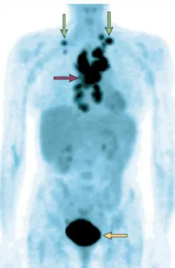
Figure 6. Positron-emission tomography imaging with 18fluorodeoxyglucose of a patient with lymphoma. The
mediastinal nodes (purple arrow) and supra clavicular nodes (green arrows) show high uptake of
18
fluorodeoxyglucose (FdG), showing that tumours in these nodes have high levels of FdG uptake. The bladder (yellow arrow) also has high activity, because of excretion of the radionuclide [Gatenby and Gillies, 2004].
1.2.1 Prevalence of aerobic glycolysis in cancer
In the 1920s, Otto Warburg made the surprising finding that tumour cells, unlike their normal counterparts, utilize glycolysis instead of mitochondrial oxidative phosphorylation for glucose metabolism even when in normoxia, a phenomenon historically known as the Warburg effect or aerobic glycolysis [65].
Introduction
12 Study of the role of monocarboxylate transporters in colorectal carcinoma
However, the proliferative advantages gained from altered glucose metabolism are far from clear. Firstly, anaerobic respiration is more than an order of magnitude less efficient than its aerobic counterpart, producing only 2 ATP per glucose in comparison to approximately 36 ATP (Figure 5). Secondly, glycolysis increases acid production resulting in a highly acidic extra-cellular environment [66]. This results in local toxicity including cell death and extra-cellular matrix degradation due to release of proteolytic enzymes [67]. Thus, the dynamics that leads to adoption of aerobic glycolysis as a typical component of the malignant phenotype remains unknown.
Recently, Gatenby and Gillies [68] proposed that the glycolytic phenotype initially arises as an adaptation to local hypoxia due to disordered angiogenesis and blood flow (Figure 7). Persistent or cyclical hypoxia subsequently induces constitutive upregulation of glycolysis even in the presence of oxygen, through mutations or epigenetic changes. Increased acid production from upregulation of glycolysis results in microenvironmental acidosis and requires further adaptation to phenotypes resistant to acid-induced toxicity. Cell populations that emerge from this evolutionary sequence have a powerful growth advantage, as they alter their environment through increased glycolysis in a way that is toxic to normal populations, but harmless to themselves. The environmental acidosis also facilitates invasion through destruction of adjacent normal cells, degradation of the extracellular matrix and promotion of angiogenesis (Figure 7) [68].
Increased glycolysis may also confer adaptive advantages if it allows excess pyruvate to be available for lipid synthesis or providing essential anabolic substrates, such as ribose for nucleic acid synthesis [69]. Glucose consumption through the pentose pathway may also provide essential reducing equivalents (NADPH) to reduce the toxicity of reactive oxygen species conferring resistance to senescence [70, 71].
These evolutionary advantages can explain the significant prevalence of aerobic glycolysis in human cancers. This conceptual model is supported by empirical studies that demonstrate constitutive upregulation of glycolysis is consistently observed during the transition from premalignant lesions and invasive cancer [63, 72, 73, 74]. Additionally, Smallbone et al. [75] developed a hybrid cellular automation approach to investigate the cell-microenvironmental interactions that mediate somatic evolution of cancer cells. That study supported the hypothesis that regional variations in oxygen, glucose and H+ levels drive the final stages of somatic evolution during carcinogenesis.
Introduction
They proposed that the phenotypic adaptations to the sequence of hypoxia-glycolysis-acidosis are necessary to form an invasive cancer [75].
Figure 7. Model for cell–environment interactions during carcinogenesis, giving the stages of tumour growth and their associated physiological states. Normal epithelial (grey), hyperplastic (pink), hypoxic (blue), glycolytic (green) and motile (yellow) cells [Gatenby and Gillies, 2004].
These results suggest that tumour prevention strategies aimed at interrupting glycolysis and the resulting cellular adaptations could be explored to delay or prevent transition of in situ to invasive cancer. Furthermore, in advanced metastatic cancers, understanding of the molecular and physiological causes and consequences of upregulated glycolysis may lead to targeted therapies [76].
1.2.2 Molecular mechanisms
The increase in tumour glucose uptake is achieved by up-regulating the expression of glycolytic enzymes and glucose transporters [77]. The molecular basis for evolution of the glycolytic phenotype has been clarified by recent advances in
Introduction
14 Study of the role of monocarboxylate transporters in colorectal carcinoma
understanding the hypoxia-inducible factor, HIF-1, system [78]. Hypoxia and HIF-1 increase virtually all the enzymes in the glycolytic pathway, as well as the glucose transporters 1 and 3 (GLUT1, GLUT3) [79], and other hypoxia-related genes such as those involved in angiogenesis, iron metabolism and cell proliferation/survival (Figure 8) [80].
Figure 8. A summary of the HIF system demonstrating several factors which govern the level of HIF-1α and some of its protean effects on cell metabolism, survival, proliferation and their microenvironment [Gatenby and Gillies, 2007].
HIF-1 is a heterodimeric complex consisting of a hypoxically inducible subunit HIF-1α and a constitutively expressed subunit HIF-1β [81]. Although HIF-1β is constitutively expressed and its mRNA and protein are maintained at constant levels regardless of oxygen availability [82], HIF-1α protein has a short half-life (t1/2 ~ 5 min)
and is highly regulated by oxygen [83]. The transcription and synthesis of HIF-1α are constitutive and seem not to be affected by oxygen [81, 82, 84]. However, in normoxia, the HIF-1α proteins are rapidly degraded, resulting in essentially no detectable HIF-1α protein [81]. During hypoxia, HIF-1α becomes stabilized and translocates from the cytoplasm to the nucleus, where it dimerizes with HIF-1β, and the HIF complex formed becomes transcriptional active [82, 85]. The activated HIF complex then associates with hypoxia response elements (HREs) in the regulatory regions of target genes and binds the transcriptional coactivators to induce gene expression [86].
Introduction
Furthermore, the glycolysis metabolic products, such as lactate and pyruvate, have been reported to cause HIF-1α accumulation under normoxia and regulate hypoxia-inducible gene expression, establishing a potential positive feedback loop which may be critical to aerobic glycolysis [87]. However, HIF activity can also be stabilized in the presence of oxygen by growth factors (Figure 8) that also participate in carcinogenesis, including RAS, HSP 90, Cox 2, HER, and the AKT/mTOR pathway [88]. Because of this, HIF can be characterized as being reversibly increased by physiologic stress (e.g., hypoxia) or by hormonal growth factor stimulation or as being constitutively active under normoxic conditions through heritable alterations, such as activated oncogenes.
1.2.3 Consequences of increased glucose metabolism
One consequence of this dependence on glycolysis is the need for metabolically active tumours to efflux the accumulating lactic acid to the tumour microenvironment to prevent intracellular acidosis [89, 90]. Cellular acidosis has been shown to be a trigger in the early phase of apoptosis and leads to activation of endonucleases inducing DNA fragmentation. In addition, cellular pH is crucial for biological functions such as cell proliferation [90], invasion [91], metastasis [92] and drug resistance [93]. The observation that the intracellular pH of tumours is more alkaline than that of normal tissues [94, 95], even in the presence of an acidic tumour microenvironment, suggests the existence of an active mechanism for lactate/H+ efflux. Although several transporters including Na+/H+ exchangers (NHE1), vacuolar adenosine triphosphatases, anion exchangers, and lactate/H+ symporters (monocarboxylate transporters [MCTs]) are involved in pH homeostasis in mammalian cells [89], MCTs are thought to be most important for pH regulation within rapidly metabolizing, highly glycolytic tumour cells [96, 97].
1.3 Monocarboxylate Transporters (MCTs)
MCTs are members of the SLC16 gene family [98]. This family includes 14 proteins related to each other by sequence homology. The first four members,
MCT1-Introduction
16 Study of the role of monocarboxylate transporters in colorectal carcinoma
MCT4 have been experimentally demonstrated to transport aliphatic monocarboxylates, a diverse group of compounds which includes many important metabolites such as lactate, pyruvate and ketone bodies. This transport is controlled by hydrogen ion and substrate gradients, thus facilitating transport of MCTs either in (influx) or out (efflux) of the cell. Lactate efflux is especially important for cells and tissues which generate large quantities of lactic acid as a result of glycolysis (e.g. red and white blood cells, skeletal muscle, and most tumour cells). Lactate influx is of value to tissues where it is being utilized (via pyruvate) as a respiratory fuel (brain, heart, red skeletal muscle), as a gluconeogenic substrate in liver, or under fasting conditions, as a glycerol-neogenic substrate in white adipose tissue [99]. Since lactate is a substrate for MCT1-MCT4 transporters, considerable attention has been given to the potential role of these transporters in the handling of lactate in tumour cells.
MCTs have 12 transmembrane domains with the N- and C-termini located in the cytoplasm [98, 100]. The transmembrane domains (TMDs) are highly conserved between isoforms with the greatest sequence variations observed in the C-terminus and the large intracellular loop between TMDs 6 and 7, which has a range of 29–105 amino acid residues (Figure 9) [100]. This observed variability is common to transporters with 12 TMDs and it is thought that these sequence variations are related to substrate specificity or regulation of transport activity [100, 101].
Figure 9. Proposed membrane topology of the MCT family. Adapted from Halestrap and Meredith, 2004.
Regulation of MCTs has been demonstrated to occur via transcriptional, translational and post-transcriptional mechanisms [102-104]. These regulatory pathways
Introduction
appear to be age- and tissue dependent, which further complicates the understanding of these pathways [103, 104]. Some MCTs require an ancillary protein which can be involved in cellular localization [105] or protein–protein interactions [106]; however, the role of these accessory proteins in overall transporter function is not yet completely understood [105].
1.3.1 MCT1
MCT1 is the most well-studied and functionally characterized member of the MCT family, largely due to the fact that it is the only monocarboxylate transporter expressed in human erythrocytes, and it also has the widest tissue distribution. MCT1 is ubiquitously expressed but is especially prominent in heart and red muscle where it is upregulated in response to increased work, suggesting an important role in lactic acid oxidation [101, 107, 108, 109].
Transport kinetics have been thoroughly explored using lactate for this isoform and have demonstrated that it functions as a proton-dependent cotransporter/exchanger [110, 111]. Transport occurs by ordered sequential binding with association of a proton followed by lactate binding. The complex is translocated across the membrane and the lactate and proton are released sequentially. Since the transporter functions as an exchanger, transport can occur bidirectionally; however, it is primarily responsible for the uptake of substrates [101].
Relatively few studies have been conducted to assess the regulation of MCTs. Studies have indicated that altered physiological conditions and the presence of xenobiotics may alter the regulation of MCTs, in addition to altered expression at different developmental stages (112–114). MCT1 expression undergoes transcriptional, post-transcriptional and post-translational regulation and appears to be regulated in a tissue-specific manner [102-104]. In colonic epithelium, exposure to butyrate resulted in a concentration- and time dependent increase in MCT1 mRNA, protein expression and a corresponding increase in butyrate transport [115]. These data suggest the possibility of altered transcriptional regulation; however, the authors further demonstrated increased transcript stability indicating additional post-transcriptional regulation mechanisms [115]. High concentrations of lactate have also been demonstrated to increase MCT1
Introduction
18 Study of the role of monocarboxylate transporters in colorectal carcinoma
mRNA and protein levels in L6 cells [116]. In contrast, treatment with testosterone resulted in increased skeletal muscle MCT1 protein expression and lactate transport in the absence of mRNA changes suggesting the importance of post-transcriptional regulation [103].
MCT1 is further regulated by its association with the cell surface glycoprotein CD147 (Basigin), which has a single transmembrane domain with the C-terminus located in the cytosol (Figure 10) [117, 118]. Topology studies suggest that one MCT1 molecule interacts with a single CD147 molecule with subsequent dimerization with another MCT1/CD147 pair (Figure 11) [111]. The initial association of CD147 and MCT1 is required for the translocation of MCT1 to the plasma membrane [117]. Furthermore, more recent studies indicate that CD147 is also required for catalytic activity of MCT1 [119].
1.3.2 MCT2
MCT2 is a high-affinity transporter for monocarboxylates and its expression under normal conditions is restricted to tissues such as the liver, kidney, and neurons that take up lactate rather than release it. In humans, expression of MCT2 is more restricted than MCT1, with the greatest expression observed in the testis [120]. In both rodents and humans, MCT2 splice variants have been detected in a species and
Figure 10. The proposed topology of CD147, the
ancillary protein that associates with MCT1 and MCT4. Adapted from Halestrap and Meredith, 2004.
Figure 11. Hypothetical model of the topology of
CD147 and MCT1 in the plasma membrane [Wilson et al, 2002].
Introduction
dependent manner suggesting that transcriptional and post-transcriptional regulation pathways play an important role in the tissue specificity of this isoform [120-122]. Similar to MCT1, MCT2 requires an accessory protein for translocation to the plasma membrane. However, MCT2 requires gp70 (EMBIGIN), not CD147 [105]. MCT2 possesses high affinity for lactate and most likely functions in the import of lactate into cells.
1.3.3 MCT3
MCT3 is believed to have the most restricted distribution of any MCT with expression in the basolateral membrane of the retinal pigment epithelium (RPE) and the choroid plexus epithelium (CPE) in humans, rodents and chickens [123-125]. However, recent studies demonstrated MCT3 expression in vascular smooth muscle cell lines [126], human aorta [126] and human kidney [127], suggesting that MCT3 mRNA may be more widely distributed than originally thought.
1.3.4 MCT4
In contrast to MCT1, MCT4 is predominantly expressed in highly glycolytic cells such as white muscle and white blood cells suggesting that its physiological function is lactate efflux [101, 128, 129]. MCT4 localization at the plasma membrane is dependent on CD147 expression, which is consistent with results obtained for MCT1 [114]. The role of MCT4 in lactate efflux is further supported by its high expression in the placenta where it is involved in the transfer of lactate into the maternal circulation [98]. While there is a great degree of overlap in the substrate specificity of MCT1 and MCT4, these two isoforms differ in their substrate affinities with MCT4 having lower affinities for a range of monocarboxylates.
1.3.5 Regulation of MCTs in cancer
Introduction
20 Study of the role of monocarboxylate transporters in colorectal carcinoma
including breast, head and neck, and lung cancers [130] as well as neuroblastoma [113], brain [131], alveolar sarcoma of soft tissues [132], colon [130, 133, 134], melanoma [135], pancreatic [136], cervical [137], and gastric [138] cancers.
MCT4 is expressed under normal conditions in tissues that are glycolytic in nature with resultant increased production of lactate [98]. It has low affinity for lactate, thus making it an ideal transporter for export of lactate when this metabolite is generated at high levels as occurs in glycolytic tissues. In spite of the fact that the kinetic features of MCT4 are suitable for lactate export, the expression of this transporter in cancer is controversial. Some studies have found no evidence of cancer-associated expression of MCT4 [133, 139]. However, more recent studies have demonstrated that MCT4 is upregulated in cancer and in tumour cells [114, 134, 137]. In accordance with these recent findings, the gene coding for MCT4 has been found to be upregulated by hypoxia through a HIF-1α-dependent mechanism [140].
The expression of MCT2 is reduced in tumour cells [133, 134]. Since tumour cells release lactate rather than import it, downregulation of MCT2 in tumour cells correlates well with the metabolic phenotype of tumour cells. Thus, MCT2, which is involved in the uptake of monocarboxylates into the cells in normal metabolism [141], does not appear to have an important role in highly glycolytic cancer cells. Furthermore, the gene coding for MCT2 lacks binding sites for HIF-1 [140].
Broad MCT distribution among human cancers opens promising therapeutic perspectives for the development and clinical evaluation of pharmacological MCT inhibitors. Furthermore, a number of reports have attributed to MCTs a role in lactate efflux in tumour cells with active aerobic glycolysis [113, 142, 143, 144, 145]. These studies showed further evidence of lethal intracellular acidification upon MCT inhibition in vitro. Thus, MCTs can constitute attractive targets for cancer therapy.
1.3.6 Inhibitors of MCTs
Since MCTs are transmembrane proteins exposed to the extracellular milieu, they are prone to targeting by systemic application of small-molecule inhibitors. Several chemicals are known to inhibit the function of MCTs (Table 1) [110]. Those inhibitors fall into four broad categories: (1) bulky or aromatic monocarboxylates which act as
Introduction
competitive inhibitors (e.g. phenyl-pyruvate and α-cyano-4-hydroxycinnamate (CHC)); (2) amphiphilic compounds with divergent structures (e.g. quercetin and phloretin); (3) some 4,4’-substituted stilbene-2,2’-disulphonates (e.g. DIDS); and (4) miscellaneous inhibitors including thiol reagents such as p-chloromercuribenzene sulphonate (pCMBS) and amino reagents (e.g. pyridoxal phosphate and phenylglyoxal [98].
Table 1. Inhibitors of monocarboxylate transporters.
MCT Isoform Inhibitor References
MCT1 CHC pCMBS Phloretin Quercetin DIDS AR-C155858 [147] [105] [110] [110] [150] [149] MCT2 CHC Phloretin [147] [110] MCT4 CHC pCMBS Phroretin Fluvastatin Atorvastatin Lovastatin [147] [105] [110] [151] [151] [151] Simvastatin [151]
1.3.6.1 α-cyano-4-hydroxycinnamic acid (ACCA; CHC)
Some "classic" inhibitors of monocarboxylate transporters are derivatives of cinnamic acid, first identified by Halestrap and co-workers for their effect on isolated mitochondrial pyruvate transport [146] and by Lehninger and co-workers on intact Ehrich ascites tumour [147]. Others studies [142, 144] have indicated cinnamic acid derivates to be competitive inhibitors of lactate transport in tumours, with α-cyano-4-hydroxycinnamic acid (ACCA; CHC) (Figure 12) as one of the most potent inhibitors of lactate transport, with a Ki of 0.5 mM. A study [148] evaluating its therapeutic
efficacy against malignant glioma indicates CHC to be an effective cytotoxic and cytostatic agent both in vitro and in vivo, but at mM concentrations (≥ 10 mM) due to its high ki against lactate. This study on glioma cells also indicated that CHC remains
22 Study of the role of
extracellular. Thus, the inhibitory effect of CHC was restricted to the glioma plasma membrane monocarboxylate transport
inhibited by CHC, i.e., the mitochondrial pyruvate carrier (MPC) were not affected. A recent in vivo study [130] demonstrated that the inhibitory effect of CHC is restricted to tumour cells expressing MCT1
concentrations of CHC up to 125 mM
Figure 12.
1.3.6.2 Other inhibitors
The stilbene derivatives such (DIDS) act as reversible
eventually becomes irreversible on prolonged incubation, reflecting covalent modification of the transporter
thiol and amino reagents, especially the organomercurial thiol reagent p chloromercuribenzene sulphonate
inhibition is not of MCT1 and MCT4
In contrast to other MCTs, lactate transport via MCT4 is inhibited by a range of statin drugs which may play a role in cytotoxicities obser
1.3.6.3 New MCT1 specific inhibitors
A group of small molecular weight compounds, synthesized by AstraZeneca, were recently identified as potent and selective inhibitors of MCT1 in activated human T cells. These compounds produced specific inhibition of human T lymphocyte
Introduction
Study of the role of monocarboxylate transporters in colorectal carcinoma
extracellular. Thus, the inhibitory effect of CHC was restricted to the glioma plasma e monocarboxylate transport. Intracellular transporters known to be strongly inhibited by CHC, i.e., the mitochondrial pyruvate carrier (MPC) were not affected. A ] demonstrated that the inhibitory effect of CHC is restricted to r cells expressing MCT1 at the plasma membrane. That study also showed that concentrations of CHC up to 125 mM were not toxic in mice (animal models).
. Structure of α-cyano-4-hydroxy cinnamic acid (CHC).
inhibitors
The stilbene derivatives such as 4,4’-diisothiocyanostilbene-2,2
inhibitors of MCT1 in erythrocytes. Inhibition by DIDS eventually becomes irreversible on prolonged incubation, reflecting covalent modification of the transporter [150]. In addition, MCT1 and MCT4 are
reagents, especially the organomercurial thiol reagent p chloromercuribenzene sulphonate (pCMBS). Interestingly it has been shown that this
and MCT4 directly, but of the ancillary protein
In contrast to other MCTs, lactate transport via MCT4 is inhibited by a range of statin drugs which may play a role in cytotoxicities observed with statin administration [
New MCT1 specific inhibitors
small molecular weight compounds, synthesized by AstraZeneca, were recently identified as potent and selective inhibitors of MCT1 in activated human T cells. These compounds produced specific inhibition of human T lymphocyte
monocarboxylate transporters in colorectal carcinoma
extracellular. Thus, the inhibitory effect of CHC was restricted to the glioma plasma . Intracellular transporters known to be strongly inhibited by CHC, i.e., the mitochondrial pyruvate carrier (MPC) were not affected. A ] demonstrated that the inhibitory effect of CHC is restricted to plasma membrane. That study also showed that
mice (animal models).
2,2’-disulphonate Inhibition by DIDS eventually becomes irreversible on prolonged incubation, reflecting covalent and MCT4 are inhibited by reagents, especially the organomercurial thiol reagent
p-(pCMBS). Interestingly it has been shown that this rotein CD147 [105]. In contrast to other MCTs, lactate transport via MCT4 is inhibited by a range of statin ved with statin administration [151].
small molecular weight compounds, synthesized by AstraZeneca, were recently identified as potent and selective inhibitors of MCT1 in activated human T cells. These compounds produced specific inhibition of human T lymphocyte
Introduction
proliferation in vitro with nanomolar potency [149]. This study also demonstrated that the effects of MCT1 inhibitors in activated T cells, which rely on aerobic glycolysis for energy, seem to be mediated by the blockade in lactate efflux rather than by effects on pH, by transport of alternative substrates or via CD147 [149].
1.4 MCT1 and MCT4 as potential targets for CRC therapy
CRCs, in common with many other malignancies, have enhanced glucose utilization and glycolytic metabolism [152-154], producing high amounts of lactic acid, which is effluxed to the tumour microenvironment via lactate transporters, namely monocarboxylate transporters (MCTs).
There are evidences for the upregulation of MCTs in colorectal carcinomas [130, 133, 134]. Koukourakis et al. [133] reported an increase in MCT1 expression in tumour cells, which is supported by the metabolic alterations induced by anaerobic glycolysis. That study also assessed the expression of MCT2 and MCT4, finding a strong cytoplasmic expression of MCT2 in cancer cells, but a weak expression of MCT4 in the tumour environment. Pinheiro et al. [134] reported an increase in expression of MCT1, MCT2 and MCT4 in tumour cells when comparing with the adjacent normal epithelium. In that study, the authors also found a significant gain of membrane expression for MCT1 and MCT4 and loss of plasma membrane expression for MCT2 in tumour cells. Taking into consideration the lactate affinities to the different MCT isoforms, MCT1 and MCT4 expression in the membrane is in agreement with the metabolic alterations observed in colorectal cancer cells. That is, MCT1 and MCT4 need to be at the plasma membrane to export the accumulating acids in highly glycolytic tumour cells [134].
Those findings point to MCT1 and MCT4 as playing an important role in highly glycolytic CRC cells. Thus, targeting MCT1 and MCT4 activity may debilitate such tumours via disruption of aerobic glycolysis and promoting apoptosis, suggesting that MCT inhibitors may be particularly effective against CRC. Moreover, inhibiting MCTs would not only induce apoptosis due to cellular acidosis, but would also lead to reduction in tumour angiogenesis [155], invasion [91], and metastasis [92].
Introduction
24 Study of the role of monocarboxylate transporters in colorectal carcinoma
1.5 Aims
The first purpose of this work was to explore the role of MCTs in CRC, by evaluating the effects of MCT inhibitors in human colon carcinoma cell viability, migration, lactate efflux, and MCT expression. The second purpose was to investigate the effects of combination of CHC (MCT inhibitor) with 5-FU (conventional chemotherapy) in human colon carcinoma cell viability and migration.
Thus, the specific aims were:
– Characterization of MCT1, MCT4 and CD147 expression in colorectal carcinoma cell lines by Western blotting and immunocytochemistry. – Evaluate the effect of MCT inhibitors on:
a) Cell viability by MTT assay and/or trypan blue assay; b) Cell migration by wound-healing assay;
c) MCT activity;
d) MCT expression by Western blotting and immunocytochemistry. – Investigate the effect of the combined treatment with CHC and 5-FU on:
a) Cell viability by trypan blue assay; b) Cell migration by wound-healing assay.
Material and methods
2 Materials and methods
2.1 Cell lines
The human colon carcinoma-derived cell lines HCT-15 and Co-115 were kindly provided by Dr. Raquel Seruca, IPATIMUP, Oporto.
2.2 Chemicals
5-FU, a commonly used anti-cancer drug for the treatment of colorectal carcinoma, and CHC, a competitive inhibitor of lactate/pyruvate transporters, were purchased from Sigma-Aldrich (St. Louis, MO, USA). AR-C155858, a specific monocarboxylate transporter-1 (MCT-1) inhibitor, was synthesized and gently provided by AstraZeneca (R&D Charnwood, UK).
5-FU, CHC and AR-C155858 were dissolved in 100% dimethylsulfoxide (DMSO) at the concentrations of 2 M, 5 M and 10 mM, respectively, and further diluted in RPMI 1640 medium (Gibco, Invitrogen, USA) to the desired final concentrations. The pH of the CHC solutions were adjusted to 7.4 with NaOH and all solutions were sterilized by filtration (0.20 µm syringe filter units, Starstedt, Germany) before use. Control cells were incubated with vehicle (DMSO). The final concentration of DMSO was maintained at 0.5%.
2.3 Cell culture conditions
HCT-15 and Co-115 colon carcinoma cells were cultured in RPMI 1640 medium (Gibco) supplemented with 10% (v/v) fetal bovine serum (FBS, Gibco, Invitrogen, USA) and 1% (v/v) penicillin-streptomycin solution (Invitrogen, USA) and incubated at 37 ºC in a humidified atmosphere containing 5% CO2. Cells were subcultured every
Materials and methods
26 Study of the role of monocarboxylate transporters in colorectal carcinoma
2.4 Cell viability assays
Cell viability was measured with both MTT (3-(4,5-dimethylthiazol-2-yl)-2,5-diphenyl tetrazolium bromide) dye reduction [Cell Proliferation Kit I (MTT), Roche, Mannheim, Germany] and Trypan Blue dye exclusion assays.
2.4.1 MTT dye reduction assay
This assay is based on the cleavage of the yellow tetrazolium salt MTT to purple formazan crystals by metabolic active cells. This cellular reduction involves the pyridine nucleotide cofactors NADH and NADPH.
HCT-15 and Co-115 cells were seeded in 96-well plates (6×10³/100 µl/well) and incubated at 37 ºC in a 5% CO2 atmosphere for 24 hours. To examine the effects of the
treatment with 5-FU, CHC or AR-C155858, the spent media were removed and 0.01-10 mM 5-FU, 5-25 mM CHC, or 1-10 µM AR-C155858 were added. Controls were performed with DMSO alone. After 24 hours of incubation, 10 µl MTT labelling reagent (final concentration 0.5 mg/ml) was added to each well and microplates were incubated for 4 hours at 37ºC. Subsequently, 100 µl of solubilisation solution (10% SDS in 0.01 M HCl) were added to each well for solubilisation of the purple formazan crystals and the mixture incubated at 37 ºC overnight. Spectrophotometrical absorbances were measured using a microplate reader (Model 450, Bio-Rad). The wavelength to measure absorbance of the formazan product and the reference wavelength were 570 nm and 750 nm, respectively. To determine cell viability, percent viability was calculated as (OD experiment/OD control) × 100 (%). Results are presented as mean ± SD of three independent experiments.
2.4.2 Trypan Blue dye exclusion assay
Trypan Blue is a dye which cannot enter healthy cells, but can permeate compromised membranes of dying cells. Dye exclusion was measured with 0.4% trypan blue in 0.85% saline (Invitrogen).
Material and methods
HCT-15 and Co-115 cells were seeded in 24-well plates (2.5×10⁴/500 µl/well) and incubated at 37 ºC in a 5% CO2 atmosphere for 24 hours. Spent media were
removed and to examine the effects of the treatment with CHC, cells were treated with 5-25 mM CHC for 24 hours. To determine the effect of CHC exposure time, HCT-15 cells were treated with 10 mM CHC for 6-48 hours. To address the effects of 5-FU and CHC co-treatment, cells were treated with 1 mM 5-FU (IC50) and increasing concentrations of CHC (5-25 mM) for 24 hours. Controls were performed with DMSO alone. The supernatant from each well was recovered and 75 µl trypsin (0.05% trypsin, 0.53 mM ethylenediamine tetra-acetic acid·4Na, Invitrogen) was added to each well and incubated at 37ºC for 10-20 minutes. Subsequently, 150 µl of fresh medium was added to quench the trypsin activity, and cells were recovered and pooled with the culture supernatant. Aliquots of the cell suspensions were diluted 1:1 in trypan blue solution. Clear and blue cells were counted with a hemocytometer under a bright-field microscope, and the viable fraction was estimated by dividing the number of clear cells by the total number of cells. Results are presented as mean ± SD of three independent experiments.
2.5 Western blotting
The characterization of MCT1, MCT4 and CD147 protein expression in untreated HCT-15 and Co-115 cells and HCT-15 CHC treated cells were evaluated by western blotting.
HCT-15 and Co-115 cells were cultured in 6-well plates at a density of 3 × 10⁵ cells per well and incubated at 37 ºC in a 5% CO2 atmosphere for 48 hours. To evaluate
the effect of CHC on MCT1, MCT4 and CD147 protein expressions, HCT-15 cells were treated with 10 mM CHC and incubated for further 24 hours. Controls were performed with DMSO alone. Cells were washed with PBS and quickly scraped from the well with 50 µl lysing buffer (150 mM NaCl, 50 mM Tris-HCl, pH 7.5, 0.1 mM EDTA, 1% Triton X-100, 1% Nonidet P-40), supplemented with protease inhibitor cocktail (Roche, Mannheim, Germany), and homogenized 10 minutes on ice. Cells were then centrifuged at 3000 rpm, for 15 minutes at 4ºC, to remove cell debris. Supernatants were collected and protein concentrations were quantified using a Bio-Rad DC protein assay (Bio-Rad
Materials and methods
28 Study of the role of monocarboxylate transporters in colorectal carcinoma
Laboratories, Inc., Hercules, CA, USA). BSA was used as a protein standard. Twenty micrograms of total protein from each cell lysate were separated on a 10% (w/v) polyacrylamide gel and electrotransferred to nitrocellulose membranes (Amersham Pharmacia Biotech, Piscataway, NJ, USA). Membranes were blocked with 0.1% Tween-20, 5% (w/v) non-fat dry milk in TBS for 1 hour at room temperature. Membranes were then incubated overnight at 4ºC with primary polyclonal antibodies for MCT1 (AB3538P, Chemicon International, Temecula, CA, USA), MCT4 (AB3316P, Chemicon International), and CD147 (18-7344, Zymed, Invitrogen, Carsbad, CA, USA) diluted 1:200 for MCT1 and MCT4, and 1:750 for CD147, in TBS, 0.1% Tween-20 and 1% (w/v) non-fat dry milk. After washing, membranes were incubated 1 hour at room temperature with secondary antibody conjugated with IgG horseradish peroxidise (Santa Cruz Biotechnology, Santa Cruz, CA, USA), diluted 1:10 000 in TBS, 0.1% Tween-20 and 1% (w/v) non-fat dry milk. Subsequently, immunoreactive bands were visualized by chemiluminescence (Supersignal West Femto kit, Pierce, Rockford, IL, USA). Band area intensity was quantified using ImageJ/Java, a free software downloaded from http://rsb.info.nih.gov/ij/. ß-actin was used as loading control.
2.6 Immunocytochemistry
Characterization of MCT1 and MCT4 protein expression in untreated HCT-15 and Co-115 cells and HCT-15 CHC treated cells were evaluated by immunocytochemistry.
HCT-15 and Co-115 cells were grown on coverslips at 30% confluence. To evaluate the effect of CHC in MCT1 and MCT4 protein expression, HCT-15 cells were treated with 10 mM CHC for 24 hours at 37 ºC. Controls were performed with DMSO alone. Untreated and treated cells grown on coverslips were fixed with PBS containing 4% (w/v) paraformaldehyde for 15 minutes at room temperature and then permeabilized in PBS containing 0.05% Triton X-100 for 4 minutes. Cells were incubated with 0.3% hydrogen peroxide in methanol for 30 minutes, to inactivate endogenous peroxidases, and washed in PBS. Cells were then incubated for 20 minutes with Normal Horse Serum (Vector, Burlingame, CA, USA), to block non-specific binding, and incubated
![Figure 1. Progression model of colorectal cancer. The changes that occur in the outlined in this figure [Garnis et al, 2004].](https://thumb-eu.123doks.com/thumbv2/123dok_br/17954391.853972/18.892.136.762.137.395/figure-progression-colorectal-cancer-changes-outlined-figure-garnis.webp)
![Figure 2. Overview of the Wnt signaling pathway [BIOCARTA, www.biocarta.com].](https://thumb-eu.123doks.com/thumbv2/123dok_br/17954391.853972/19.892.189.716.538.1123/figure-overview-wnt-signaling-pathway-biocarta-www-biocarta.webp)
![Figure 3. Adenocarcinoma progression [portal de oncologia português, www.pop.eu.com]](https://thumb-eu.123doks.com/thumbv2/123dok_br/17954391.853972/21.892.222.671.417.750/figure-adenocarcinoma-progression-portal-oncologia-português-www-pop.webp)
![Figure 4. Mechanisms of action of 5-FU and its metabolites [Longley et al, 2003].](https://thumb-eu.123doks.com/thumbv2/123dok_br/17954391.853972/23.892.268.626.337.827/figure-mechanisms-action-fu-metabolites-longley-et-al.webp)
![Figure 5. Transport of glucose and its metabolites in mammalian cells [Gatenby and Gillies, 2007]](https://thumb-eu.123doks.com/thumbv2/123dok_br/17954391.853972/25.892.192.706.608.896/figure-transport-glucose-metabolites-mammalian-cells-gatenby-gillies.webp)

![Figure 8. A summary of the HIF system demonstrating several factors which govern the level of HIF-1α and some of its protean effects on cell metabolism, survival, proliferation and their microenvironment [Gatenby and Gillies, 2007].](https://thumb-eu.123doks.com/thumbv2/123dok_br/17954391.853972/29.892.269.629.346.588/figure-demonstrating-metabolism-survival-proliferation-microenvironment-gatenby-gillies.webp)

![Figure 11. Hypothetical model of the topology of CD147 and MCT1 in the plasma membrane [Wilson et al, 2002]](https://thumb-eu.123doks.com/thumbv2/123dok_br/17954391.853972/33.892.523.699.563.790/figure-hypothetical-model-topology-mct-plasma-membrane-wilson.webp)