Article
ISSN 0102-695X http://dx.doi.org/10.1590/S0102-695X2011005000164 Received 24 Oct 2010 Accepted 23 Mar 2011 Available online 9 Sep 2011
from
Garcinia mangostana
fruit rinds on HCT
116 human colorectal carcinoma cells
Abdalrahim F.A. Aisha,
1Khalid M. Abu-Salah,
2Zeyad D.
Nassar,
1Mohammad J. Siddiqui,
1Zhari Ismail,
2Amin Malik
Shah Abdul Majid
*,11School of Pharmaceutical Sciences, University Sains Malaysia, Malaysia,
2The Chair of Cancer Targeting and Treatment, Biochemistry Department and King
Abdulla Institute for Nanotechnology, King Saud University, Saudi Arabia.
Abstract: This study aimed to investigate the antitumorigenicity of xanthones-rich extract from Garcinia mangostana L., Clusiaceae, fruit rinds which was obtained
by a simple recrystallization of 75% ethanolic extract. α-Mangostin content of the
extract was determined qualitatively by TLC and quantitatively by HPLC, and total xanthones content was quantified by UV spectrophotometry. The extract was evaluated for cytotoxicity, apoptosis and antitumorigenicity on HCT 116 human
colorectal carcinoma cells. α-Mangostin was found to be the main constituent of the
extract which was 71.2±0.1%, and the total xanthones content was 95±4.8% (wt/ wt). The extract showed potent dose dependent cytotoxicity with IC50 value 9.2 µg/ mL. Apoptosis studies revealed activation of caspases 3 and 7, DNA fragmentation, chromatin condensation and loss of mitochondrial membrane potential. Studies on cell migration and colony formation indicate reduced cell migration ability and clonogenicity of treated HCT 116 cells at sub-inhibitory concentrations. Taken together, the cytotoxic effect of the xanthones extract is mediated through the mitochondrial pathway of apoptosis. The reduced cell migration and clonogenicity of HCT 116 cells might prevent both primary and metastatic tumor growth in vivo which will be the topic of our future work using the metastatic orthotopic colon cancer model.
Keywords:
apoptosis cytotoxicity
Garciniamangostana α-mangostin
tumorigenicity xanthones
Introduction
Garcinia mangostana L. or mangosteen is a tropical tree from the family Clusiaceae. The tree is cultivated for centuries in the tropical rainforests of some Asian countries. The tree can be found now in Northern Australia, Brazil, Central America, Hawaii, Southern India, Indonesia, Malaysia, Thailand and other tropical countries (Ji et al., 2007). The fruit is dark purple or reddish with soft, white and juicy edible pulp. The pericarps of the fruit have been used in folk medicine for the treatment of several human diseases including skin and wound infections, hemorrhoids, arthritis, tuberculosis, inflammation, mycosis, ulcers, genitourinary tract infections, fever, amoebic dysentery and abdominal pain (Baxter et al., 1999; Caius, 2003; Moongkarndi et al., 2004; Salguero, 2003; Suksamrarn et al., 2006). Mangosteen is also used worldwide as an ingredient of several commercial products including nutritional supplements, herbal cosmetics and pharmaceutical products (Ji et al., 2007).
The presence of high levels of xanthones in mangosteen fruit rinds was reported by several research groups (Ee et al., 2006; Peres et al., 2000; Zhang et al., 2010). α-mangostin was the first xanthone isolated from G. mangostana and several other xanthones were isolated as well (Pedraza-Chaverri et al., 2008). Previous studies on different xanthones demonstrated remarkable pharmacological activities including analgesic (Cui et al., 2010), antioxidant (Jung et al., 2006), antiinflammatory (Chen et al., 2008; Tewtrakul et al., 2009), anticancer (Akao et al., 2008; Doi et al., 2009; Hung et al., 2009; Matsumoto et al., 2004), antiallergy (Nakatani et al., 2002), antibacterial (Chomnawang et al., 2009; Sakagami et al., 2005), antituberculosis (Suksamrarn et al., 2003), antifungal (Kaomongkolgit et al., 2009) and antiviral (Chen et al., 1996).
behind this study was to produce a cost effective but pharmacologically potent extract from the fruit rinds of G. mangostana using a simple and commercially applicable extraction method. This extract can be a good candidate for developing new noninvasive chemopreventive and therapeutic agent targeting colon cancer and other cancers as well.
Material and Methods
Plant material
Ripened fruits of Garcinia mangostana L., Clusiaceae, were obtained from a local fruit farm from the Island of Penang, Malaysia, in June 2009. Taxonomic authentication was done by Mr. Shanmugan A/C Vellosamy, Herbarium of School of Biology, USM, where a voucher specimen (11155) was deposited. The fruit rinds were separated from the edible pulp, dried at 45-50 °C for 24 h and grinded into fine powder. 60 g of the powdered material was extracted on a magnetic stirrer with 600 mL 75% ethanol: 25% water at 60 °C for 48 h with continuous shaking. After filtration ethanol was evaporated at 50 °C by rotavapor under vacuum, and the liquid residue was kept at 2-8 °C for 24 h. A yellow precipitate was formed which was collected by filtration and dried at 45-50 °C. The percent yield to the dried plant material was found to be 7.5% (wt/wt).
Chemicals
α-Mangostin (97% purity) was purchased from Chroma DEX Inc. Wizard® SV Genomic DNA
Purification System and Caspase Glo 3/7 kit were purchased from Promega. 2,3-Bis (2-methoxy-4-nitro-5-sulfophenyl)-2H-tetrazolium-5-carboxanilide inner salt (XTT) reagent, phenazine methosulfate (PMS), betulinic acid (90% purity), agarose, ethidium bromide, dimethyl sulfoxide (DMSO) were purchased from Sigma-Aldrich. HPLC grade solvents were purchased from Merck. Fetal bovine serum (FBS), penicillin/ streptomycin solution (PS) and RPMI-1640 cell culture medium were purchased from GIBCO BRL. TLC plates precoated with 0.25 mm layer of silica gel 60 F254 was obtained from Merck.
Cell culture
Human colorectal carcinoma HCT 116 cells (ATCC) were grown and maintained in RPMI 1640 medium with L-glutamine supplemented with 10% FBS, 100 µg/mL streptomycin, and 100 U/mL penicillin at 37 °C in a humidified atmosphere of 5% CO2.
Determination of α-mangostin and total xanthones
Firstly, the extract was analyzed on TLC as the following: 10 µL α-mangostin or the xanthone extract at 1 mg/mL in methanol was spotted and allowed to dry. The chromatograms were developed under chamber saturation conditions with n-hexane: ethyl acetate: methanol at 7:3:0.5 v/v. After drying, the plates were visualized at 254 and 366 nm. The identity of the band corresponding to α-mangostin in the sample was confirmed by comparing the Rf value with that of the reference compound.
Then α-mangostin content of the extract was determined quantitatively by HPLC (Agilent 1100) as the following; 10 µL of the extract or α-mangostin at 1 mg/mL was separated on Nucleosil C18 (5 µm, 4.6x250 mm) column at 25 ºC. The mobile phase was isocratic consisting of 90% acetonitrile and 10% (0.1% H3PO4 in water) at 1 mL/min. Separation time was 10 min and detection was performed at 254 nm. The data acquisition was performed by ChemStation version A.08.03. The percentage of α-mangostin in the extract was calculated based on the peak area. The total xanthones content of the extract was determined by UV spectrophotometry. Briefly, a calibration curve of α-mangostin was prepared by plotting the absorbance at 260 nm versus the concentration. The absorbance of the extract was taken at 50 µg/mL in methanol and then the concentration was calculated by applying the linear regression equation of the standard calibration curve. The wt/wt percentage of total xanthones was calculated using the formula: (the calculated concentration µg/ mL/50 µg/mL)x100%.
Cell viability
To determine cell viability, XTT assay was performed in 96-well plate format as described previously (Jost et al., 1992). Briefly, 1x10⁴ cells
were seeded in each well in 100 µL culture medium. After overnight incubation, additional 100 µL of fresh medium containing different concentrations of the xanthones extract or the positive controls (α-mangostin and betulinic acid) were added and further incubated for 48 h. Afterwards, 20 µL of a mixture of XTT and PMS at 1 mg/mL and 10 µg/mL was added to each well and incubated at 37 °C for 4 h. The OD was measured at 450 nm by microplate reader (Thermolab Systems 354, Finland). The cell viability was calculated using the formula: ((OD450 samples-blank)/(OD450 negative control-blank))x100%. IC50 values were determined from the respective dose response curves±SD (n=3).
Effect on caspase 3 and 7 was measured using caspase 3/7 assay system according to supplier’s instructions. Briefly, cells were seeded at 2x10⁴/well
in 100 µL medium in a white 96-well plate. After overnight incubation, cells were treated with the xanthones extract at 25 µg/mL for three time intervals viz. 45 min, 90 min and 180 min. α-mangostin was used as a positive control and the vehicle (DMSO) was used as a negative control. At the end of incubation, 100 µL of caspase 3/7 reagent was added to each well and further incubated at room temperature for 30 min. Luminescence was measured by a microplate reader (Multitechnology Plate Reader type, 425-100 Finland). The results are presented as a mean of relative light units (RLU)±SD (n=4).
DNA laddering assay
2x10⁶ cells at 70-80% confluency were treated
with the xanthones extract or α-mangostin at 25 µg/mL for different intervals. At the end of each incubation period, cells were collected by centrifugation at 3000 rpm for 5 min and washed once with PBS. Total genomic DNA was extracted using Wizard® SV genomic DNA
purification system and analyzed by electrophoresis on 1.2% agarose gel stained with 0.5 µg/mL ethidium bromide.
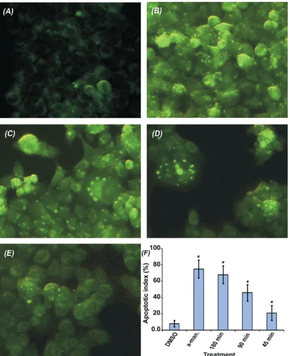
Effect on mitochondrial membrane potential and nuclear morphology
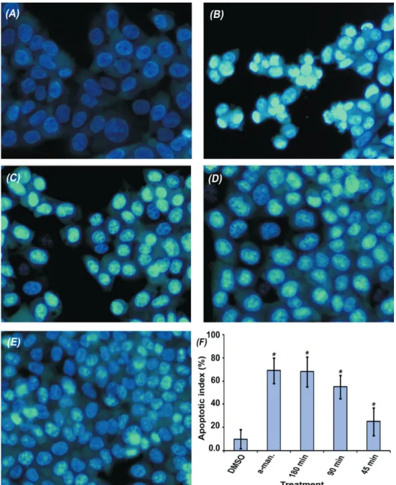
Mitochondrial staining with rhodamine 123 was performed in order to study the effect on mitochondrial membrane potential (Johnson et al., 1980), and nuclear morphology was examined by staining with Hoechst 33258 as previously described (Cheah et al., 2006; Chen et al., 2008). Briefly, 60-70% confluent cultures of HCT 116 cells were treated with α-mangostin or the xanthone extract at 25 µg/mL for 45, 90 and 180 min. Then the cells were washed with PBS and fixed with 4% paraformaldehyde for 20 min. After washing with PBS, the cells were stained simultaneously for 20 min with rhodamine 123 and Hoechst 33258 at 1 µg/mL and 10 µg/mL, respectively. The stained cells were washed extensively with PBS and the morphology of cells was examined immediately under x20 magnification of fluorescent microscope supplied with digital camera (IX71, Olympus). The morphological changes of treated cells were evaluated by studying 25 randomly selected microscopic fields. The apoptotic index was calculated for each microscopic field using the formula: (the number of apoptotic cells/ the total cell count)x100% and the results are presented as mean±SD.
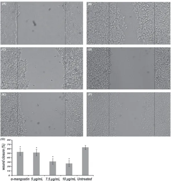
Effect on migration of HCT 116 cells
Effect on migration ability of HCT 116 cells was studied by wound healing assay as described previously (Liang et al., 2007). Briefly, cells were prepared in a single cell suspension and seeded in 6-well plate at 1x10⁶ cells/well in 3 mL medium. After
48 h when cells reach 100% confluency, a scratch was made along the middle line of each well with sterile 200 µL micropipette tip. The edges of the scratch were smoothened by cell culture medium and the detached cells were washed out with PBS. Afterwards, 3 mL cell migration medium containing 2% FBS and the treatment compounds was immediately added. The scratches were photographed at zero time at x4 magnification in inverted light microscope with digital camera and after 18 h. Leica Quin software was used to measure the width of the cell-free area of minimum 100 points/ well. The average percentage of wound closure of three independent experiments was calculated relatively to zero time using the formula: (1- (the width at 18 h/ the width at zero time)) x 100%.
Effect on clonogenicity of HCT 116 cells
Effect on clonogenicity of HCT 116 cells was evaluated by the colony formation assay as previously described (Franken et al., 2006). 1000 single suspended cells were seeded in 6-well plate at 2.5 mL/well. After overnight incubation, the old medium was replaced with a fresh one containing different concentrations of the extract 10, 7.5, 5 and 2.5 µg/mL. After 48 h, the medium containing the treatment compounds was removed and the cells were washed twice with PBS and a fresh medium was added. Then the cells were incubated for 12 days to allow colonies to form. At the end of incubation, the colonies were fixed in 4% paraformaldehyde and stained with 0.5% crystal violet. The number of colonies of more than 50 cells was counted using a stereomicroscope. The plating efficiency (PE) of untreated cells was determined as the following: (the number of formed colonies/ the number of seeded cells) x 100%. The survival fraction (SF) of treated cells was calculated using the formula: ((the number of formed colonies after treatment/(the number of seeded cells x PE)) X 100% (n=3). α-mangostin at 5 µg/mL was used as a positive control.
Statistical analysis
Results
Determination of α-mangostin and total xanthones
The UV spectra of α-mangostin and the xanthone extract were collected in the wavelength range of 500-200 nm (Figure 1a). The figure indicates almost identical spectra which provide the basis for quantitative analysis of total xanthones. Analysis in TLC indicates α-mangostin as the main constituent of the extract (Figure 1b). In HPLC analysis the retention time was found to be 5.52±0.02 min for both α-mangostin standard and that from the extract which further confirms the identity of the compound (Figure 1c and 1d).Quantitative analysis in HPLC shows that α-mangostin is present at 71.2±0.1%. Concentration of xanthones in the extract was quantified by applying the linear regression equation of α-mangostin calibration curve (y=0.0662x–0.0205, R2=0.9935). The calculated
wt/wt percentage of total xanthones in the extract was 95±4.8% (n=4).
The xanthones extract exhibited potent cytotoxic effect on HCT 116 cells
Cell viability was measured following 48 h treatment at seven concentration points 25, 20, 15, 12.5, 10, 7.5 and 5 µg/mL. α-Mangostin and betulinic acid were used as positive control. Figure 2a shows that the test compounds exhibit strong cytotoxic activity in a dose dependent manner. Analysis of the dose response curves indicates low IC50 values of α-mangostin 6.6±0.1 µg/mL. The IC50 of the xanthone extract was 9.2±0.09 µg/mL and was equivalent to that of betulinic acid which was 8.7±0.9 µg/mL (n=3).
The xanthones extract enhanced caspases 3 and 7 activity
Activity of the executioner caspases was significantly induced by treating HCT 116 cells with both the extract and the positive control at 25 µg/mL in time dependent manner (Figure 2b). Statistical analysis
by One way ANOVA and Dunnett test using untreated cells as reference indicates significant enhancement of caspases 3 and 7 activity as early as 45 min of treatment,
p values=0.0
The xanthones extract induced DNA fragmentation
We looked into the ability of the xanthones extract to cause DNA fragmentation of HCT 116 cells as a late marker of apoptosis. Treatment of cells with the extract caused apparent DNA degradation. A characteristic DNA ladder was produced on agarose gel electrophoresis which was detected early after 6 h of treatment. Untreated cells show the presence of intact DNA characterized by the presence of a single band (Figure 2c).
Effect on mitochondrial membrane potential and nuclear morphology
In order to study the apoptotic pathway induced by the extract, we investigated effect of the extract on mitochondrial membrane potential of HCT 116 cells. Microscopic examination revealed altered mitochondrial potential of treated cells compare to untreated cells. Figure 3 shows more brightly stained and swollen mitochondria of treated cells (apoptotic) which is lacking in untreated cells. The bar chart analysis reveals a time dependent and significant increment in the apoptotic index of treated cells compare to untreated cells, p values=0.0.
Staining of HCT 116 cells with Hoechst 33258 resulted in differential morphological appearance of treated cells. Cells treated with the vehicle only (DMSO) showed normal nuclei morphology characterized by weak intensity fluorescence with uniformly stained DNA. On the other hand, the treated cells showed clear apoptotic morphology characterized by either single intense fluorescence or multiple strong fluorescence signals in the cell nuclei (Figure 4). The apoptotic index was calculated in each interval and compared to untreated cells, the results indicate that the treatment compounds caused a significant and time dependent induction of chromatin condensation, p values=0.0.
The xanthones extract inhibited cell migration
We examined the effect of the extract on migration ability of HCT 116 cells by wound healing assay. The percent of wound closure in the presence and absence of treatment compounds was calculated after 18 h relatively to the zero time. The wound of untreated cells was closed by 64±5%. Wound closure was significantly reduced by treatment with the extract in a dose dependent manner, the percent wound closure
Figure 2. Effect on cell viability and apoptosis markers. Both treatments reduced cell viability in a dose dependent manner (a), enhancement of caspases 3 and 7 in time dependent manner (b) and induction of DNA fragmentation of HCT 116 cells (c), L
(DNA ladder), 1 (untreated cells), 2 (α-mangostin), 3, 4 and 5
for the xanthones crude extract (6, 12 and 24 h treatments).
was 27±7% at 10 µg/mL, 32±7% at 7.5 µg/mL and 52±7% at 5 µg/mL (p=0.0). No significant difference was found when the pure compound (α-mangostin) was compared with the extract at the same concentration point (p=0.532).
The xanthones extract decreased clonogenicity of HCT 116 cells
µg/mL was 2.2±0.3, 26±1.7, 48±7.5 and 76±7.3%, respectively. The SF of α-mangostin treated cells at 5 µg/mL was 48.8±3.6%. Statistical analysis indicate that clonogenicity of HCT 116 cells was significantly reduced in all treatments compare to untreated cells (p=0.0). No significant difference was found between α-mangostin and the extract at 5 µg/mL, (p=0.158).
Discussion
In this study, we report antitumorigenic effects
of xanthones-rich extract from fruit rinds of Garcinia mangostana L., Clusiaceae. Phytochemical analysis shows α-mangostin as the main constituent of the extract and total xanthones was almost 100% (wt/wt).
Anti-colon cancer effects of the extract were investigated on HCT 116 colorectal carcinoma cell line using α-mangostin and betulinic acid as positive control. HCT 116 was selected because it was previously reported as a model of invasive and metastatic colon cancer (Rajput et al., 2008). α-mangostin was selected as a positive control because it is the main constituent of
Figure 3. Effect on mitochondrial membrane potential. Untreated cells (a), and the treated cells with; α-mangostin (b), the extract for
the extract and it was reported previously as apoptotic compound (Matsumoto et al., 2004; Matsumoto et al., 2003). Betulinic acid was selected as positive control for the cytotoxicity study because it is a cytotoxic compound from natural origin. Dose dependent cytotoxicity was obtained at seven doses of all treatments, the IC50 value of the extract was achieved at 9.2±0.09 µg/mL. Cytotoxicity of the extract was equivalent to that of betulinic acid which shows IC50 value of 8.7±0.9 µg/ mL
On the basis of cytotoxicity results we carried
out more advanced experiments to investigate the effect on both early stage and late stage apoptosis markers. The apoptosis experiments were performed at 25 µg/mL since this concentration was found to induce apoptotic cell death after short period of treatment and since the experiments were carried out in short term manner. Lower concentrations but above IC50 could be used, however the time required for detection of early signals of apoptosis activation will be increased as well. We studied the effect on the executioner caspases 3 and 7 as early markers of apoptosis; the results show
Figure 4. Effect on nuclear morphology of HCT 116 cells. Untreated cells (a), and the treated cells with; α-mangostin (b), the extract
that these proenzymes were activated as early as 45 min after treatment (Figure 2b). Following that, we studied the effect on late stage events of apoptosis including DNA fragmentation and nuclear condensation. DNA fragmentation was clearly obvious after 6 h incubation (Figure 2c). Induction of nuclear condensation was also observed in a time dependent manner providing further confirmation on the activation of apoptotic cell death machinery (Figure 4). In order to explore which apoptotic pathway is involved, we looked into the effect on mitochondrial membrane potential. Treated cells appeared more brightly stained than untreated cells (Figure 3), which indicate lower concentration of the cationic dye in mitochondria relatively to the cytoplasm. This indicates loss of mitochondrial membrane potential which considered as early stage
in the activation of mitochondrial pathway of apoptosis that precedes cytochrome c release and activation of different caspases (Scarlett et al., 2000). Based on these findings, we can conclude that the apoptotic potential of the xanthones crude extract and α-mangostin was mediated through activation of the intrinsic (mitochondrial) pathway of apoptosis.
Since treatment of HCT 116 cells with high concentrations of the xanthone extract resulted in potent cytotoxic and apoptotic effects, we found it essential to investigate the effects at sub-cytotoxic concentrations. The treated cells at these concentrations did not reveal any sings of cytotoxicity but showed a reduced migration ability (Figure 5). This result highlights the anti-metastatic potential of the xanthone extract. In studies that looked into reversibility of cytotoxic effect
Figure 5. Effect on cell migration ability of HCT 116 cells. Zero time (a), and the cells after 18 h; untreated cells (b), α-mangostin at
of the extract, we observed irreversible cell damage and significant reduction in the clonogenic potential of HCT 116 cells (Figure 6). Overall, we report a novel xanthone extract from G. mangostana fruit rinds with cytotoxic, apoptotic, with potential anti-metastatic effect. This extract might provide effective prevention
and treatment of the metastatic colon cancer. However, this extract suffers the low aqueous solubility (<0.5 µg/ mL) and probably low oral bioavailability as reported previously (Li et al., 2011). Therefore, reliable in vivo results could be obtained only after improving its aqueous solubility which will be the topic of our future work.
Figure 6. Effect on clonogenicity of HCT 116 cells. Untreated cells (a), α-mangostin at 5 µg/mL (b), the extract at 10 µg/mL (c), the
extract at 7.5 µg/mL (d), the extract at 5 µg/mL (e) and the extract at 2.5 µg/mL (f).
Acknowledgements
This work was supported by University Sains Malaysia, science fund number 305/ PFARMASI/613219 and by FRGS-MOE fund number 203/PFARMASI/61154; and partially by the research chair of "Drug Targeting and Treatment of Cancer using Nanoparticles" at king Saud University, Riyadh, Saudi Arabia. The first and the third authors would like to acknowledge with thanks Universiti Sains Malaysia for providing scholarship under the fellowship program for 2010. Finally, we would like to thank Mr. Shanmugan A/C Vellosamy for authentication of the plant.
References
Akao Y, Yoshihito N, Munekazu L, Yshinori N 2008. Anti-cancer Effects of Xanthones from Pericarps of Mangosteen. Int J Mol Sci 9: 355-370.
Baxter H, Harborne J, Moss G 1999. Phytochemical Dictionary - A Handbook of Bioactive Compounds from Plants. Taylor & Francis 590.
Caius JF 2003. The Medicinal and Poisonous Plants of India. Scientific Publishers, India.
Cheah Y, Azimahtol H, Abdullah N 2006. Xanthorrhizol exhibits antiproliferative activity on MCF-7 breast cancer cells via apoptosis induction. Anticancer Res 26: 4527-4534.
Chen J, Peng H, Ou-Yang X, He X 2008. Research on the antitumor effect of ginsenoside Rg3 in B16 melanoma cells. Melanoma Res 18: 322-329.
Chen L-G, Yang L-L, Wang C-C 2008. Anti-inflammatory activity of mangostins from Garcinia mangostana.
Food Chem Toxicol 46: 688-693.
Chen S, Wan M, Loh B 1996. Active constituents against HIV-1 protease from Garcinia mangostana. Planta Med 62: 381-382.
Chomnawang M, Surassmo S, Wongsariya K, Bunyapraphatsara N 2009. Antibacterial Activity of Thai Medicinal Plants against Methicillin-resistant Staphylococcus aureus. Fitoterapia 80: 102-104. Cui J, Hu W, Cai Z, Liu Y, Li S, Tao W, Xiang H 2010. New
medicinal properties of mangostins: analgesic activity and pharmacological characterization of active ingredients from the fruit hull of Garcinia mangostana
Doi H, Shibata M, J Shibata E M, Akao Y, Iinuma M, Tanigawa N, Otsuki Y 2009. Panaxanthone isolated from pericarp of Garcinia mangostana L. suppresses tumor growth and metastasis of a mouse model of mammary cancer. Anticancer Res 29: 2485-2495. Ee G, Daud S, Taufiq-Yap Y, Ismail N, Rahmani M 2006.
Xanthones from Garcinia mangostana (Guttiferae).
Nat Prod Res 12: 1067-1073.
Franken NP, Rodermond H, Stap J, Haveman J, Bree C 2006. Clonogenic assay of cells in vitro. Nat Protoc 1: 2315-2319.
Hung S, Shen K, Wu C, Liu C, Shih Y 2009. Alpha-mangostin suppresses PC-3 human prostate carcinoma cell metastasis by inhibiting matrix metalloproteinase-2/9 and urokinase-plasminogen expression through the JNK signaling pathway. J Agric Food Chem 57: 1291-1298.
Ji X, Avula B, Khan IA 2007. Quantitative and qualitative determination of six xanthones in Garcinia mangostana
L. by LC-PDA and LC-ESI-MS. J Pharmaceut Biomed
43: 1270-1276.
Johnson L, Walsh M, Chen L 1980. Localization of mitochondria in living cells with rhodamine 123. Proc Natl Acad Sci 77: 990-994.
Jost L, Kirkwood J, Whiteside T 1992. Improved short- and long-term XTT-based colorimetric cellular cytotoxicity assay for melanoma and other tumor cells. J Immunol Methods 4: 153-165.
Jung H, Su B, Keller W, Mehta R, Kinghorn A 2006. Antioxidant xanthones from the pericarp of Garcinia mangostana (Mangosteen). J Agric Food Chem 54: 2077-2082.
Kaomongkolgit R, Jamdee K, Chaisomboon N 2009. Antifungal activity of alpha-mangostin against
Candida albicans. J Oral Sci 51: 401-406.
Li L, Brunner I, Han AR, Hamburger M, Kinghorn AD, Frye R, Butterweck V 2011. Pharmacokinetics of alpha-mangostin in rats after intravenous and oral application. Mol Nutr Food Res.
Liang C-C, Park A, Guan J-L 2007. In vitro scratch assay: a convenient and inexpensive method for analysis of cell migration in vitro. Nat Protoc 2: 329-333. Matsumoto K, Akao Y, Yi H, Ohguchi K, Ito T, Tanaka T,
Kobayashi E, Iinuma M, Nozawa Y 2004. Preferential target is mitochondria in [alpha]-mangostin-induced apoptosis in human leukemia HL60 cells. Bioorgan Med Chem 12: 5799-5806.
Matsumoto K, Akao Y, Kobayashi E, Ohguchi K, Ito T, Tanaka T, Iinuma M, Nozawa Y 2003. Induction of apoptosis by xanthones from mangosteen in human leukemia cell lines. J Nat Prod 66: 1124-1127. Moongkarndi P, Kosem N, Kaslungka S, Luanratana O,
Pongpan N, Neungton N 2004. Antiproliferation, antioxidation and induction of apoptosis by Garcinia
mangostana (mangosteen) on SKBR3 human breast cancer cell line. J Ethnopharmacol 90: 161-166. Nakatani K, Atsumi M, Arakawa T, Oosawa K, Shimura
S, Nakahata N, Ohizumi Y 2002. Inhibitions of histamine release and prostaglandin E2 synthesis by mangosteen, a Thai medicinal plant. Biol Pharm Bull 25: 1137-1141.
Pedraza-Chaverri J, Cárdenas-Rodríguez N, Orozco-Ibarra M, Pérez-Rojas J 2008. Medicinal properties of mangosteen (Garcinia mangostana). Food Chem
Toxicol 46: 3227-3239.
Peres V, Nagem T, Oliveira Fd 2000. Tetraoxygenated naturally occurring xanthones. Phytochemistry 55: 683-710.
Rajput A, Dominguez San Martin I, Rose R, Beko A, Levea C, Sharratt E, Mazurchuk R, Hoffman RM, Brattain MG, Wang J 2008. Characterization of HCT116 human colon cancer cells in an orthotopic model. J
Surg Res 147: 276-281.
Sakagami Y, Iinuma M, Piyasena K, Dharmaratne H 2005. Antibacterial activity of alpha-mangostin against vancomycin resistant Enterococci (VRE) and synergism with antibiotics. Phytomedicine 12: 203-208.
Salguero C 2003. A Thai Herbal. Findhorn Press 118. Scarlett J, Sheard P, Hughes G, Ledgerwood E, Ku H-H,
Murphy M 2000. Changes in mitochondrial membrane potential during staurosporine-induced apoptosis in Jurkat cells. FEBS Lett 475: 267-272.
Suksamrarn S, Komutiban O, Ratananukul P, Chimnoi N, Lartpornmatulee N, Suksamrarn A 2006. Cytotoxic prenylated xanthones from the young fruit of Garcinia mangostana. Chem Pharm Bull 54: 301-305.
Suksamrarn S, Suwannapoch N, Phakhodee W, Thanuhiranlert J, Ratananukul P, Chimnoi N, Suksamrarn A 2003. Antimycobacterial activity of prenylated xanthones from the fruits of Garcinia mangostana. Chem Pharm Bull 51: 857-859.
Tewtrakul S, Wattanapiromsakul C, Mahabusarakam W 2009. Effects of compounds from Garcinia mangostana on inflammatory mediators in RAW264.7 macrophage cells. J Ethnopharmacol 121: 379-382.
Zhang Y, Song Z, Hao J, Qiu S, Xu Z 2010. Two new prenylated xanthones and a new prenylated tetrahydroxanthone from the pericarp of Garcinia mangostana. Fitoterapia 81: 595-599.
*Correspondence
Dr. Amin Malik Shah Abdul Majid
School of Pharmaceutical Sciences, University Sains Malaysia (USM).
Minden 11800, Pulau Pinang, Malaysia. [email protected]