Synthesis, Swelling Properties and Evaluation of Genotoxicity of Hydrogels Based on
(Meth)acrylates and Itaconic Acid
Dijana Takić Miladinova*, Simonida Tomićb, Sanja Stojanovićc,d, Jelena Najdanovićc,d, Jovanka Filipovićb, Miroslav Trajanoviće, Stevo Najmanc,d
Received: March 15, 2016; Revised: June 8, 2016; Accepted: July 25, 2016
In this study we prepared hydrogels based on 2-hydroxyethyl methacrylate (HEMA): PHEMA homopolymer and two terpolymers of HEMA, itaconic acid (IA) and two poly(alkylene glycol) (meth) acrylates (PAGM): poly(ethylene glycol)6 acrylate (P(HEMA/IA/PAGM1)) and poly(propylene glycol)5 methacrylate (P(HEMA/IA/PAGM2)). Hydrogels were synthesized by gamma-irradiated radical polymerization and subjected to swelling measurements and genotoxicity evaluation. Swelling studies
conirmed that these hydrogels deserve consideration as biomaterials due to their ability to swell in phosphate bufer but maintaining physical integrity for a prolonged contact time after equilibrium state has been reached. Comet assay showed certain genotoxic efect following cell exposure to
extracts of hydrogels, which was dependent on the concentration of extracts, chemical composition
of hydrogels and the degree of crosslinking. The inluence of concentration on genotoxicity was the
most pronounced. The synthesis of these novel HEMA-based hydrogels should be optimized so as to reduce their toxicity and enable the use in clinical practice.
Keywords: HEMA-based hydrogels, itaconic acid, swelling, genotoxicity, Comet assay
* e-mail: [email protected]
1. Introduction
Hydrogels are three-dimensional crosslinked polymer networks
that exhibit the ability to swell and retain a signiicant amount of water or biological luids within their structure1,2. In the swollen state, hydrogels are soft and rubbery, resembling natural living tissue more than any other class of synthetic biomaterials3-6. Therefore, hydrogels have found widespread applications in medicine as wound dressings6,7, contact lenses8 and artiicial skin9; in tissue engineering for reparation and regeneration of organs and tissues such as bones10,11 and cartilages12, and in pharmacy as controlled drug delivery systems6,13-15.
Hydrogels based on 2-hydroxyethyl methacrylate (HEMA)
are very commonly studied for use as biomaterials in diferent
applications because of theirs excellent physicochemical properties16. HEMA is generally prepared in the form of copolymeric hydrogels with ionic or more hydrophilic monomers17. Copolymers of HEMA with methacrylic18, acrylic19 and itaconic acid20, as pH sensitive components, have been reported previously as stimuli-responsive hydrogels for use in drug delivery systems9. Although many HEMA-based hydrogels are generally considered to be non-toxic and have been used in biomedical applications21-23, the information
on their safety is still incomplete. HEMA, as (meth)acrylate
monomer, is capable to induce various adverse efects at
cellular level, such as oxidative stress, cell cycle disturbance and apoptosis24-27. Table 1 shows the results of diferent in
vitro studies which have demonstrated that HEMA is a potent mediator of DNA damage, at concentrations ranging from micromolar to millimolar. On the other side, only few studies tested the genotoxic potential of polymerized hydrogels, and the results were controversial.
Most of the problems associated with hydrogels regarding toxicity are unreacted compounds such as monomers,
oligomers, initiators, stabilizers, inhibitors, emulsiiers and
crosslinkers used in hydrogel synthesis. The process of hydrogel polymerization is almost always incomplete, resulting in the presence of unreacted compounds, which in turn can
cause cytotoxic and genotoxic efects leading to irreversible
disturbance of basic cellular functions13. Moreover, Samuelsen et al.36 suggested that, if released at low concentrations for a prolonged period of time, HEMA could reduce the cellular proliferation rate and lead to apoptosis probably due to DNA damage. Since genotoxicity can limit or completely disable the use of materials in clinical practice, it is very important to evaluate potential genotoxicity of any novel material intended for implantation or long term exposure.
aDepartment of Biology and Ecology, Faculty of Sciences and Mathematics, University of Niš, Niš, Serbia
bDepartment of Organic Chemical Technology, Faculty of Technology and Metallurgy, University of
Belgrade, Belgrade, Serbia
cDepartment for Cell and Tissue Engineering, Faculty of Medicine, University of Niš, Niš, Serbia
dDepartment of Biology and Human Genetics, Faculty of Medicine, University of Niš, Niš, Serbia
eLaboratory for Intelligent Production Systems, Faculty of Mechanical Engineering, University of Niš,
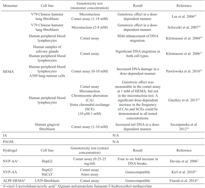
Table 1: Genotoxic potential of HEMA monomer and extracts of polymerized hydrogels assessed in various cell types by employing
diferent genotoxicity tests.
Monomer Cell line Genotoxicity test
(monomer concentration) Result Reference
HEMA
V79 Chinese hamster
lung ibroblasts Comet assay (1-18 mM)Micronucleus
Genotoxic efect in a
dose-dependent manner. Lee et al. 2006
28
V79 Chinese hamster
lung ibroblasts Micronucleus (2-8 mM) Genotoxic efect in a dose-dependent manner. Schweikl et al. 2007
29
Human peripheral blood
lymphocytes Comet assay
Mild enhancement of DNA
migration. Kleinsasser et al. 2004
30
Human samples of salivary glands Human peripheral blood
lymphocytes
Comet assay Signiicant DNA migration in
both cell types. Kleinsasser et al. 2006
31
Human peripheral blood lymphocytes A549 lung-tumour cells
Comet assay (0-10 mM) Increased DNA damage in a
dose-dependent manner Pawlowska et al. 2010
32
Human peripheral blood lymphocytes
Comet assay Micronucleus Chromosome aberration
(CA)
Sister chromatid exchange (SCE)
(10 μM-1 mM)
Genotoxic efect was
measurable in the comet assay at 1 mM of HEMA, but not
in the micronucleus test. A
signiicant dose-dependent increase in the frequency
of CAs and SCEs could be demonstrated in all tested
concentrations.
Ginzkey et al. 201533
Human gingival
ibroblasts Comet assay (1-10 mM) Increased tail DNA in a dose-dependent manner.
Szczepanska et al. 201234
IA N/A
PAGM N/A
Hydrogel Cell line Genotoxicity test (extract
concentration) Result Reference
NVP-AAa HepG2 Comet assay (0.25-25
mg/ml)
Four to six fold increase in
DNA breaks. Devine et al. 2006
5
NVP-AA HepG2
HaCaT
Comet assay
Ames assay Genocompatible Kirf et al. 2010
23
ALPF-HEMAb L929 ibroblasts Comet assay Genocompatible Finosh et al. 201435 aN-vinyl-2-pyrrolidone/acrylic acid b Alginate-polypropylene fumarate/2-hydroxyethyl methacrylate
Over the past decade, Comet assay was developed as
a rapid, simple, and sensitive technique for analyzing and quantifying DNA damage in individual cells37,38. The Comet assay, also called single cell gel electrophoresis (SCGE),
combines DNA gel electrophoresis with luorescence
microscopy in order to visualize migration of DNA strands from individual agarose-embedded cells39. The resulting image resembles a “comet” with a distinct head consisting of intact DNA, and a tail which contains damaged or broken pieces of DNA. The amount of DNA liberated from the head of the comet during electrophoresis depends on genotoxic potential of tested compound37. Over time this method has been improved and today it is suitable for detection of DNA damage caused by double and single strand breaks, alkali labile sites, DNA crosslinking with DNA or protein and oxidative base damage. The advantages of Comet assay, relative to the other genotoxicity tests, include its high
sensitivity for detecting low levels of both single and double stranded breaks in damaged DNA, small number of cells
per sample, lexibility, low cost and ease of application40,41. Comet assay is increasingly used to test genotoxicity of hydrogels’ extracts42 and other biomaterials13,30.
In our previous investigation we reported the radiation-induced synthesis of copolymeric hydrogels composed of 2-hydroxyethyl methacrylate (HEMA), itaconic acid (IA) and
diferent poly(alkylene glycol) (meth)acrylates (PAGM)43.
The PAGM components and itaconic acid were used to improve the hydrophilicity of PHEMA. Itaconic acid, as a
ionic component, imparts pH sensitivity and inluences the
swelling and mechanical properties of hydrogels20. PHEMA and P(HEMA/IA/PAGM) hydrogels were characterized by Fourier transform infrared (FT-IR) spectroscopy, scanning electron microscopy (SEM) and thermogravimetric (TG)
chemical structure, porosity and thermal properties to be used as multifunctional hydrogels in biomedical applications, while in vitro citotoxicity test showed that none of the tested hydrogels were cytotoxic43.
The aim of the present study was to further characterize these novel HEMA-based hydrogels through evaluation of their swelling properties and genotoxic potential in vitro. The examined hydrogels were prepared in our laboratory by gamma-irradiated free radical polymerization and included: PHEMA, poly(2-hydroxyethyl methacrylate/itaconic acid/ poly(ethylene glycol)6 acrylate (P(HEMA/IA/PAGM1)) and poly(2-hydroxyethyl methacrylate/itaconic acid/poly(propylene glycol)5 methacrylate (P(HEMA/IA/PAGM2)). To the best
of our knowledge, these are the irst studies referring to
the genotoxic potential of hydrogels composed of above mentioned components, evaluated by Comet assay.
2. Materials and Methods
2.1. Chemicals
2-Hydroxyethyl methacrylate (HEMA), itaconic acid (IA) (both from Sigma-Aldrich, St. Louis, MO, USA), poly(alkylene glycol) (meth)acrylates: poly(ethylene glycol)6 acrylate (PAGM1) and poly(propylene glycol)5 methacrylate (PAGM2) (both from Laporte Chemicals, Luton, UK) were used as components for hydrogel preparation. Dulbecco’s
Modiied Eagle Medium (DMEM), fetal bovine serum
(FBS), stable glutamine, antibiotic-antimycotic solution, Trypsin–EDTA solution were purchased from Biological Industries (Kibbutz Beit-Haemek, Israel). Low melting point agarose and ethidium bromide (EtBr) were obtained from SERVA (Heidelberg, Germany) while regular agarose was obtained from Applied Biosystems (Foster city, CA, USA). Trypan blue dye, disodium EDTA, Tris and Triton X-100 were obtained from Sigma-Aldrich, St. Louis, MO, USA.
2.2. Preparation of hydrogels
The PHEMA and P(HEMA/IA/PAGM) hydrogels were prepared by gamma-irradiated free radical polymerization. The feed composition for each sample is listed in Table 2. According to the PAGM component samples were designated as P(HEMA/IA/PAGM1) and P(HEMA/IA/PAGM2). The same conditions were used to prepare the PHEMA hydrogel. The reaction mixture was degassed prior to polymerization and placed between two glass plates sealed with a PVC spacer (2 mm thick). The reaction solution was irradiated in a 60Co radiation source, under ambient conditions, at a dose rate of 0.5 kGy/h, to absorbed dose of 25 kGy. After the reaction, the hydrogels were cut into discs and immersed in water for one week. Water was changed daily and collected. After 7 days, the collected water was concentrated to a smaller volume by evaporation and was used for determination of unreacted monomers.The
amount of unreacted IA was determined by titration of extract against NaOH (0.05 mol/L) to phenolphthalein end point. On the other hand, the amount of unreacted HEMA and PAGM components was determined using UV spectroscopy43.
In all cases, the processes indicate that the conversion during polymerization/crosslinking reaction was high as demonstrated in Table 2.
2.3. Swelling study
Dynamic swelling measurements were performed in
phosphate bufer at pH 7.4 and temperature of 37 °C. Swollen
gels were removed from the swelling medium at regular
intervals, dried supericially with ilter paper, weighed and
placed in the same bath until constant weight was reached. The
amount of luid absorbed was monitored gravimetrically. The equilibrium degree of swelling (qe) was calculated as follows:
/
( )
q
e=
Q
m
e-
m
oV
m
o1
where me is the weight of swollen hydrogel at equilibrium
and mo is the weight of xerogel44,45. All swelling experiments were performed in triplicate.
The most important parameters characterizing a hydrogel network structure are the molecular weight between crosslinks (Mc), and the efective crosslinking density (v
e). Caykara et
al.46 described the molecular weight of the polymer chain between two neighboring crosslinks for ionic polymer networks by following relation:
( )
ln
IV
V X
K
K K
K K
K
M
V
4
2 10
10
2
10
1
2
, , , , ,/ ,/ r s pH pHa a a
a a
pH a
s s s
c
r s
2
1 2 22
2
1 1 2
1 2 1 2
2 2 22
1 2 2 3 2 1 3
z
z
z
|z
t
z z
+
+
+
=
-
+
+
+
--Q
T Q
T
V
V
Y
Y
" %where Ka1and Ka2 are the irst and second dissociation
constants of a diprotic acid, X is the weight fraction of ionisable polymer in the system, I is ionic strength of the swelling medium, φ2,S is the polymer volume fraction in the swollen gel, φ2,r is the polymer volume fraction in the relaxed state, V1
is the molar volume of water, ρ is the polymer density, Vr is
the average molar volume of polymer repeating units, and χ is the Flory polymer-solvent interaction parameter. Polymer volume fraction in the relaxed state (φ2,r) is determined according to the following formula:
/
( )
V V
3
,r d r
2
z
=
Q V
where Vd is volume of the polymer sample in dry state and Vr is the volume of the polymer sample in relaxed state, immediately after synthesis. The volumes were calculated
by measuring dimensions of hydrogels discs. The efective
crosslinking density (ve) was calculated using the relation:
/
( )
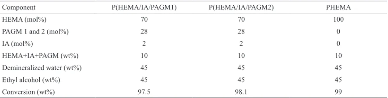
Table 2: Feed compositions for P(HEMA/IA/PAGM) and PHEMA hydrogels.
Component P(HEMA/IA/PAGM1) P(HEMA/IA/PAGM2) PHEMA
HEMA (mol%) 70 70 100
PAGM 1 and 2 (mol%) 28 28 0
IA (mol%) 2 2 0
HEMA+IA+PAGM (wt%) 10 10 10
Demineralized water (wt%) 45 45 45
Ethyl alcohol (wt%) 45 45 45
Conversion (wt%) 97.5 98.1 99
2.4. Preparation of hydrogels extracts
Individual hydrogel discs were weighed (0.2 g in total) and immersed in 5 ml of complete DMEM (DMEM supplemented with 10% fetal bovine serum, antimycotic-antibiotic solution and 2 mM stable glutamine). Extraction of hydrogels was performed under sterile conditions in
a water bath at 37 °C during 3 days. Extracts were then discarded and diluted with complete DMEM to give inal
concentrations of 10% and 50%.
2.5. Cell culture
Genotoxic potential of hydrogels’ extracts was tested using HeLa cell line (ATCC, Manassas, USA). Cells were
grown in complete DMEM at 37 °C in humidiied atmosphere
containing 5% CO2. Medium was replaced every 2–3 days.
After reaching approximately 70% conluence, cells were
detached by using Trypsin–EDTA solution, centrifuged at
4 °C for 10 min at 1000 rpm, washed and appropriate cell
density was set up by using Trypan Blue Dye.
2.6. Treatment of cells
HeLa cells were seeded in 12-well culture plate at a density of 3 × 10⁴ cells per ml. After 24 hours medium was replaced with extracts of hydrogels in concentrations of 10% and 50%. Incubation of cells with extracts was continued for the next 24 hours. In parallel, the control cells were treated with 200 μM H2O2 for 15 min as positive control. Negative control were cells incubated only with complete DMEM (untreated cells). All samples, as well as controls were examined in triplicate.
2.7. Comet assay
Comet assay was performed according to procedure described by Dhawan et al.47 with some modiications. Following treatment, hydrogels’ extracts and control media
were removed and cell viability was determined as quickly as
possible by using Trypan Blue Dye Exclusion Method to avoid
false positive responses due to cytotoxicity. Subsequently,
100 µl of the cell suspension was mixed with low-melting
point agarose (0.75% wt), and 50 µl cell-aliquots were spread
on slides precoated with normal-melting point agarose (1% wt). The slides were immersed into cold, freshly made lysis solution (2.5 M NaCl, 100 mM disodium EDTA, 10 mM Tris, 1% Triton X-100; pH 10). Lysis of cells was performed in
dark and cold room (5 °C) for 2 hours. Following alkaline
unwinding for 40 min by immersion into cold electrophoresis
bufer (300 mM NaOH, 1 mM EDTA; pH ≥ 13), the slides
were subjected to electrophoresis at 30 V and 300 mA for
40 min. Slides were then neutralized in 400 mM Tris bufer
(pH 4) for 5 min and stained with EtBr (20 μg/mL). DNA
migration was observed by using luorescence microscope
LEICA DMR (Wetzlar, Germany) and images were taken
from approximately 10 ields of the each slide by using
LEICA DC 300 camera. Image analysis system CometScore v1.5 (TriTek Corp., USA) was employed to determine the percentage of DNA in comet tail (%DNAt) and the tail moment (Mt). These parameters were calculated as follows:
%
DNAt
=
Ic
It
#
100
( )
5
%
( )
Mt
=
DNAt
#
Lt
6
where It is the total comet tail intensity, Ic is the total comet intensity and Lt is the tail length.
2.8. Statistical analysis
The Comet assay was conducted on duplicate slides per concentration of extract of each hydrogel, as well as controls, with approximately 100 cells scored per slide. Normality of data was evaluated with the Kolmogorov-Smirnov test. The
statistical diference between control and treated cells was
analyzed with the nonparametric Mann-Whitney test using %DNAt and Mt values. The signiicance level was set at p ≤ 0.01. Multiple correlation analysis was performed in order
to estimate the combined inluence of independent variables
(extract concentration, crosslinking density (νe) and swelling
equilibrium (qe)) on the dependent variable (%DNAt). Regression
analysis was employed to further deine degree and type of
relationship between independent and dependent variables,
which showed signiicant correlation. Statistical analysis was
3. Results and Discussion
3.1. Swelling properties
The physical behavior of hydrogels is dependent on
their equilibrium and dynamic swelling behavior in aqueous
media. For application of hydrogels, swelling and shrinking kinetics are very important, e.g. in controlled release drug delivery systems, where the kinetics determine the rate of
difusion of the active component from the gel matrix and
in gel extraction where the gel is swollen and shrunk several times48. Swelling kinetics of synthesized samples was determined by monitoring the swelling process in phophate
bufer mimicking physiological conditions (pH 7.4 and 37
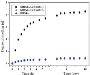
oC). The equilibrium swelling degree (q
e) values were in
the range of 0.42 - 4.28 (Figure 1). After the equilibrium swelling was reached all samples were kept in bufer solution
for additional 10 days. Figure 1 shows that in that period qe
values remained practically constant. Furthermore, at the end of this period, all samples had a soft consistency and exhibited a transparent and colorless appearance.
According to the potential biomedical application, the calculations of crosslinking densities were done for the
results obtained in pH 7.4, at 37 °C. The values of efective
crosslinking densities for PHEMA, P(HEMA/IA/PAGM1) and P(HEMA/IA/PAGM2) were calculated as 48.18, 0.187 and 47.51 mol/dm3, respectively. It is evident that in case of terpolymers (P(HEMA/IA/PAGM1) and P(HEMA/IA/
PAGM2)), efective crosslinking density depends very
much on PAGM component, i.e. alkylene glycol pendant chains in the polymeric network. Theνe values follow the expected trend in accordance with the hydrophilic character of the PAGM component and the crosslinking degree of the
sample. The sample with the highest equilibrium degree of
swelling (P(HEMA/IA/PAGM1)) has the lowest νe value.
Due to the higher sensitivity of propylene glycol units in P(HEMA/IA/PAGM2) hydrogel to gamma radiation, in comparison to ethylene glycol units in P(HEMA/IA/ PAGM1) hydrogel, a higher crosslinking density in the case of P(HEMA/IA/PAGM2) hydrogel was obtained. Therefore, the crosslinking density values along with the hydrophobic/ hydrophilic character of monomer residues in hydrogel are in good accordance with their qevalues.
3.2. Comet assay
Table 3 represents the results of Trypan Blue Dye Exclusion Method, employed to evaluate cytotoxicity concurrently with Comet assay.
Although a dose-dependent decrease in HeLa cell viability was observed, more than 70% of cells were viable after treatment with hydrogels’ extracts, regardless the hydrogel type and extract concentration. According to ISO standard
(ISO 10993-12), preparation of luid extracts of the device materials is the most appropriate technique when there is a
need to determine toxicity of possible chemical leachables, especially by Comet assay5,38. Because DNA damage is associated with cell death, evaluation of genotoxicity is only relevant at sub-cytotoxic concentrations of examined samples. It is crucial to evaluate cytotoxicity at the end of the exposure period and general approach is to exclude concentrations that decrease cell viability by more than 30%50,51. Since the results of the Trypan Blue assay performed in this study show that cell viability, after exposure to the both concentrations of tested extracts, is higher than 70%, none of the hydrogels’ extracts can be considered cytotoxic to HeLa cells, and these extracts concentrations (10% and 50%) are suitable for further genotoxicity examination. Also, cell viability determined in this study was similar with the results obtained by using neutral red method in our previous characterization of these hydrogels43.
Figure 2 shows the representative images of HeLa cells after staining with EtBr in Comet assay.
The results of the Comet assay show that extracts of all tested hydrogels are capable to induce certain genotoxic
Figure 1: Swelling proiles and equilibrium degree of swelling (qe) of HEMA-based hydrogels in phosphate bufer (pH 7.4) at 37 °C.
The swelling properties depend on many factors such as network density, solvent nature and polymer-solvent interaction parameter4,49. P(HEMA/IA/PAGM1) hydrogel swells more than P(HEMA/IA/PAGM2) terpolymer and PHEMA homopolymer network. Terpolymer hydrogels
contain diferent PAGM components, with pendent chains of diferent length which inluenced the swelling process.
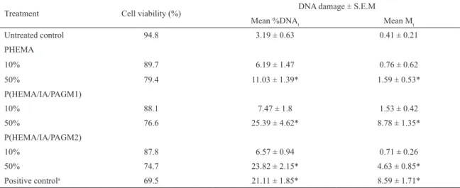
Table 3. Cell viability and Comet assay data for HeLa cells exposed to the extracts of HEMA-based hydrogels for 24 h.
Treatment Cell viability (%) DNA damage ± S.E.M
Mean %DNAt Mean Mt
Untreated control 94.8 3.19 ± 0.63 0.41 ± 0.21
PHEMA
10% 89.7 6.19 ± 1.47 0.76 ± 0.62
50% 79.4 11.03 ± 1.39* 1.59 ± 0.53*
P(HEMA/IA/PAGM1)
10% 88.1 7.47 ± 1.8 1.53 ± 0.42
50% 76.6 25.39 ± 4.62* 8.78 ± 1.35*
P(HEMA/IA/PAGM2)
10% 87.8 6.57 ± 0.94 0.71 ± 0.26
50% 74.7 23.82 ± 2.15* 4.63 ± 0.85*
Positive controla 69.5 21.11 ± 1.85* 8.59 ± 1.71*
a Positive control agent: 200 μM H
2O2; * Denotes a signiicant diference from the untreated control (p ≤ 0.01).
Figure 2: Representative images of HeLa cells in Comet assay;
untreated cells (a), cells treated with 200 μM H2O2 (b) and cells
treated with two concentrations (10% and 50%) of PHEMA (c, d), P(HEMA/IA/PAGM1) (e, f) and P(HEMA/IA/PAGM2) (g, h)
hydrogels’ extracts. Magniication ×400.
efects, as measured by %DNAt and Mt parameters (Table
3, Figure 2). Extracts of tested HEMA-based hydrogels in
concentration of 10% induced no signiicant increase in
%DNAt and Mt compared with untreated cells. Signiicant augmentation of DNA migration (p < 0.01) was evident only
after treatment of HeLa cells with higher extract concentration (50%).Extracts of PHEMA, P(HEMA/IA/PAGM1) and P(HEMA/IA/PAGM2) hydrogels in concentration of 50%
induced 1.8 (p ≤ 0.05), 3.4 (p ≤ 0.01) and 3.6 (p ≤ 0.01)
times higher %DNAt compared with %DNAt induced by lower concentration (10%) of the same extracts. Also, higher extracts’ concentrations induced up to 3.6 times higher Mt than same extracts of lower concentration. Mt parameter is also presented as Tukey box plot diagram (Figure 3). The outliners that can be seen in untreated control as well as in treated cells, are probably the cells that underwent spontaneous apoptosis/necrosis events29 and were not included in the analysis.
Figure 3: Tukey box plot diagram for tail moment parameter of HeLa cells treated with 10% and 50% extracts of HEMA-based
hydrogels, 200 μM H2O2 as positive control and untreated cells.
Whiskers extend to the 1.5 box heights. If minimum and maximum values are out of this range, then they are shown as outliners (dots
above boxes). *Denotes a statistically signiicant diferences
between concentrations of extracts and compared with untreated
control (p ≤ 0.01).
(extract concentration, νe and qe) on the dependent variable (%DNAt), we performed multiple correlation analysis.
As presented in Figure 4a, extract concentration showed the strongest positive impact on genotoxicity as determined
by correlation coeicient (0.82). We further analized the
relationship between extract concentration and genotoxicity by employing regression analysis. As can be seen in Figure
4b, coeicient of determination (R2) equals 0.68, which
means that 68% of the variation in genotoxicity is explained
by the extract concentration. According to the coeicients, the equation of regression line is:
extract. The extent of DNA damage induced by P(HEMA/
IA/PAGM1) is about 1.1 (p ≤ 0.05) time higher than in the
case of P(HEMA/IA/PAGM2) hydrogels’ extract. Therefore, it is obvious that properties of hydrogel such as its degree of
crosslinking, swelling equilibrium and genotoxic potential, are
related. Impact of these independent variables on genotoxicity was also tested by employing multiple correlation analysis (Figure 4a). Crosslinking density of the hydrogel and its
swelling equilibrium are in almost perfect negative correlation
- increasing of crosslinking density reduces the swelling
equilibrium. These two variables have opposite inluence on
genotoxicity (%DNAt) – increasing of crosslinking density
reduces while increasing of swelling equilibrium increases the genotoxic efect of hydrogel extracts. However, the inluence of crosslinking density and swelling equilibrium
on overall genotoxicity is relatively weak as measured
by correlation coeicients (≈ 0.26, Figure 4a). PHEMA
homopolymer and P(HEMA/IA/PAGM2) terpolymer have
higher efective crosslinking density (νe), lower equilibrium degree of swelling (qe), and show less pronounced genotoxic
efect than P(HEMA/IA/PAGM1) terpolymer. The generally
accepted property of highly crosslinked polymers is that they are more resistant to degradative processes and elution of unreacted components, based on more limited space and
pathways available for solvent molecules to difuse within
.
.
( )
y
=
3 41
+
0 33
x
7
In other words, for each unit increase in extract concentration (x), genotoxicity (y) increases with 0.33 units. This could be valuable information when there is a need to predict the
genotoxic efect of diferent concentrations of extract.
The %DNAt values of the cells treated with the same concentration (50%) of extracts of tested hydrogels were compared. The results indicate that genotoxic potential increases in this order: PHEMA<P(HEMA/IA/PAGM2)<P(HEMA/IA/ PAGM1). In other words, extracts of P(HEMA/IA/PAGM1) and P(HEMA/IA/PAGM2) hydrogels induced about 2.2
(p ≤ 0.01) times higher %DNAt than PHEMA hydrogel’s
Figure 4: (a) Multiple correlation analysis employed to determine the strength and direction of the association between the independent variables (extract concentration, crosslinking density (νe) and swelling equilibrium (qe)) and the one dependent variable (%DNAt). Correlation coeicient ranges between – 1 to +1, and quantiies the stenght (the closer coeicient is to 1, the stronger linear association
is) and the direction (positive or negative) of the association. (b) Regression analysis of the relationship between extract concentration
structure13,52,53. The elution of unreacted components from
hydrogels is inluenced by several factors including the
chemical composition of components in hydrogel synthesis, the conversion during polymerization reaction and the degree of crosslinking of the polymeric network13. Due to the lowest degree of crosslinking, extract of P(HEMA/IA/ PAGM1), which showed the highest level of genotoxicity, probably contains more unreacted components comparing with extracts of PHEMA and P(HEMA/IA/PAGM2) samples. In the case of tested hydrogels, these unreacted monomers are IA, methacrylates HEMA and PAGM2, and acrylate PAGM1. It is well known that unreacted monomers in hydrogels can cause the living tissue damage13,54. Itaconic acid is a component of natural origin and it is supposed that will not induce genotoxicity. (Meth)acrylate monomers are reported
in the literature to exhibit genotoxic efects24-30. Furthermore,
Dearield et al.55 showed that acrylates are generally more potent to induce mutations, abberations and micronuclei than methacrylates, although this appears to be structure-related (dependent upon number of functional vinyl groups and the length of oxyethylene chains). PAGM1 has functional vinyl group and longer oxyethylene chain than PAGM2 and this
structural diference may be responsible for its more potent genotoxic efect detected in Comet assay. However, this study is the irst step in biocompatibility assessment of these
novel HEMA-based hydrogels and it is early to comment on the mechanisms underlying the increased DNA migration observed in the alkaline version of Comet assay.
4. Conclusion
Swelling studies conirmed that hydrogels based
on 2-hydroxyethyl methacrylate, poly(alkylene glycol) (meth)acrylates and itaconic acid swell in phosphate
bufer but maintain physical integrity and have soft and
rubbery consistency even when the swelling experiments
were conducted for a long time after the equilibrium state
was reached. The results of the Comet assay showed that extracts of all tested hydrogels are capable to induce
certain genotoxic efects, which depended on chemical
composition, extract concentration as well as on degree of crosslinking of examined hydrogels. Future research would be directed toward optimization of the synthesis of these novel HEMA-based hydrogels in order to obtain hydrogels that are not genotoxic at all and, as such, may have application in clinical practice.
5. Acknowledgments
This work was supported by the Ministry of Education, Science and Technological Development of the Republic of Serbia under Grant [III 41017] and Grant [OI 172062].
6. Conlict of interests
Authors declare that they have no conlict of interests.
7. References
1. Chaterji S, Kwon IK, Park K. Smart polymeric gels: Redeining
the limits of biomedical devices. Progress in Polymer Science. 2007;32(8-9):1083-1122.
2. Urban MW. Stratiication, stimuli-responsiveness, self-healing,
and signaling in polymer networks. Progress in Polymer Science. 2009;34(8):679-687.
3. Drury JL, Mooney DJ. Hydrogels for tissue engineering:
scafold design variables and applications. Biomaterials. 2003;24(24):4337-4351.
4. Zhu J, Marchant RE. Design properties of hydrogel
tissue-engineering scafolds. Expert Review of Medical Devices. 2011;8(5):607-626.
5. Devine DM, Devery SM, Lyons JG, Geever LM, Kennedy JE, Higginbotham CL. Multifunctional polyvinylpyrrolidinone-polyacrylic acid copolymer hydrogels for biomedical applications.
International Journal of Pharmaceutics. 2006;326(1-2):50-59. 6. Caló E, Khutoryanskiy VV. Biomedical applications of hydrogels:
A review of patents and commercial products. European Polymer Journal. 2015;65:252-267.
7. Yates DW, Hadield JM. Clinical experience with a new hydrogel
wound dressing. Injury. 1984;16(1):23-24.
8. Maldonado-Codina C, Efron N. Hydrogel lenses - Materials and Manufacture: A Review. Optometry in Practice. 2003;4:101-115. 9. Young CD, Wu JR, Tsou TL. Fabrication and characteristics of
polyHEMA artiicial skin with improved tensile properties.
Journal of Membrane Science. 1998;146(1):83-93.
10. Çetin D, Kahraman AS, Gümüşderelioğlu M. Novel scafolds based on poly(2-hydroxyethyl methacrylate) superporous hydrogels for bone tissue engineering. Journal of Biomaterials Science. Polymer Edition. 2011;22(9):1157-78.
11. Costa VC, Costa HS, Vasconcelos WL, Pereira MM, Oréice
RL, Mansur HS. Preparation of hybrid biomaterials for bone tissue engineering. Materials Research.2007;10(1):21-26. 12. Keeney M, Lai JH, Yang F. Recent progress in cartilage tissue
engineering. Current Opinion in Biotechnology. 2011;22(5):734-740.
13. Bakopoulou A, Papadopoulos T, Gareis P. Molecular Toxicology
of Substances Released from Resin–Based Dental Restorative Materials. International Journal of Molecular Sciences. 2009;10(9):3861-3899.
14. Tomić SLj, Dimitrijević SI, Marinković AD, Najman S, Filipović JM. Synthesis and characterization of
poly(2-hydroxyethylmethacrylate/itaconic acid) copolymeric hydrogels.
Polymer Bulletin.2009;63(6):837-851.
15. Fathi M, Entezami AA, Arami S, Rashidi MR. Preparation of N-isopropylacrylamide/itaconic acid magnetic nanohydrogels
by modiied starch as a crosslinker for anticancer drug carriers.
16. Brahim S, Narinesingh D, Giuseppi-Elie A. Release characteristics of novel pH-sensitive p(HEMA-DMAEMA) hydrogels containing 3-(trimethoxy-silyl) propyl methacrylate. Biomacromolecules. 2003;4(5):1224-1231.
17. Ozay O. Synthesis and characterization of novel pH-responsive poly(2-hydroxylethyl methacrylate-co-N-allylsuccinamic acid) hydrogels for drug delivery. Journal of Applied Polymer Science. 2014;131(1):39660. doi: 10.1002/app.39660.
18. García DM, Escobar JL, NoaY, Bada N, Hernáez E, Katime I. Timolol maleate release from pH-sensible poly (2-hydroxyethyl methacrylate-co-methacrylic acid) hydrogels. European Polymer Journal. 2004;40(8):1683-1690.
19. Johnson BD, Beebe DJ, Crone WC. Efects of swelling on the
mechanical properties of a pH-sensitive hydrogel for use in
microluidic devices. Materials Science and Engineering: C. 2004;24(4):575-581.
20. Tomić SLj, Mićić MM, Dobić SN, Filipović JM, Suljovrujić
EH. Smart poly(2-hydroxyethyl methacrylate/itaconic acid) hydrogels for biomedical application. Radiation Physics and Chemistry. 2010;79(5):643-649.
21. Nasr FH, Khoee S, Dehghan MM, Chaleshtori SS, Shaiee A.
Preparation and evaluation of contact lenses embedded with polycaprolactone-based nanoparticles for ocular drug delivery.
Biomacromolecules. 2016;17(2):485-495.
22. Omidian H, Park K, Kandalam U, Rocca J. Swelling and
mechanical properties of modiied HEMA-based superporous
hydrogels. Journal of Bioactive and Compatible Polymers. 2010;25(5):483-497.
23. Kirf D, Higginbotham CL, Rowan NJ, Devery SM. Cyto- and genotoxicological assessment and functional characterization of N-vinyl-2-pyrrolidone-acrylic acid-based copolymeric hydrogels with potential for future use in wound healing applications.
Biomedical Materials (Bristol, England). 2010;5(3):35002. 24. Chang HH, Guo MK, Kasten FH, Chang MC, Huang GF, Wang
YL, et al. Stimulation of glutathione depletion, ROS production and cell cycle arrest of dental pulp cells and gingival epithelial cells by HEMA. Biomaterials. 2005;26(7):745-753. 25. Becher R, Kopperud HM, Al RH, Samuelsen JT, Morisbak E,
Dahlman HJ, et al. Pattern of cell death after in vitro exposure to GDMA, TEGDMA, HEMA and two compomer extracts.
Dental Materials.2006;22(7):630-640.
26. Spagnuolo G, D’Antò V, Cosentino C, Schmalz G, Schweikl
H, Rengo S. Efect of N-acetyl-L-cysteine on ROS production
and cell death caused by HEMA in human primary gingival
ibroblasts. Biomaterials. 2006;27(9):1803-1809.
27. Toy E, Yuksel S, Ozturk F, Karatas OH, Yalcin M. Evaluation of the genotoxicity and cytotoxicity in the buccal epithelial cells of patients undergoing orthodontic treatment with three light-cured bonding composites by using micronucleus testing.
Korean Journal of Orthodontics. 2014;44(3):128-135. 29. Lee DH, Lim BS, Lee YK, Ahn SJ, Yang HC. Involvement
of oxidative stress in mutagenicity and apoptosis caused by dental resin monomers in cell cultures. Dental Materials. 2006;22(12):1086-1092.
29. Schweikl H, Hartmann A, Hiller KA, Spagnuolo G, Bolay C,
Brockhof G, Schmalz G. Inhibition of TEGDMA and
HEMA-induced genotoxicity and cell cycle arrest by N-acetylcysteine.
Dental Materials.2007;23(6):688-695.
30. Kleinsasser NH, Wallner BC, Harréus UA, Kleinjung T, Folwaczny M, Hickel R, et al. Genotoxicity and cytotoxicity of dental materials in human lymphocytes as assessed by the single cell microgel electrophoresis (comet) assay. Journal of Dentistry. 2004;32(3):229-234.
31. Kleinsasser NH, Schmid K, Sassen AW, Harreus UA, Staudenmajer
R, Folwaczny M, et al. Cytotoxic and genotoxic efects of resin
monomers in human salivary gland tissue and lymphocytes as assessed by the single cell microgel electrophoresis (comet) assay. Biomaterials. 2006;27(9):1762-1770.
32. Pawlowska E, Poplawski T, Ksiazek D, Szczepanska J, Blasiak J. Genotoxicity and cytotoxicity of 2-hydroxyethyl methacrylate.
Mutation Research. 2010;696(2):122-129.
33. Ginzkey C, Zinnitsch S, Steusslof G, Koehler C, Hackenberg S,
Hagen R, et al. Assessment of HEMA and TEGDMA induced DNA damage by multiple genotoxicological endpoints in human lymphocytes. Dental Materials. 2015;31(8):865-876. 34. Szczepanska J, Poplawski T, Synowiec E, Pawlowska E,
Chojnacki CJ, Chojnacki J, et al. 2-hydroxylethyl methacrylate (HEMA), a tooth restoration component, exerts its genotoxic
efects in human gingival ibroblasts trough methacrylic acid,
an immediate product of its degradation. Molecular Biology Reports. 2012;39(2):1561-1574.
35. Thankam FG, Muthu J. Iniltration and sustenance of viability
of cells by amphiphilic biosynthetic biodegradable hydrogels.
Journal of Materials Science: Materials in Medicine. 2014;25(8):1953-1965.
36. Samuelsen JT, Holme JA, Becher R, Karlsson S, Morisbak E, Dahl JE. HEMA reduces cell proliferation and induces apoptosis in vitro. Dental Materials. 2008;24(1):134-140.
37. Liao W, McNutt MA, Zhu WG. The comet assay: a sensitive method for detecting DNA damage in individual cells. Methods. 2009;48(1):46-53.
38. Tice RR, Agurell E, Anderson D, Burlinson B, Hartmann A, Kobayashi H, et al. Single cell gel/comet assay: guidelines for
in vitro and in vivo genetic toxicology testing. Environmental and Molecular Mutagenesis. 2000;35(3):206-221.
39. Olive PL, Banáth PJ. The comet assay: a method to measure DNA damage in individual cells. Nature Protocols. 2006;1(1):23-29. 40. Collins AR, Dobson VL, Dusinská M, Kennedy G, Štětina R. The comet assay: what can it really tell us? Mutation Research/ Fundamental and Molecular Mechanisms of Mutagenesis. 1997;375(2):183-193.
41. Collins AR, Oscoz AA, Brunborg G, Gaivão I, Giovannelli L, Kruszewski M, et al. The comet assay: topical issues.
Mutagenesis. 2008;23(3):143-151.
43. Tomić SLj, Babić MM, Antić KM, Vuković JSJ, Malešić NB, Filipović JM. pH sensitive hydrogels based on (meth)acrylates and
itaconic acid. Macromolecular Research. 2014;22(11):1203-1213. 44. Bell CL, Peppas NA. Measurement of the swelling force in
ionic polymer networks. III. Swelling force of interpolymer complexes. Journal of Controlled Release. 1995;37(3):277-280. 45. Peppas NA. Analysis of Fickian and non-Fickian drug release from polymers. Pharmaceutica Acta Helvetiae. 1985;60(4):110-111. 46. Çaykara T, Doǧmuş M, Kantoǧlu Ö. Network structure and swelling-shrinking behaviours of pH-sensitive
poly(acrylamide-co-itaconic acid) hydrogels. Journal of Polymer Science Part B Polymer Physics. 2004;42(13):2586-2594.
47. Dhawan A, Bajpayee M, Pandey AK, Parmar D. Protocol for the single cell gel electrophoresis/comet assay for rapid genotoxicity assessment. Lucknow: Industrial Toxicology Research Centre; 2003. p 1-10. Available from: <http://www. cometassayindia.org/protocol%20for%20comet%20assay.pdf>. Access in: 30/7/2016.
48. Andersson M, Axelsson A, Zacch G. Swelling kinetics of poly(N-isopropylacrylamide) gel. Journal of Controlled Release. 1998;50(1-3):273-281.
49. Hofman AS. Hydrogels for biomedical applications. Advanced Drug Delivery Reviews. 2002;54(1):3-12.
50. Henderson L, Wolfreys A, Fedyk J, Bourner C, Windebank S. The ability of the Comet assay to discriminate between genotoxins and cytotoxins. Mutagenesis. 1998;13(1):89-94.
51. Anderson D, Yu TW, McGregor DB. Comet assay responses as indicators of carcinogenic exposure. Mutagenesis. 1998;13(6):539-555.
52. Krongauz VV. Difusion in polymers dependence on crosslink
density. Journal of Thermal Analysis and Calorimetry. 2010;102(2):435-445.
53. Wu Y, Joseph S, Aluru NR. Efect of cross-linking on the difusion of water, ions, and small molecules in hydrogels. TheJournal of Physical Chemistry B. 2009;113(11):3512-3520. 54. Das N. Preparation methods and properties of hydrogel: a
review. International Journal of Pharmacy and Pharmaceutical Sciences. 2013;5(3):112-117.
55. Dearield KL, Milis CS, Harrington-Brock K, Doerr CL, Moore