Icariin reduces human colon carcinoma cell growth
and metastasis by enhancing p53 activities
Meili Tian
1, Shuang Yang
2and Xinpeng Yan
31Department of Health Care, Shengli Oil
field Central Hospital, Dongying, Shandong, China
2Department of Health Management, Shengli Oil
field Central Hospital, Dongying, Shandong, China
3Department of Traditional Chinese Medicine, Shengli Oil
field Central Hospital, Dongying, Shandong, China
Abstract
Icariin has been reported to possess high anticancer activity. Colon carcinoma is one of the leading causes of cancer-related mortality worldwide. Here, the anticancer activity of icariin against HCT116 colon carcinoma cells and the possible underlying mechanism were studied. The trypan blue staining assay, wound healing assay, clonogenic assay, CCK-8 assay, and Annexin V-FITC/PI double staining method were carried out to determine the changes of HCT116 cell growth and migration. mRNA and protein expressions were determined by quantitative real-time PCR and western blot, respectively. Moreover, small interfering RNA (siRNA) plasmid was used to examine the role of p53 in icariin-induced apoptosis in HCT116 cells. Icariin significantly suppressed colon carcinoma HCT116 cells by decreasing migration and viability, and simultaneously promoting apoptosis. Icariin exerted the anti-tumor effect in a dose-dependent manner by up-regulating p53. During treatment of icariin, p-p53, p21, and Bax levels increased, and Bcl-2 level decreased. Short time treatment with icariin induced DNA damage in HCT116 cells. Furthermore, the cytotoxicity of icariin was decreased after p53 knockdown or by using caspase inhibitors. p53 was involved in activities of caspase-9 and caspase-3. Icariin repressed colon carcinoma cell line HCT116 by enhancing p53 expression and activating p53 functions possibly through Bcl-2/Bax imbalance and caspase-9 and -3 regulation. Icariin treatment also induced DNA damage in HCT116 cells.
Key words: Colon carcinoma; Icariin; Mechanism; p53; DNA damage; Caspases
Introduction
Colon carcinoma is a common type of malignant tumor of the alimentary system, with high morbidity and mortality. In recent years, the incidence of colon carcinoma has increased worldwide. Due to increasing resistance to conventional treatment, new therapies should be urgently explored (1).
Icariin, a flavonol glycoside and a major constituent of the extract from leaf and stem of Herba epimedii (Berberidaceae) plant, has been found to have antineo-plastic activities against a variety of human malignancies (2,3). As a tumor inhibitor, icariin has been shown to inhibit cell growth by arresting cells in G1 phase and decreasing mitochondrial transmembrane potential in prostate carci-noma cells (4). Icariin also exerted its negative effects on human gastric cancer cell invasion and migration by vasodilator-stimulated phosphoprotein via Rac1 pathway (5), and regulated the proliferation and apoptosis of human ovarian cancer cells through microRNA-21 by targeting some tumor suppressor genes (6). Icariin showed high potential of anti-tumor effect on many cancer cells and the anticancer mechanisms have been widely researched.
However, the biological role of icariin in colon carci-noma and its underlying molecular mechanism remain undefined.
Some studies reported that the transcriptional factor p53 played an indispensable role in active function of icariin (7,8). p53 is one of the most important tumor sup-pressors in cells, which can protect normal cell growth and initiate malignant cell death. In unstressed cells, the level and activity of p53 is strictly controlled especially by the ubiquitin E3 ligase mdm2 (9). Blocking the mdm2-p53 inter-action and reactivating p53 function is a promising ther-apeutic strategy for the treatment of cancers (10). p53 can be activated when cells suffer toxic stresses, inducing cell growth arrest, cell senescence, and apoptosis (11,12). Thus the functions of p53 in icariin-treated cells were analyzed.
In this study, the anti-tumor effect of icariin in human colon carcinoma cells was assessed. The interaction between icariin and p53 was also investigated in order to reveal the underlying action mechanisms of icariin and the role of p53 in the anti-tumor effect of icariin.
Correspondence: Xinpeng Yan:<yanxinpeng815@126.com>
Material and Methods
Cell culture and transient transfection
Human colon carcinoma HCT116 cells and normal colon epithelial FHC cells were obtained from the American Type Culture Collection (USA). Cells were cultured in Dulbecco’s modified Eagle medium (Hyclone, USA) and supplemented with 10% fetal calf serum (Hyclone), in a humidified incubator containing 95% air and 5% CO2at
37°C. The specific siRNA against p53 was purchased from Santa Cruz Biotech (USA). Transfections were carried out using Lipofectamine 2000 reagent (Invitrogen, USA) according to instructions. After 48 h of transfection, cells were harvested for analyses.
Cell viability assay
HCT116 cells were plated in 24-well plates at a density of 1105 cells per well and then treated with various
doses of icariin (Shanghai U-sea Biotech Co., Ltd., China). Wells added with DMSO were used as negative controls. Cells were trypsinized and stained with trypan blue dye, and viable cells were counted using a cell counting chamber every day for a total of 5 days. Finally, cell growth curves were plotted according to the viable cell numbers of each group. The viabilities of the HCT116 and FHC cells after icariin treatment were assessed by Cell Counting Kit-8 (CCK-8, Dojindo Molecular Technologies, USA), and the difference between them was analyzed. HCT116 cells were seeded in a 96-well plate at a density of 5103cells
per well. After icariin treatment, 20 mL CCK-8 solution was added to the culture medium and the cultures were further incubated for 1 h at 37°C in humidified 95% air and 5% CO2. After incubation, the absorbance was measured
at 450 nm using a Microplate Reader (Bio-Rad, USA).
Wound healing assay
A wound healing assay was conducted to evaluate the migratory capacity of HCT116 cells in each group. Equal numbers of cells were cultured to 95% confluence in 6-well plates. The wound gaps were created by scratching cell sheets with a sterile 200 mL-pipette tip. Wells were washed twice with PBS to remove thefloating cells and added with fresh mediums containing DMSO or icariin with final concentrations of 50 and 100 nM. Changes of the scratched areas were observed by an inverted micro-scope (Leica, Germany) every 24 h. Wound widths were measured to calculate the relative widths.
Clonogenic assay
HCT116 cells were plated in 60-mm dishes (1000 cells per well) and cultured in a medium containing DMSO or icariin. After 2 weeks, cell clones werefixed with a 4% paraformaldehyde solution (Beyotime Biotechnology, China) and stained with 0.1% crystal violet (Beyotime Biotechnology). Visible colonies on plates with different treatments were captured by ChemiDoc XRS+imaging system (Bio-Rad).
The surviving fraction was calculated as a ratio of the number of colonies to the number of plated cells (plating efficiency), divided by the same ratio calculated for the non-treated group.
Apoptosis assay
Annexin V-FITC/PI apoptosis detection kit (Beijing Biosea Biotechnology, China) was used for apoptosis analysis. Briefly, cells were seeded on 6-well plates (1105cells/well). Treated cells were washed twice with
PBS and resuspended in a binding buffer (KeyGEN Biology, China). The adherent andfloating cells were collected and treated following the manufacturer’s protocol, and sam-ples were immediately analyzed on a flow cytometer (Beckman Coulter, USA) to differentiate apoptotic cells (Annexin-V positive and PI-negative) from necrotic cells (Annexin-V and PI-positive).
Real-time PCR
HCT116 cells were treated with various concentrations of icariin for 24 h. Total RNA was then extracted using the RNAiso Plus Reagent (TaKaRa, China) according to the manufacturer’s instructions. Total RNA with 500 ng was reversely transcribed to cDNA using PrimeScriptTM RT Master Mix (Perfect Real Time (TaKaRa). Real-time PCR was performed on the CFX 96 Real-time PCR detection system (Bio-Rad) using SYBRs Premix Ex TaqTM II
(Tli RnaseH Plus) and specific primers (TaKaRa). The mRNA level of each gene was normalized tob-actin with 2-DDCtmethod using Bio-Rad CFX Manager V1.1.308.1111 software.
Western blot
Bio-Rad ChemiDoct XRS system, with an addition of 200 mL Immobilon western chemiluminescent HRP sub-strate (Millipore, USA) to cover the membrane surface. The signals were captured and the intensity of the bands was quantified using Image LabtSoftware (Bio-Rad).
Statistical analysis
All experiments were repeated three times. Results are reported as means ±SD. Statistical analyses were
performed using Graphpad statistical software (GraphPad Software Inc., USA) and P values were calculated using a one-way analysis of variance (ANOVA). Po0.05 was
considered to be significant.
Results
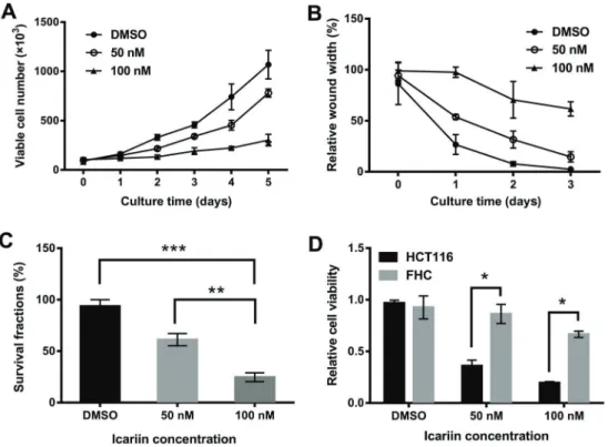
Icariin inhibited the growth and migration of colon carcinoma cells
The effect of icariin on HCT116 cell viability was spe-cifically analyzed. The results showed that viability was inhibited, with an enhanced effect after an increase in icariin concentration and extension in treatment time (Figure 1A). Next, the effect of icariin on cell migration was detected by
a wound healing assay. As shown in Figure 1B, icariin at 50 and 100 nM effectively suppressed cell migration in a time-dependent manner, compared with DMSO control. Meanwhile, a clonogenic assay was carried out to analyze the effect of icariin on HCT116 cell growth. Compared with the number of colonies in the DMSO control group, those in icariin-treated groups decreased significantly, suggest-ing that icariin markedly suppressed HCT116 cell growth (Figure 1C). Moreover, according to the CCK-8 assay, icariin significantly reduced the viability of HCT116 cells compared to normal colon FHC epithelial cells (Po0.05;
Figure 1D), indicating that HCT116 cells were more sensi-tive to icariin.
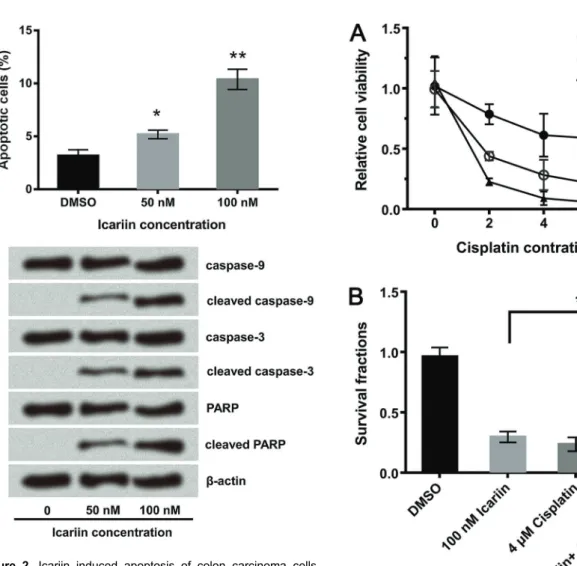
Icariin promoted apoptosis of colon carcinoma cells
The anti-tumor efficacy of icariin was further evidenced by the detection of cell apoptosis. The flow cytometry analysis given in Figure 2A displayed that apoptotic cell rates significantly increased after icariin treatments of 50 nM (Po0.05) and 100 nM (Po0.01), which is
con-sistent with the western blot assay data (Figure 2B). Some apoptosis-promoting proteins were activated by icariin, including cleaved caspase-9 and -3 and cleaved PARP.
Icariin enhanced the anti-tumor effect of cisplatin
Since cisplatin is one of the main clinical therapies for cancer, we evaluated if it could be used in combination with icariin to treat colon carcinoma, or if icariin could enhance the sensitivity of colon carcinoma cells to cisplatin. As shown in Figure 3A, the combination of icariin and cisplatin inhibited tumor cell viability more effectively than their respective treatments separately. In the clonogenic survival assay, the combination significantly enhanced HCT116 cell mortality (Po0.05; Figure 3B).
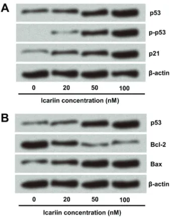
p53 was up-regulated in icariin-treated cells
The anti-colon carcinoma effect of icariin was verified, but the underlying mechanism was unclear. HCT116 cells
express p53, an important tumor suppressor, the over-expression of which can lead to cell cycle arrest and apop-tosis. Therefore, we speculated that p53 might be involved in the carcinostatic role of icariin. According to the results, p53 was indeed up-regulated, and mRNA and protein levels of p53 were enhanced along with the increase in icariin concentration (Figure 4A and B). Phosphorylation of the NH2-terminal residues of p53 could mediate its
stabilization and nuclear accumulation after treatment with anticancer drugs (13). Thus, p-p53 level gradually increased when treated with equally increasing concentrations of icariin. In normal, non-transformed cells, p53 levels are Figure 2. Icariin induced apoptosis of colon carcinoma cells.
A, Apoptotic analysis of HCT116 cells. HCT116 cells were treated with different doses of icariin and incubated with AV-FITC and PI. Percentage of apoptotic cells (AV-FITC +/PI ) was obtained using flow cytometry. B, Western blot analysis of caspase-9 and -3 and PARP proteins in icariin-treated cells. b-actin was used as internal control. Assays were performed in triplicate. Data are reported as means±SD. *Po0.05, **Po0.01 (ANOVA)
kept low through activities of negative regulators, such as mdm2 (14). Some studies reported that under stress and with the inhibition of mdm2/p53 interaction, p53 protein levels rapidly increased (15,16). In this study, we found that mdm2 was down-regulated after icariin treatment, which was accordingly disproportionate to p53 level. Therefore, p53 might be targeted by mdm2 in icariin-treated cells.
Effect of icariin on cell growth-related proteins in colon carcinoma cells
p21 was the key p53 target protein, which was enhanced after icariin treatment (Figure 5). Next, levels of important apoptosis-related proteins Bcl-2 and Bax were also eval-uated. The anti-apoptosis protein Bcl-2 was down-regulated by icariin in a dose-dependent manner, while the pro-apoptosis protein Bax level was increased (Figure 5). These data
suggested that icariin not only enhanced p53 expression, but possibly also regulated other important genes inter-fering with cell growth.
Icariin induced DNA damage in colon carcinoma cells
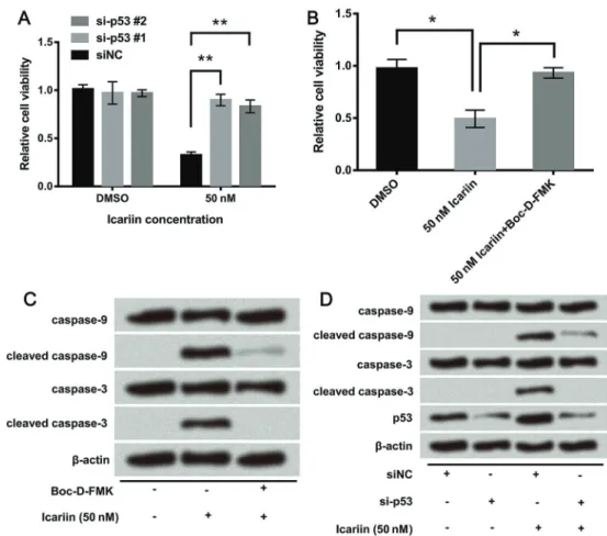
p53 is a key regulator not only of the cell cycle and apoptosis, but also of DNA repair. Due to the abnormal expression of p53, we hypothesized that icariin might induce DNA damage. We evaluated the effect of icariin on the level of g-H2AX (phosphorylated histone H2AX on serine 139, a sensitive DNA damage marker specifically induced by DNA double-strand breaks). Firstly, we con-structed the specific siRNA against p53, to silence p53 in HCT116 cells. p53 was successfully knocked down after transfection with siRNA #1 and siRNA #2 (Figure 6A). The si-p53 #1 was applied in the next experiment. As shown in Figure 6B,g-H2AX was up-regulated by icariin after treat-ment for 6 h, confirming the DNA damage property of icariin. Subsequently, the effect of icariin on DNA damage after p53 knockdown was investigated, and results showed thatg-H2AX expression was not enhanced. This indicated that p53 knockdown inhibited icariin-induced DNA damage and DNA damage was an outcome of p53-induced apoptosis (Figure 6B).
Figure 4. Expression of p53 was enhanced by icariin. A, The mRNA level of p53 was analyzed by real-time PCR.BandC, Protein levels of p53, p-p53, and mdm2 were detected by western blot analysis.b-actin was used as internal control. Assays were performed in triplicate. Data are reported as means±SD. *Po0.05 (ANOVA).
Icariin inhibited viability and promoted apoptosis of colon carcinoma cells through p53-activated caspase-9 and -3
In this study, the role of p53 in icariin-treated colon carcinoma cells was assessed further. Firstly, cell viability was detected when p53-targeted siRNA was transfected into icariin-treated cells. As shown in Figure 7A, the si-p53 #1 and si-p53 #2 abolished the viability-inhibiting effect of icariin. Similar results were found in the treatment group with the addition of Boc-D-FMK (a caspase inhibitor) (Figure 7B), which abolished icariin-induced decrease in cell viability. The western blot assay displayed that high levels of cleaved caspase-9 and -3 induced by icariin were decreased by the addition of Boc-D-FMK, indicating that icariin promoted apoptosis of HCT116 cells by activating caspase-9 and -3 (Figure 7C). As shown in Figure 7D, caspase-9 and -3 were not activated by icariin after p53 knockdown. The data identified p53-dependent caspase-mediated mechanisms leading to apoptosis during treat-ment period with icariin. In summary, caspase-9 and -3 played an important role in the anti-tumor effect of icariin and were regulated by p53.
Discussion
Colorectal cancer is among the most common malig-nancies, and has been rising as one of the main diseases threating public health and quality of life. Its mortality rate is only inferior to gastric carcinoma, esophageal carcinoma, and primary colon cancer, ranking forth in malignant tumors of the digestive system. Although colorectal cancer diagnosis and treatment with chemoradiation (alone or in combination with surgery) has greatly improved, it remains a problem that would benefit from more effective ther-apeutic strategies.
In the present study, we evaluated the anticancer activity of icariin against colon cancer cell line HCT116. The results indicated that growth, migration, colony forma-tion, and viability of HCT116 cells were inhibited by icariin
and the suppressing effect was improved with the increase of icariin concentration. Moreover, icariin was found to promote cell apoptosis in a dose-dependent manner, and some apoptosis-promoting proteins were enhanced after icariin treatment. The obtained results were consistent with other reports. Research showed that icariin induced cytotoxicity with an IC50of 40 mM in esophageal cancer
cells, induced G2/M cell cycle arrest, and inhibited esoph-ageal cancer cell migration, invasion, and metastasis (17). It also exerted negative effects on human ovarian and gastric cancer, among others (3,6).
Our results showed that the combination of icariin with cisplatin provided more effective anti-tumor activity com-pared to treatment with icariin or cisplatin alone. Previous studies demonstrated that icariin enhanced the in vitro antitumor activity of arsenic trioxide against acute pro-myelocytic leukemia (18), as well as the cytotoxicity of doxorubicin in human multidrug-resistant osteosarcoma cells (19). These findings suggest that a combination of icariin with other anticarcinogens might offer a therapeutic benefit to patients with cancer.
Icariin was considered a potential drug for colorectal cancer therapy based on its strong anti-tumor effect, which led to the assessment of the possible underlying mech-anisms. Ourfindings showed that p53 played an important role in the action mechanism of icariin. p53 expression was enhanced both in mRNA and protein levels when HCT116 cells were treated with icariin. Furthermore, p53 overexpression might be caused by abnormal expres-sion of icariin-induced mdm2. The mdm2 can directly bind to p53 and continuously mediate p53 ubiquitination and proteasomal degradation. Besides, p53 level and activity is strictly controlled by mdm2 (20,21). The present study showed that nutlin-bound mdm2 disturbed the p53-mdm2 interaction, and then showed anti-tumor activity by acti-vating p53 (22).
known to be a regulator of Bax, because a p53-binding site has been found in the Bax gene promoter (24). There-fore, p53 is responsible for regulating cell death through Bcl-2/Bax imbalances, which was consistent with our data; down-regulation of Bcl-2 and up-regulation of Bax were found in our study. Additionally, previous studies also reported the role of p53 in HCT116 cells, which showed that cell-cycle arrest in G1 and G2 were regulated by p53 (25), and p53-wild type participated in HCT116 colon carcinoma cell apoptosis, leading to caspase activation and cell death (26). These studies indicated that target-ing p53 might be an effective strategy to fight colon carcinoma.
Cancer therapy drugs such as cisplatin, mitomycin C, etoposide, and a number of other compounds are known to act on cellular DNA (27). Thus, the effect of icariin on cellular DNA was also investigated. Results displayed DNA damage occurring at 6 h after icariin treatment (50 nM), but the damage was inhibited after p53 silencing. It might be possible that p53 knockdown inhibited the
cytotoxicity of icariin and impaired the harmful effect on HCT116 cells, including DNA damage. However, the detailed underlying mechanism requires further study.
Based on our results, icariin exerted its anti-tumor effect by regulating caspase-9 and caspase-3, but the caspases could not be activated by icariin after p53 knockdown. Therefore, caspase-9 and caspase-3 might be directly regulated by p53, and p53 by icariin. Some studies reported that icariin inhibited colorectal cancer cell growth by mainly suppressing NF-kB activity (28,29). However, the pharmacological effects of anti-cancer drugs are particularly complex and their effects might occur in more than one way. Therefore, more studies are needed in future.
In summary, our study showed that icariin effectively inhibited migration and viability of the colon carcinoma cell line HCT116 and promoted apoptosis. Icariin had an anti-tumor effect in a p53-dependent manner and p53 might be regulated by mdm2. Icariin may represent a promising therapeutic strategy for the treatment of human colon carcinoma.
References
1. Li C, Chen J, Wang Y, Song G, Xiao C, Yan D, et al. Correction: High-level expression of P21-Cdc/Rac-activated kinase 7 is closely related to metastatic potential and poor prognosis of colon carcinoma.Oncotarget2017; 8: 45031, doi: 10.18632/oncotarget.18923.
2. Zhang H, Li P, Li J, Song T, Wang L, Li E, et al. Icariin induces apoptosis in acute promyelocytic leukemia by targeting PIM1.Pharmacol Rep2017; 69: 1270–1281, doi: 10.1016/j.pharep.2017.06.005.
3. Wang Y, Dong H, Zhu M, Ou Y, Zhang J, Luo H, et al. Icariin exterts negative effects on human gastric cancer cell inva-sion and migration by vasodilator-stimulated phosphoprotein via Rac1 pathway. Eur J Pharmacol 2010; 635: 40, doi: 10.1016/j.ejphar.2010.03.017.
4. Huang X, Zhu D, Lou Y. A novel anticancer agent, icaritin, induced cell growth inhibition, G1 arrest and mitochondrial transmembrane potential drop in human prostate carcinoma PC-3 cells.Eur J Pharmacol2007; 564: 26–36, doi: 10.1016/ j.ejphar.2007.02.039.
5. Wang Y, Dong H, Zhu M, Ou Y, Zhang J, Luo H, et al. Icariin exterts negative effects on human gastric cancer cell inva-sion and migration by vasodilator-stimulated phosphoprotein via Rac1 pathway. Eur J Pharmacol 2010; 635: 40–48, doi: 10.1016/j.ejphar.2010.03.017.
6. Li J, Jiang K, Zhao F. Icariin regulates the proliferation and apoptosis of human ovarian cancer cells through microRNA-21 by targeting PTEN, RECK and Bcl-2.Oncol Rep2015; 33: 2829, doi: 10.3892/or.2015.3891.
7. Qian ZQ, Wang YW, Li YL, Li YQ, Ling Z, Yang DL. Icariin prevents hypertension-induced cardiomyocyte apoptosis through the mitochondrial apoptotic pathway.Biomed Pharmacother 2017; 88: 823–831, doi: 10.1016/j.biopha.2017.01.147. 8. Huang X, Xiang T, Xu S, Lu S, Liu H, Zhang WC, et al.
[Icariin reduces S-nitrosogultathione induced endothelial cell apoptosis through modulating AKT/P53 pathway].Zhonghua Xin Xue Guan Bing Za Zhi2016; 44: 707–713, doi: 10.3760/ cma.j.issn.0253-3758.2016.08.013.
9. Meek DW. Tumour suppression by p53.Nat Rev Cancer 2009; 10: 714–723, doi: 10.1038/nrc2716.
10. Shangary S, Wang S. Small-molecule inhibitors of the MDM2-p53 protein-protein interaction to reactivate p53 func-tion: a novel approach for cancer therapy.Ann Rev Pharmacol Toxicol2009; 49: 223–241, doi: 10.1146/annurev.pharmtox.48. 113006.094723.
11. Junttila MR, Evan GI. p53 - a Jack of all trades but master of none.Nat Rev Cancer2009; 9: 821, doi: 10.1038/nrc2728. 12. Zhao F, Huang W, Ousman T, Zhang B, Han Y, Clotaire DZJ, et al. Triptolide induces growth inhibition and apoptosis of human laryngocarcinoma cells by enhancing p53 activities and suppressing E6-mediated p53 degradation.PloS One 2013; 8: e80784, doi: 10.1371/journal.pone.0080784. 13. Batchu RB, Gruzdyn OV, Qazi AM, Kaur J, Mahmud EM,
Weaver DW, et al. Enhanced phosphorylation of p53 by microRNA-26a leading to growth inhibition of pancreatic cancer.Surgery2015; 158: 981–986; discussion 6–7, doi: 10.1016/j.surg.2015.05.019.
14. Enge M, Bao W, Hedstrom E, Jackson SP, Moumen A, Selivanova G. MDM2-dependent downregulation of p21 and hnRNP K provides a switch between apoptosis and growth arrest induced by pharmacologically activated p53.Cancer Cell2009; 15: 171–183, doi: 10.1016/j.ccr.2009.01.019. 15. Nayak SK, Khatik GL, Narang R, Monga V, Chopra HK.
p53-Mdm2 interaction inhibitors as novel nongenotoxic anticancer agents. Curr Cancer Drug Targets 2017; doi: 10.2174/1568009617666170623111953.
16. Ganeshan L, Jin XL, O’Neill C. The induction of tumour sup-pressor protein P53 limits the entry of cells into the pluri-potent inner cell mass lineage in the mouse embryo.Exper Cell Res 2017; 358: 227–233, doi: 10.1016/j.yexcr.2017. 06.020.
17. Gu ZF, Zhang ZT, Wang JY, Xu BB. Icariin exerts inhibitory effects on the growth and metastasis of KYSE70 human esophageal carcinoma cells via PI3K/AKT and STAT3 pathways. Environ Toxicol Pharmacol 2017; 54: 7, doi: 10.1016/j.etap.2017.06.004.
18. Zhi W, Hong Z, Dai L, Song T, Ping L, Liu Y, et al. Arsenic Trioxide and Icariin Show Synergistic Anti-leukemic Activity. Cell Biochem Biophysi 2015; 73: 213–219, doi: 10.1007/ s12013-015-0660-2.
19. Wang Z, Yang L, Xia Y, Guo C, Kong L. Icariin enhances cytotoxicity of doxorubicin in human multidrug-resistant osteosarcoma cells by inhibition of ABCB1 and down-regulation of the PI3K/Akt pathway.Biol Pharm Bull2015; 38: 277–284, doi: 10.1248/bpb.b14-00663.
20. Wang S, Zhao Y, Aguilar A, Bernard D, Yang CY. Targeting the MDM2-p53 protein-protein interaction for new cancer therapy: progress and challenges.Cold Spring Harb Perspec Med2017; 7.
21. Zhao F, Huang W, Ousman T, Zhang B, Han Y, Clotaire DZ, et al. Triptolide induces growth inhibition and apoptosis of human laryngocarcinoma cells by enhancing p53 activities and suppressing E6-mediated p53 degradation.PloS One 2013; 8: e80784, doi: 10.1371/journal.pone.0080784. 22. Vassilev LT, Vu BT, Graves B, Carvajal D, Podlaski F,
Filipovic Z, et al.In vivoactivation of the p53 pathway by small-molecule antagonists of MDM2.Science2004; 303: 844–848, doi: 10.1126/science.1092472.
23. Symonds H, Krall L, Remington L, Saenz-Robles M, Lowe S, Jacks T, et al. p53-dependent apoptosis suppresses tumor growth and progressionin vivo.Cell 1994; 78: 703, doi: 10.1016/0092-8674(94)90534-7.
24. Miyashita T, Reed JC. Tumor suppressor p53 is a direct transcriptional activator of the human bax gene.Cell1995; 80: 293–299, doi: 10.1016/0092-8674(95)90513-8. 25. Hermeking H, Lengauer C, Polyak K, He T-C, Zhang L,
Thiagalingam S, et al. 14-3-3sIs a p53-regulated inhibitor of G2/M progression.Mol Cell1997; 1: 3–11, doi: 10.1016/ S1097-2765(00)80002-7.
27. Fritsche M, Haessler C, Brandner G. Induction of nuclear accumulation of the tumor-suppressor protein p53 by DNA-damaging agents.Oncogene1993; 8: 307–318.
28. Shi DB, Li XX, Zheng HT, Li DW, Cai GX, Peng JJ, et al. Icariin-mediated inhibition of NF-kB activity enhances the in vitro and in vivo antitumour effect of 5-fluorouracil in
colorectal cancer.Cell Biochem Biophys2014; 69: 523–530, doi: 10.1007/s12013-014-9827-5.