online | memorias.ioc.fiocruz.br
Optimisation of a quantitative polymerase chain reaction-based strategy
for the detection and quantification of human herpesvirus 6 DNA in
patients undergoing allogeneic haematopoietic stem cell transplantation
Miriam YH Ueda1/+, Paulo G Alvarenga2, Juliana M Real3, Eloisa de Sá Moreira4, Aripuanã Watanabe1,
Ana Maria Passos-Castilho1, Matheus Vescovi2, Yana Novis3, Vanderson Rocha3, Adriana Seber5,
Jose SR Oliveira2, Celso A Rodrigues2,3, Celso FH Granato1
1Disciplina de Doenças Infecciosas e Parasitárias 2Disciplina de Hematologia e Hemoterapia, Universidade Federal de São Paulo,
São Paulo, SP, Brasil 3Centro de Oncologia, Instituto de Ensino e Pesquisa, Hospital Sírio Libanês, São Paulo, SP, Brasil 4Dendrix Research, São Paulo, SP, Brasil 5Instituto de Oncologia Pediátrica, São Paulo, SP, Brasil
Human herpesvirus 6 (HHV-6) may cause severe complications after haematopoietic stem cell transplantation (HSCT). Monitoring this virus and providing precise, rapid and early diagnosis of related clinical diseases, consti-tute essential measures to improve outcomes. A prospective survey on the incidence and clinical features of HHV-6 infections after HSCT has not yet been conducted in Brazilian patients and the impact of this infection on HSCT outcome remains unclear. A rapid test based on real-time quantitative polymerase chain reaction (qPCR) has been optimised to screen and quantify clinical samples for HHV-6. The detection step was based on reaction with Taq-Man® hydrolysis probes. A set of previously described primers and probes have been tested to evaluate efficiency, sensitivity and reproducibility. The target efficiency range was 91.4% with linearity ranging from 10-106 copies/re-action and a limit of detection of five copies/recopies/re-action or 250 copies/mL of plasma. The qPCR assay developed in the present study was simple, rapid and sensitive, allowing the detection of a wide range of HHV-6 loads. In conclusion, this test may be useful as a practical tool to help elucidate the clinical relevance of HHV-6 infection and reactivation in different scenarios and to determine the need for surveillance.
Key words: human herpesvirus 6 - real-time PCR - viral load
doi: 10.1590/0074-02760150004
Financial support: FAPESP (2008/57498-2)
+ Corresponding author: [email protected] Received 8 January 2015
Accepted 7 May 2015
Haematopoietic stem cell transplantation (HSCT) has become an important treatment modality of malignant and nonmalignant haematological diseases (Kernan et al. 1993, Chapenko et al. 2012), but immunosuppression may allow the development of infections that remain a concerning cause of post-transplant morbidity and mor-tality (Cordonnier 2008). Viral infections are frequent after HSCT and may be life threatening, especially when affecting the lung, liver or central nervous system in al-logeneic HSCT and solid organ recipients (Boutolleau et al. 2003, de Pagter et al. 2008a, Schonberger et al. 2010, Pollack et al. 2011, Gotoh et al. 2014). Human her-pesvirus (HHV) appears to play an important a role in this setting (Jenkins et al. 2002, Razonable & Paya 2003, Kalpoe et al. 2006, Ogata 2009, Al Fawaz et al. 2014).
HHV-6 was first isolated by Salahuddin et al. (1986) from peripheral blood mononuclear cells of patients with lymphoproliferative disorders and has been identified as the causative agent of exanthem subitum (roseola
infan-tum) (Yamanishi et al. 1988). HHV-6 is still considered to be emergent and is commonly associated with reactiva-tion in patients undergoing HSCT (Ljungman 2002, Yo-shikawa 2004, Sakai et al. 2011, Robles et al. 2014), but its clinical presentations in this setting remain obscure (Wang et al. 2006, Ogata 2012, Jeulin et al. 2013, Illiaquer et al. 2014) due to a lack of standardised diagnostic meth-ods and appropriate follow-up, which make the definition and interpretation of HHV-6 infection symptoms difficult (Zerr et al. 2005, Leibovitch et al. 2014).
SUBJECTS, MATERIALS AND METHODS
Samples - Peripheral blood samples from 98 patients undergoing HSCT were collected in sterile EDTA k3 treated tubes. In total, 1,082 plasma samples were in-cluded in the study. Samples were collected starting on D0 (transplantation day), weekly until D+100, unless otherwise requested by the assistant physician.
HSCT patients’ samples were used to validate the technique, as they were part of a clinical study to evalu-ate the impact of HHV-6 infection in these patients.
DNA - DNA extraction was performed from 400 µL of plasma using the QIAamp DNA Blood Mini QIAcube Kit and the QIAcube robot (Qiagen) following the manu-facturer’s protocol. Purified DNA was eluted in 100 µL nuclease free water and stored at -20ºC until use.
Positive control - Frozen MOLT-3 cells infected with HHV-6 (Z29 strain) were kindly provided by the HHV-6 Foundation. The infected cells (1 x 10⁶) were thawed, washed twice and suspended in phosphate-buffered saline (400 µL) for DNA extraction, following the manufactur-er’s protocol (QIAamp DNA Blood Mini Kit) and eluted in 100 µL of nuclease free water (Promega P1193).
Primers and probe sequence - The set of primers and TaqMan® probe for the hydrolysis approach has been described by Sugita et al. (2008). The primers (Fw: GA-CAATCACATGCCTGGATAATG, Rv: TGTAAGCGT-GTGGTAATGTACTAA) amplify a 173-176 bp sequence within the U65-U66 genes of HHV-6A and HHV-6B and the probe was labelled with 5’carboxyfluorescein (FAM) and carboxytetramethylrhodamine (TAMRA) at the 3’ (P: FAM-AGCAGCTGGCGAAAAGTGCTGTGC-TAM-RA) (Gautheret-Dejean et al. 2002, Sugita et al. 2008).
Standard curve - PCR products were purified with MinElute PCR Purification kit (Qiagen) and 10-fold se-rial dilutions (10-106 copies) were used as standard curve (Table I). To avoid and reduce the loss of amplicons, nuclease-free water with carrier (100 ng Yeast tRNA-Invitrogen, 15401-011) was used as the diluent.
In house PCR - Template DNA was obtained by con-ventional PCR reaction using Platinum Taq DNA Poly-merase Kit (Invitrogen, 10966-026). Each 50 µL ampli-fication reaction contained: HHV-6 Z29 DNA 5 µL, 10X PCR Buffer without MgCl2 (total volume 5 µL), 10 mM dNTP mixture 1 µL, 50 mM MgCl2 2 µL, 10 µM Fw and Rv primers 1 µL (Life Technologies), Platinum Taq DNA Polymerase 0.4 µL and water nuclease free 34.6 µL (Promega, P1193).
A Veriti® Thermal Cycler (Applied Biosystems) was used to perform the reactions as follows: 95ºC for the 5 min initial denaturation step, 40 cycles (95ºC for 45 s, 55ºC for 45 s and 72ºC for 1 min) and 72ºC for the 7 min final extension.
PCR product purification - The PCR product (40 µL) was purified with MinElute Kit (Qiagen), which allows recuperation of 70 bp-4 kb fragments and was eluted in 10 µL of nuclease free water (Promega, P1193). After purification step, the PCR product (1 µL) was submitted to electrophoresis for integrity evaluation (Bioanalyzer-Agilent) using Agilent DNA 1000 kit. The purification and electrophoresis were made according to the manu-facturer protocol.
PCR product quantification - The PCR product quantification (Qubit® 2.0 Fluorometer) was made after purification, using Qubit® dsDNA HS (High Sensitivity)
TABLE I
Standard curve dilution
HHV-6 copies/µL
HHV-6 PCR product amount
(µL)
Dilution H2O amount
(µL)
Final volume
(µL)
Final concentration
(copies/µL)
42.5 x 10¹⁰ 2.4 47.6 50 2 x 10¹⁰
2 x 10¹⁰ 100 900 1,000 2 x 10⁹
2 x 10⁹ 100 900 1,000 2 x 10⁸
2 x 10⁸ 100 900 1,000 2 x 10⁷
2 x 10⁷ 100 900 1,000 2 x 10⁶
2 x 10⁶ 100 900 1,000 2 x 10⁵
2 x 10⁵ 100 900 1,000 2 x 10⁴
2 x 10⁴ 100 900 1,000 2 x 10³
2 x 10³ 100 900 1,000 2 x 10²
2 x 10² 100 900 1,000 2 x 101
2 x 101 100 100 200 1 x 101
2 x 101 100 900 1,000 2 x 100
2 x 100 100 100 200 1 x 100
2 x 100 100 900 1,000 2 x 10⁻¹
1 x 100 100 100 200 5 x 10⁻¹
HHV-6 positive reactions were confirmed by a sec-ond independent analysis in duplicates using the same detection method.
Ethics - The study protocol was approved by the Re-search Ethical Committee of Federal University of São Paulo (protocol CEP 1297/08) and of all participating centres’ local committees and written informed consent was obtained from all subjects.
RESULTS
Primers and probe - An optimisation approach was used to achieve the optimal concentration of primers and probe, starting with 500 nM from each primer (Fw/Rv) and 300 nM probe. The combination of primers (Fw/Rv) at 250 nM and probe at 200 nM was the optimal concen-tration, resulting in a decrease of Ct from 37.9-32.4.
Positive control - The PCR product electrophoresis (Bioanalyzer-Agilent) showed a unique and well de-fined band around 180 bp and the fluorometric quan-tification (Qubit® 2.0 Fluorometer) was 80.8 ng/µL or 42.5 x 10¹⁰ copies/µL.
Real-time reaction - The reaction linearity was 10-106 copies/reaction or 500-5 x 10⁷ copies/plasma mL and the Assay Kit, following manufacturer protocol and the
ap-proximate copy numbers were calculated using the fol-lowing formula:
Q x 6.022 x 1023
T x 1 x 109 x 650 Copy n° =
where Q = PCR product quantification and T= am-plicon length.
The number of copies allowed the construction of the standard curve and was used to determine the linearity, limit of detection (LoD), inter-assay reproducibility, ef-ficiency and specificity of the reaction.
Reaction linearity and LoD were determined ana-lysing 12 replicates of 1-106 copies/reaction and the inter-assay reproducibility was assessed by calculating the coefficient of variation (CV) of cycle threshold (Ct) values from 10 replicates of 10-106 copies/reaction. The efficiency was determined by the slope of the reaction and the test specificity by testing the same sample for herpes simplex virus (HSV)1, HSV2, varicella zoster vi-rus (VZV), Epstein-Barr vivi-rus (EBV), cytomegalovivi-rus (CMV), HHV-7 and HHV-8 qPCRs.
Internal control - A commercial internal positive con-trol (IPC) (Life Technologies) was used as an internal control. The virus probe was labelled with FAM™, while the IPC probe was labelled with VIC® dye. 2 μL IPC DNA 50X (1:20 water diluted) was spiked in 400 μL of plasma
sample before the extraction and its detection was per-formed in a duplex reaction with HSV1 (data not shown).
Real-time reaction - The qPCR reaction (25 μL reac -tion) contained: extracted DNA solution or water in the
no template control (NTC) reactions 5 μL, 2X TaqMan Universal Master Mix 12.5 μL (Life Technologies), 10 nM primer (Fw and Rv) 0.625 μL (Life Technologies), 10 nM probe 0.5 μL (Life Technologies) and water 5.75 μL. All qPCR reactions were set up in a semi-automated
workflow Qiagility (Qiagen) and performed in a 7900HT (Life Technologies). Thermal cycling consisted of 2 min at 50ºC, 10 min at 95ºC, followed by 40 cycles of 15 s at 95ºC and 60 s at 60ºC.
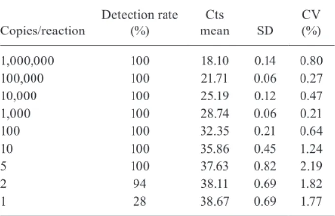
TABLE II
Detection rate and cycle threshold (Ct) values of standard curve
Copies/reaction
Detection rate (%)
Cts mean SD
CV (%)
1,000,000 100 18.10 0.14 0.80
100,000 100 21.71 0.06 0.27
10,000 100 25.19 0.12 0.47
1,000 100 28.74 0.06 0.21
100 100 32.35 0.21 0.64
10 100 35.86 0.45 1.24
5 100 37.63 0.82 2.19
2 94 38.11 0.69 1.82
1 28 38.67 0.69 1.77
CV: coefficient of variation; SD: standard deviation.
LoD found was approximately five copies of target DNA/ reaction, representing 250 copies/plasma mL (Table II).
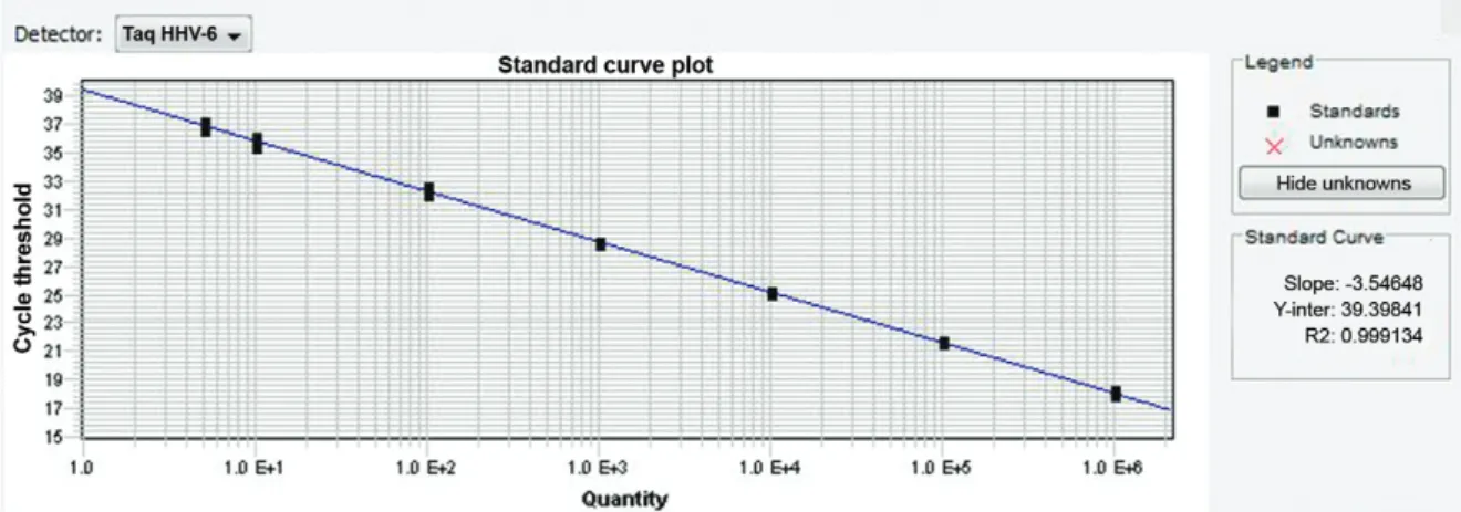
The reaction efficiency was 78.3% (slope -3.98) when the primers and probe concentration was 500 nM and 200 nM, respectively (Fig. 1). After primers optimisation (250 nM of primers and 200 nM of probe concentrations), the efficiency result was 91.4% and slope -3.546 (Fig. 2).
The inter-assay variation (reproducibility) range was 0.78-2.82% (Table III) and the test resulted to be specific as no false positive were detected even in the presence of other herpesviruses fragments (HSV1, HSV2, VZV, EBV, CMV, HHV-7 and HHV-8).
Quantitation of HHV-6 DNA in plasma samples - HHV-6 genome sequences were detected in 2% (18/1,082) of plasma samples. Among these samples, 39% (7/18) yielded more than 250 HHV-6 copies/plasma mL (LoD value) and the highest HHV-6 mean viral load (VL) detected was 69,163 copies/mL.
Despite the low LoD, samples with results lower than 250 copies/mL were retested a second time and all re-sults were confirmed.
Four patients (P1, P2, P3 and P4) presented persistent vi-raemia after transplant and all cases are illustrated in Fig. 3.
DISCUSSION
Monitoring HHV-6 and providing precise, rapid and early diagnosis of related clinical diseases constitutes an essential measure to improve disease outcome on several backgrounds, such as HHV-6 reactivation after umbilical cord blood transplantation (Chevallier et al. 2010, Mori et al. 2010, Ogata et al. 2013, Scheurer et al. 2013, Illiaquer et al. 2014, Le Bourgeois et al. 2014), and the unexplained presence of HHV-6 DNAemia associated to specific clin-ical conditions (Zerr et al. 2005, Ogata 2009, Meyding-Lamadé & Strank 2012, Al Fawaz et al. 2014, Shoji et al. 2014, Ahluwalia et al. 2015). Additionally, it has potential to elucidate the clinical significance of recent reported presence of genomic integration (Pellett et al. 2012, Pan-try et al. 2013, Bell et al. 2014, Hill et al. 2015).
In this study a strategy to optimise a qPCR technique using a standard curve constructed with PCR product was applied, which seemed to be quite effective when compared
Fig. 2: human herpesvirus (HHV)-6 standard curve after optimisation.
to the results of a qPCR standardisation reaction using a standard curve constructed with clones inserted into the plasmid, which is a well-described technique in the litera-ture (Locatelli et al. 2000, Collot et al. 2002, Gautheret-De-jean et al. 2002, Ogata et al. 2006, Isegawa et al. 2007).
The first tests with the primers and probe showed that the linearity between 10-100 copies/reaction (500-5,000 copies/plasma mL) were negatively affected when us-ing 500 nM primers and 300 nM probe concentrations. The results of primers and probe test optimisation using 100 HHV-6 copies/reaction (target) with 500 nM of each primer and 300 nM of probe concentrations, initially showed a 37.9 Ct reaction. When these concentrations were modified to 250 nM of each primer and 200 nM of probe, the Ct reaction decreased to 32.4, showing an ef-ficiently improvement of primers and probe. Other target concentrations (50 and 10 HHV-6 copies/reaction) were tested with 250 nM and 200 nM of primers and probe concentrations, respectively and similar Ct reaction re-ductions were observed. Gautheret-Dejean et al. (2002) have used 200 nM of each primer and 100 nM probe.
As previously mentioned, a purified PCR product was used to build a standard curve. The purified PCR product was submitted to electrophoresis for integrity evaluation (Bioanalyzer-Agilent) and a 180 bp ampli-con was found, close to 176 bp ampliampli-con founded by Gautheret-Dejean et al. (2002). The fluorometric PCR product quantitation (80.8 ng/µL) was used to estimate the copy number/µL (42.5 x 10¹⁰ copies/µL) that allowed the generation of a standard curve.
The 10-fold dilution curve presented linearity rang-ing from 10-106 copies/reaction (500 to 5 x 10⁷ copies/ plasma mL). Collot et al. (2002) and Isegawa et al. (2007) have founded linearity ranging 101-108 copies/reaction.
The LoD determined was five copies/reaction or 250 copies/plasma mL, which means the lowest analyte amount present in a sample that could be detected with 95% probability, although not quantified as an exact value. All results between two-five copies/reaction com-prised a “gray zone” (linearity range out) and have been tested for a second time. These results have all been con-firmed. Our results were similar to the LoD reported by de Pagter et al. (2008b) of 250 copies/mL approximately. Tavakoli et al. (2007) detected five gene copies/reaction
or 200 gene copies/sample mL, and the LoD related by Yao et al. (2009) was 10 copies/reaction in a nested PCR reaction. Sugita et al. (2008) defined a cut-off of 50 cop-ies per tube and the sensitivity described by Gautheret-Dejean et al. (2002) was 10 genomic equivalent copies/ reaction. The lack of HHV-6 quantification standardisa-tion represents a substantial issue on this scenario, which makes comparisons difficult.
The reaction efficiency before primers optimisation was 78.3% (slope -3.98) and after optimisation a good improvement could be observed; the target efficiency obtained was 91.4% (slope -3.546).
The slope values presented by Watzinger et al. (2004) and Wada et al. (2009) were slope -3.374 and slope -3.135, respectively.
Dilutions from 101-106 copies/reaction have been evaluated to provide the reproducibility or inter-assay variation and the CV mean found was 1.39 (range 0.78-2.82). Collot et al. (2002) found similar results, range of 0.80-0.96 and Watzinger et al. (2004) presented a CV = 1.60% in inter-assay variation whereas Isegawa et al. (2007) related CV = 1.36% of intra-assay variation. No cross-reaction or false positive results were detected.
All samples with results lower than 250 copies/mL were tested a second time. The samples with more than five copies/reaction (or 250 copies/mL) on the second test were considered “positive” and the samples with less than five copies/reaction (or 250 copies/mL) were considered “negative”. The significance of the HHV-6 positive re-sult remains unclear, however, we believe that all patients whose VL is around 200 copies/mL should provide an-other blood sample after at least three-four days after the first sample was collected and get tested again in order to observe the VL trend (increasing or decreasing).
In conclusion, the test developed in the present study was simple, rapid and sensitive, allowing the detection of a wide range of HHV-6 loads. It may be useful as a practical tool to help elucidate the clinical relevance of HHV-6 infection and reactivation in different scenarios and to determine the need for surveillance.
REFERENCES
Ahluwalia J, Abuabara K, Perman MJ, Yan AC 2015. HHV-6 involve-ment in pediatric drug hypersensitivity syndrome. Br J Dermatol 172: 1090-1095.
Al Fawaz T, Ng V, Richardson SE, Barton M, Allen U 2014. Clinical con-sequences of human herpesvirus 6 DNAemia in peripheral blood in pediatric liver transplant recipients. Pediatr Transplant 18: 47-51.
Bell AJ, Gallagher A, Mottram T, Lake A, Kane EV, Lightfoot T, Ro-man E, Jarrett RF 2014. Germ-line transmitted, chromosomally integrated HHV-6 and classical Hodgkin lymphoma. PLoS One 9: e112642.
Betts BC, Young JA, Ustun C, Cao Q, Weisdorf DJ 2011. Human her-pesvirus 6 infection after hematopoietic cell transplantation: is routine surveillance necessary? Biol Blood Marrow Transplant 17: 1562-1568.
Boutolleau D, Fernandez C, Andre E, Imbert-Marcille BM, Mil-pied N, Agut H, Gautheret-Dejean A 2003. Human herpesvirus (HHV)-6 and HHV-7: two closely related viruses with different infection profiles in stem cell transplantation recipients. J Infect
Dis 187: 179-186.
TABLE III
Reproducibility analysis
Copies/reaction
Cts
mean SD
CV (%)
106 18.30 0.17 0.94
105 21.89 0.17 0.78
104 25.46 0.24 0.94
103 29.07 0.36 1.25
102 32.77 0.52 1.58
01 36.67 1.03 2.82
Canto CL, Sumita LM, Machado AF, Tateno A, Cunha EV, Machado CM 2008. Optimization of the sybr green real time PCR for the detection of human herpes virus type 6 (HHV-6). Rev Inst Med
Trop Sao Paulo 50: 61-63.
Cavalcanti SMB 2011. Detection of human herpesviruses 6 and 7 DNA in the saliva of renal transplanted patients and healthy indi-viduals from Rio de Janeiro, Brazil. Virus Rev Res 16: 11-15.
Chapenko S, Trociukas I, Donina S, Chistyakov M, Sultanova A, Gravelsina S, Lejniece S, Murovska M 2012. Relationship be-tween beta-herpesviruses reactivation and development of com-plications after autologous peripheral blood stem cell transplan-tation. J Med Virol 84: 1953-1960.
Chevallier P, Hebia-Fellah I, Planche L, Guillaume T, Bressolette-Bo-din C, Coste-Burel M, Rialland F, Mohty M, Imbert-Marcille BM 2010. Human herpes virus 6 infection is a hallmark of cord blood transplant in adults and may participate to delayed engraftment: a comparison with matched unrelated donors as stem cell source.
Bone Marrow Transpl 45: 1204-1211.
Collot S, Petit B, Bordessoule D, Alain S, Touati M, Denis F, Ranger-Rogez S 2002. Real-time PCR for quantification of human her-pesvirus 6 DNA from lymph nodes and saliva. J Clin Microbiol 40: 2445-2451.
Cordonnier C 2008. Infections after HSCT. In EBMT handbook on
haematopoietic stem cell transplantation, European Scholl of
Haematology, Paris, p. 198-217.
de Freitas RB, Linhares AC 1997. Prevalence of human herpesvirus 6 antibody in the population of Belém, Pará, northern Brazil. Trans
R Soc Trop Med Hyg 91: 538-540.
de Freitas RB, Linhares MI, Linhares AC 1994. Prevalence of human herpesvirus 6 antibody among isolated Amazonian Amerindian communities in Brazil. Trans R Soc Trop Med Hyg 88: 167-169.
de Pagter PJ, Schuurman R, de Vos NM, Mackay W, van Loon AM 2010. Multicenter external quality assessment of molecular methods for detection of human herpesvirus 6. J Clin Microbiol48: 2536-2540.
de Pagter PJ, Schuurman R, Meijer E, van Baarle D, Sanders EAM, Boelens JJ 2008a. Human herpesvirus type 6 reactivation after haematopoietic stem cell transplantation. J Clin Virol 43: 361-366.
de Pagter PJ, Schuurman R, Visscher H, de Vos M, Bierings M, van Loon AM, Uiterwaal CS, van Baarle D, Sanders EA, Boelens J 2008b. Hu-man herpes virus 6 plasma DNA positivity after hematopoietic stem cell transplantation in children: an important risk factor for clinical outcome. Biol Blood Marrow Transplant 14: 831-839.
Gautheret-Dejean A, Manichanh C, Thien-Ah-Koon F, Fillet AM, Mangeney N, Vidaud M, Dhedin N, Vernant JP, Agut H 2002. Development of a real-time polymerase chain reaction assay for the diagnosis of human herpesvirus 6 infection and application to bone marrow transplant patients. J Virol Methods100: 27-35.
Gerdemann U, Keukens L, Keirnan JM, Katari UL, Nguyen CT, de Pagter AP, Ramos CA, Kennedy-Nasser A, Gottschalk SM, Heslop HE, Brenner MK, Rooney CM, Leen AM 2013. Immunotherapeu-tic strategies to prevent and treat human herpesvirus 6 reactivation after allogeneic stem cell transplantation. Blood 121: 207-218.
Gotoh M, Yoshizawa S, Katagiri S, Suguro T, Asano M, Kitahara T, Akahane D, Okabe S, Tauchi T, Ito Y, Ohyashiki K 2014. Human herpesvirus 6 reactivation on the 30th day after allogeneic he-matopoietic stem cell transplantation can predict grade 2-4 acute graft-versus-host disease. Transpl Infect Dis 16: 440-449.
Guardia AC, Stucchi RSB, Milan A, Costa SCB, Boin IDFSF 2014. Human herpesvirus 6 and cytomegalovirus DNA in liver donor biopsies and their correlation with HLA matches and acute cel-lular rejection. Braz J Infect Dis 18: 220-224.
Guardia AC, Stucchi RSB, Sampaio AM, Milan A, Costa SCB, Pavan CR, Boin IDFSF 2012. Human herpesvirus 6 in donor biopsies associated with the incidence of clinical cytomegalovirus disease and hepatitis C virus recurrence. Int J Infect Dis 16: e124-e129.
Hill JA, Sedlak RH, Zerr DM, Huang ML, Yeung C, Myerson D, Jerome KR, Boeckh MJ, 2015. Prevalence of chromosomally integrated human herpesvirus 6 in patients with human herpes-virus 6-central nervous system dysfunction. Biol Blood Marrow
Transpl 1: 371-373.
Illiaquer M, Malard F, Guillaume T, Imbert-Marcille BM, Delaunay J, Le Bourgeois A, Rimbert M, Bressollette-Bodin C, Precupanu C, Ayari S, Peterlin P, Moreau P, Mohty M, Chevallier P 2014. Long-lasting HHV-6 reactivation in long-term adult survivors after double umbilical cord blood allogeneic stem cell transplan-tation. J Infect Dis 210: 567-570.
Isegawa Y, Takemoto M, Yamanishi K, Ohshima A, Sugimoto N 2007. Real-time PCR determination of human herpesvirus 6 anti-viral drug susceptibility. J Virol Methods 140: 25-31.
Jenkins FJ, Hoffman LJ, Liegey-Dougall A 2002. Reactivation of and primary infection with human herpesvirus 8 among solid-organ transplant recipients. J Infect Dis185: 1238-1243.
Jeulin H, Agrinier N, Guery M, Salmon A, Clement L, Bordigoni P, Venard V 2013. Human herpesvirus 6 infection after allogeneic stem cell transplantation: incidence, outcome and factors associ-ated with HHV-6 reactivation. Transplantation95: 1292-1298.
Kalpoe J, Kroes A, Verkerk S, Claas E, Barge R, Beersma M 2006. Clinical relevance of quantitative varicella-zoster virus (VZV) DNA detection in plasma after stem cell transplantation. Bone
Marrow Transplant38: 41-46.
Kernan NA, Bartsch G, Ash RC, Beatty PG, Champlin R, Filipov-ich A, Gajewski J, Hansen JA, Henslee-Downey J, McCullough J, McGlave P, Perkins HA, Phillips GL, Sanders J, Stroncek D, Thomas ED, Blume KG 1993. Analysis of 462 transplantations from unrelated donors facilitated by the National Marrow Donor Program. N Engl J Med 328:593-602.
Le Bourgeois A, Labopin M, Guillaume T, Delaunay J, Foucher Y, Tessoulin B, Malard F, Ayari S, Peterlin P, Derenne S, Herry P, Cesbron A, Gagne K, Lode L, Illiaquer M, Imbert-Marcille BM, Le Gouill S, Moreau P, Mohty M, Chevallier P 2014. Human her-pesvirus 6 reactivation before engraftment is strongly predictive of graft failure after double umbilical cord blood allogeneic stem cell transplantation in adults. Exp Hematol 42: 945-954.
Leibovitch EC, Brunetto GS, Caruso B, Fenton K, Ohayon J, Reich DS, Jacobson S 2014. Coinfection of human herpesviruses 6A (HHV-6A) and HHV-6B as demonstrated by novel digital droplet PCR assay. PLoS ONE 9: e92328.
Linhares MI, Eizuru Y, Tateno S, Minamishima Y 1991. Seroprevalence of human herpesvirus 6 infection in Brazilian and Japanese popula-tions in the Northeast of Brazil. Microbiol Immunol 35: 1023-1027.
Ljungman P 2002. Beta-herpesvirus challenges in the transplant re-cipient. J Infect Dis 186 (Suppl. 1): S99-S109.
Locatelli G, Santoro F, Veglia F, Gobbi A, Lusso P, Malnati MS 2000. Real-time quantitative PCR for human herpesvirus 6 DNA. J
Clin Microbiol38: 4042-4048.
Magalhães IDM, Martins RVN, Vianna RO, Oliveira SA, Cavalcanti SMB 2011. Diagnosis of human herpesvirus 6B primary infec-tion by polymerase chain reacinfec-tion in young children with exan-thematic disease. Rev Soc Bras Med Trop 44: 306-308.
Meyding-Lamadé U, Strank C 2012. Herpesvirus infections of the central nervous system in immunocompromised patients. Ther
Mori Y, Miyamoto T, Nagafuji K, Kamezaki K, Yamamoto A, Saito N, Kato K, Takenaka K, Iwasaki H, Harada N, Abe Y, Teshima T, Akashi K 2010. High incidence of human herpes virus 6-associat-ed encephalitis/myelitis following a second unrelat6-associat-ed cord blood transplantation. Biol Blood Marrow Transpl 16: 1596-1602.
Ogata M 2009. Human herpesvirus 6 in hematological malignancies.
J Clin Exp Hematop 49: 57-67.
Ogata M 2012. Human herpesvirus-6 in hematopoietic cell transplant re-cipients. Journal of Hematopoietic Cell Transplantation1: 76-92.
Ogata M, Kikuchi H, Satou T, Kawano R, Ikewaki J, Kohno K, Kashi-ma K, Ohtsuka E, Kadota J 2006. HuKashi-man herpesvirus 6 DNA in plasma after allogeneic stem cell transplantation: incidence and clinical significance. J Infect Dis 193: 68-79.
Ogata M, Satou T, Kadota J, Saito N, Yoshida T, Okumura H, Ueki T, Nagafuji K, Kako S, Uoshima N, Tsudo M, Itamura H, Fukuda T 2013. Human herpesvirus 6 (HHV-6) reactivation and HHV-6 encephalitis after allogeneic hematopoietic cell transplantation: a multicenter, prospective study. Clin Infect Dis 57: 671-681.
Oliveira SA, Siqueira MM, Camacho LA, Nogueira RM, Spinetti CC, Garcia RCC, Knowles W, Brown DW 2001. The aetiology of maculopapular rash diseases in Niterói, state of Rio de Janeiro, Brazil: implications for measles surveillance. Epidemiol Infect 127: 509-516.
Pantry SN, Medveczky MM, Arbuckle JH, Luka J, Montoya JG, Hu J, Renne R, Peterson D, Pritchett JC, Ablashi DV, Medveczky PG 2013. Persistent human herpesvirus-6 infection in patients with an inherited form of the virus. J MED Virol 85: 1940-1946.
Pellett PE, Ablashi DV, Ambros PF, Agut H, Caserta MT, Descamps V, Flamand L, Gautheret-Dejean A, Hall CB, Kamble RT, Kuehl U, Lassner D, Lautenschlager I, Loomis KS, Luppi M, Lusso P, Medveczky PG, Montoya JG, Mori Y, Ogata M, Pritchett JC, Rogez S, Seto E, Ward KN, Yoshikawa T, Razonable RR 2012. Chromosomally integrated human herpesvirus 6: questions and answers. Rev Med Virol 22: 144-155.
Pollack M, Heugel J, Xie H, Leisenring W, Storek J, Young JA, Kukreja M, Gress R, Tomblyn M, Boeckh M 2011. An interna-tional comparison of current strategies to prevent herpesvirus and fungal infections in hematopoietic cell transplant recipients.
Biol Blood Marrow Transplant 17: 664-673.
Razonable RR, Paya CV 2003. Herpesvirus infections in transplant recipients: current challenges in the clinical management of cyto-megalovirus and Epstein-Barr virus infections. Herpes 10: 60-65.
Robles JDF, Cheuk DK, Ha SY, Chiang AK, Chan GC 2014. Human herpesvirus types 6 and 7 infection in pediatric hematopoietic stem cell transplant recipients. Ann Transplant 19: 269-276.
Sakai R, Kanamori H, Motohashi K, Yamamoto W, Matsuura S, Fu-jita A, Ohshima R, Kuwabara H, Tanaka M, FuFu-jita H, Maruta A, Ishigatsubo Y, Fujisawa S 2011. Long-term outcome of human
herpesvirus 6 encephalitis after allogeneic stem cell transplanta-tion. Biol Blood Marrow Transplant 17: 1389-1394.
Salahuddin SZ, Ablashi DV, Markham PD, Josephs SF, Sturzenegger S, Kaplan M, Halligan G, Biberfeld P, Wong-Staal F, Kramarsky B, Gallo RC 1986. Isolation of a new virus, HBLV, in patients with lymphoproliferative disorders. Science 234: 596-601.
Scheurer ME, Pritchett JC, Amirian ES, Zemke NR, Lusso P, Ljung-man P 2013. HHV-6 encephalitis in umbilical cord blood trans-plantation: a systematic review and meta-analysis. Bone Marrow
Transpl 48: 574-580.
Schonberger S, Meisel R, Adams O, Pufal Y, Laws HJ, Enczmann J, Dilloo D 2010. Prospective, comprehensive and effective viral monitoring in children undergoing allogeneic hematopoietic stem cell transplantation. Biol Blood Marrow Transplant16: 1428-1435.
Shoji Y, Choo HL, Leong CO, Oo AL, Townsend G 2014. Orofacial pain of muscular origin is not associated with herpes virus-6 in-fection: a pilot study. J Oral Facial Pain Headache 28: 346-349.
Sugita S, Shimizu N, Watanabe K, Mizukami M, Morio T, Sugamoto Y, Mochizuki M 2008. Use of multiplex PCR and real-time PCR to detect human herpes virus genome in ocular fluids of patients with uveitis. Br J Ophthalmol 92: 928-932.
Tavakoli NP, Nattanmai S, Hull R, Fusco H, Dzigua L, Wang H, Du-puis M 2007. Detection and typing of human herpesvirus 6 by molecular methods in specimens from patients diagnosed with encephalitis or meningitis. J Clin Microbiol 45: 3972-3978.
Wada K, Mizoguchi S, Ito Y, Kawada J, Yamauchi Y, Morishima T, Nishiyama Y, Kimura H 2009. Multiplex real-time PCR for the simultaneous detection of herpes simplex virus, human herpesvi-rus 6 and human herpesviherpesvi-rus 7. Microbiol Immunol53: 22-29.
Wang LR, Dong LJ, Zhang MJ, Lu DP 2006. The impact of human herpesvirus 6B reactivation on early complications following allogeneic hematopoietic stem cell transplantation. Biol Blood
Marrow Transplant 12: 1031-1037.
Watzinger F, Suda M, Preuner S, Baumgartinger R, Ebner K, Baskova L, Niesters HG, Lawitschka A, Lion T 2004. Real-time quanti-tative PCR assays for detection and monitoring of pathogenic human viruses in immunosuppressed pediatric patients. J Clin
Microbiol 42: 5189-5198.
Yamanishi K, Okuno T, Shiraki K, Takahashi M, Kondo T, Asano Y, Kurata T 1988. Identification of human herpesvirus 6 as a causal agent for exanthem subitum. Lancet 1: 1065-1067.
Yao K, Honarmand S, Espinosa A, Akhyani N, Glaser C, Jacobson S 2009. Detection of human herpesvirus 6 in cerebrospinal fluid of patients with encephalitis. Ann Neurol 65: 257-267.
Yoshikawa T 2004. Human herpesvirus 6 infection in hematopoietic stem cell transplant patients. Br J Haematol 124: 421-432.