263
Vol.55, n. 2: pp.263-268, March-April 2012
ISSN 1516-8913 Printed in Brazil BRAZILIAN ARCHIVES OF
BIOLOGY AND TECHNOLOGY
A N I N T E R N A T I O N A L J O U R N A L
The Effect of Losartan on Angiotensin II-Induced Cell
Proliferation in a Rat Aorta Smooth Muscle Cell Line
Natália Tambelline
1, Karen Oliveira
1, Luiz Renato Olchanheski Junior
2, Regina Sordi
3,
Michel Fleith Otuki
2, Giovani Marino Favero
1and Daniel Fernandes
2*1
Departamento de Biologia Geral; Universidade Estadual de Ponta Grossa; Ponta Grossa - PR - Brasil.
2Departamento de Ciências Farmacêuticas; Universidade Estadual de Ponta Grossa; Ponta Grossa - PR - Brasil. 3Departamento de Farmacologia; Universidade Federal de Santa Catarina; Florianópolis - SC - Brasil
ABSTRACT
The success of revascularization procedures is limited by recurrent stenosis, which is a narrowing of a blood vessels that results from neo-intimal hyperplasia. The renin-angiotensin-aldosterone system has been implicated in the pathogenesis of neo-intimal hyperplasia, and a role for angiotensin II in vascular smooth muscle cell proliferation has been proposed. There are at least two high-affinity subtypes of angiotensin II receptors, AT1 and AT2, both of which are seven-transmembrane G protein-coupled receptors. We investigated the effect of losartan, an AT1 receptor antagonist, on vascular smooth muscle cell proliferation using the A7r5 smooth cell line derived from rat aorta. Losartan was shown to prevent angiotensin II-induced cell proliferation, thereby suggesting that the effect of angiotensin II was mediated via AT1 receptors. These data strengthen the concept that inhibitors of the renin-angiotensin system can effectively prevent recurrent stenosis.
Key words: Angiotensin II, Losartan, Cell proliferation
*Author for correspondence: [email protected]
INTRODUCTION
Coronary artery disease (CAD) is a leading cause of morbidity and mortality worldwide (Lopez et al. 2006). Advances in surgical management have improved outcomes in patients with CAD. For example, balloon angioplasty and bypass graft surgeries have been remarkably effective in improving symptoms and quality of life. However, the long-term success of these procedures is limited by recurrent stenosis, which is a reduction in vascular lumen area at the site of intervention (Schainfeld 2002; Bhargava et al. 2003).
Vascular injury produced during attempted revascularization damages the endothelial cell layer lining of the arterial wall. This injury is followed by the proliferation and/or migration of
medial smooth muscle cells resulting in the formation of intimal hyperplasia (for a review, see Ahanchi et al. 2007).
of angiotensin II, including cell growth control (for a review see Bader et al. 2010). Consequently, AT1 receptor antagonists have been studied as possible therapeutic targets for preventing stenosis (Moon et al. 2004).
The present study was designed to directly investigate the effect of a selective AT1 receptor antagonist, losartan, on angiotensin II-induced cell proliferation using the rat embryonic thoracic aorta smooth muscle cell line A7r5.
MATERIALS AND METHODS
Cell culture
The rat aortic vascular smooth muscle cell line A7r5 was obtained from the Rio de Janeiro Cell Bank (Universidade Federal do Rio de Janeiro, RJ, Brazil) and was used at passages 2–15. Cells were grown and cultured in flasks in Dulbecco’s modified Eagle’s medium (DMEM) supplemented with 10% fetal calf serum, 100 U/ml penicillin, 100 µg/ml streptomycin and 10 mM HEPES (pH 7.4) in a humidified incubator at 37 °C with a 5% CO2 atmosphere. Every 4–5 days, the cultures were confluent and were passaged using 0.25% trypsin/0.03% EDTA at a split ratio of 1:4. For experimental procedures, confluent A7r5 cells were harvested as mentioned above, resuspended in culture medium and the cell viability was determined by Trypan Blue exclusion. Cells were seeded in 200 µL of medium in 96-well plates
(approximately 3.5 x 10⁴ viable cells per well).
After 24 hours, test compounds were added in a maximum volume of 20 µL.
Cell counting
At the ends of the experimental periods, each well
was washed twice with Ca2+/Mg2+-free phosphate
buffered saline (PBS: 137 mM NaCl, 2.7 mM KCl, 1.5 mM KH2PO4 and 8.1 mM NaHPO4; pH 7.4). Cells were then detached using 100 µL of a 0.25% trypsin/0.03% EDTA solution and incubated at 37 °C for 30 minutes. Trypsin was then inactivated using 10 µL of fetal calf serum. Cells were quantified in Neubauer chambers, and the results
are expressed as cell number/mm3. Cellular
viability under all treatment conditions was determined by cell Trypan Blue exclusion.
Drugs
Tissue culture media, serum, and antibiotics were obtained from Gibco (São Paulo, SP, Brazil).
Angiotensin II and losartan were purchased from Sigma-Aldrich Co. LLC (St. Louis, MO, USA). Angiotensin II and losartan were dissolved in PBS.
Data analysis
Data are expressed as the mean ± SEM of triplicate wells. Each experiment was repeated at least twice with similar results. Statistical significance was determined using a one-way analysis of variance followed by a Bonferroni’s
post hoc t test. A p value less than 0.05 was considered statistically significant.
RESULTS
The A7r5 cell line reached confluence at 96 hours
after seeding 3.5 x 10⁴ cells in a 96-well plate.
After 96 hours, the cell numbers remained constant for 1-2 days; thereafter, the cells then began to die and detach from the substrate. Therefore, the analysis of cell numbers was performed only up to 72 hours after incubation of the compounds (96 hours after seeding of the cells in a 96-well plate).
A7r5 cell proliferation throughout the experiment duration is shown in Figure 1 (open bars). The incubation of the cells with angiotensin II (1, 10 or
100 µmol/L) resulted in increased cell
proliferation in a concentration-dependent manner. This effect was most pronounced at 100 µmol/L, and this was the only concentration that showed a significant increase in cell proliferation as early as 24 hours after incubation. Thus, only the highest concentration was used to evaluate the effect of
losartan on the proliferation induced by
angiotensin II (Fig. 2). The cellular toxicity was evaluated using a Trypan Blue exclusion test. The angiotensin II concentrations used here were not toxic, as evidenced by a cell viability of greater than 95%.
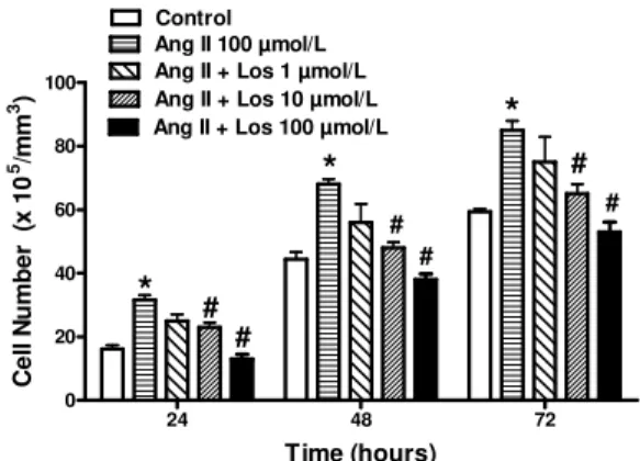
The incubation of A7r5 cells with losartan (1, 10 or 100 µmol/L) for 30 minutes resulted in a concentration-dependent decrease in angiotensin II-induced cell proliferation (Fig. 2). These data clearly indicated that AT1 receptor blockade by losartan was able to inhibit the proliferation of A7r5 cells induced by angiotensin II.
The Effect of Losartan on Angiotensin II-Induced Cell 265
24 48 72
0 20 40 60 80 100
Control Ang II 1 µmol/L Ang II 10 µmol/L
Ang II 100 µmol/L *
* *
*
*
Time (hours)
C
e
ll
N
u
m
b
e
r
(
x
1
0
5/m
m
3
)
Figure 1 - The effect of angiotensin II on A7r5 cell proliferation. Cells were seeded and incubated with different concentrations of angiotensin II or vehicle after 24 hours. Cells were quantified in Neubauer chambers at different durations of incubation. Cell viability under all treatment conditions was determined by cell Trypan Blue exclusion. Data are expressed as the mean ± SEM of triplicate wells. * p < 0.05 compared to the control.
24 48 72
0 20 40 60 80 100
Control Ang II 100 µmol/L Ang II + Los 1 µmol/L Ang II + Los 10 µmol/L
#
#
#
Ang II + Los 100 µmol/L
# #
#
* *
*
Time (hours)
C
e
ll
N
u
m
b
e
r
(
x
1
0
5/m
m
3)
Figure 2 - The effect of losartan on angiotensin II-induced A7r5 cell proliferation. Cells were seeded, and after 24 hours, they were preincubated with different concentrations of losartan or vehicle for 30 minutes, and then with angiotensin II (100 µmol/L). Cells were quantified in Neubauer chambers at the indicated incubation times. Data are expressed as the mean ± SEM of triplicate wells. * p < 0.05 compared to the control; # p < 0.05 compared to the angiotensin II group.
24 48 72
0 20 40 60 80
Control Los 1 µmol/L Los 10 µmol/L Los 100 µmol/L
*
Time (hours)
C
e
ll
N
u
m
b
e
r
(
x
1
0
5/m
m
3)
DISCUSSION
This study is the first demonstration of angiotensin II-induced cell proliferation in the A7r5 cell line. Additionally, in agreement with several other reports (for a review, see Ribichini et al. 2005), we demonstrated the role of angiotensin II in smooth muscle cell proliferation and intimal hyperplasia through AT1 receptor activation.
We chose to study A7r5 cells because A7r5 is a clonal cell line from thoracic aorta of embryonic rats and possesses many of the properties that are characteristic of smooth muscle cells. Cellular products of the A7r5 cell line include myokinase, creatine phosphokinase and myosin (Kimes and Brandt, 1976; Barisione et al. 2009).
Our in vitro results were in agreement with studies performed in animal models demonstrating that angiotensin-converting enzyme inhibitors are able to prevent restenosis after vascular injury induced by balloon barotraumas (Pratt and Dzau 1996). The seminal study by Powel and colleagues performed in a rat injury model demonstrated the antiproliferative effects of cilazapril or captopril (Powell et al. 1989). More recently, a clinical trial tested the effects of the AT1 antagonist valsartan in the setting of stent restenosis (Peters et al. 2001) and showed contradictory (Ribichini et al. 2005) but positive results. Interestingly, Tanemoto and colleagues (2007) documented the effect of losartan in preventing stenosis not only at the level of the coronary vascular bed but also at the renal artery level.
The AT1 receptor accounts for the majority of the known functions of angiotensin II in different tissues. Binding of angiotensin II to AT1 receptors results in the internalization of the complex (Bkaily et al. 2003). The receptor is then recycled
back to the plasma membrane, whereas
angiotensin II is destined for intracellular locations, such as lysosomes and the nucleus (Erdmann et al. 1996), thereby suggesting a functional role for intracellular angiotensin II (Kumar et al. 2007). More recently, studies have provided evidence of intracellular angiotensin II production (Paul et al. 2006).
In the A7r5 smooth muscle cell line, cellular growth promoted by intracellular angiotensin II showed limited sensitivity to AT1 antagonists (Filipeanu et al. 2001). This finding agreed with a more recent work that failed to show an inhibitory effect of extracellular losartan on intracrine angiotensin II-induced cell proliferation (Baker
and Kumar 2006). In contrast, other studies have suggested that losartan, after AT1 receptor binding
and internalization, blocks intracellular
angiotensin II action (Cook et al. 2001). This controversy has not yet been resolved. It has been postulated that the ability of AT1 antagonists to inhibit the intracellular functions of angiotensin II may depend on their liposolubility (Ruiz et al. 2007). Certain studies have suggested that losartan is sufficiently hydrophobic such that it may freely penetrate cell membranes (Peters et al. 1999). Candesartan, on the other hand, appears to bind tightly to AT1 but remains at the cell surface (Fierensa et al. 2001). For this reason, irbesartan, which is a highly liposoluble AT1 receptor antagonist, is thought to be an important pharmacological tool for further studies.
Losartan is an active AT1 receptor antagonist (Lo et al. 1995), but interestingly, approximately 14% of a losartan dose is converted to the pharmacologically active E3174 metabolite after oral administration (Wong et al. 1990). E3174 is a carboxylic acid metabolite in the oxidative pathway and exhibits potency and binding selectivity comparable to losartan (Wong et al. 1991). By blocking the action of angiotensin II, losartan and its metabolite cause blood vessel dilation and thereby reduce blood pressure in hypertensive patients (Oparil 2000).
Because losartan is a highly selective AT1 receptor antagonist (Smith et al. 1992), we cannot exclude the possibility that an intense AT1 blockade could permit the action of angiotensin II in AT2 receptors, which presumably mediated the observed antiproliferative effects (Stoll et al. 1995). This concept requires further investigation. Interestingly, the highest concentration of losartan (100 µmol/L) used here reduced cell proliferation even in the absence of angiotensin II (Fig. 3). This effect was not due to cytotoxicity because all analyzed concentrations of losartan yielded cell viabilities of greater than 95%. This result suggested a basal activation of AT1 receptors through intracellular angiotensin II production (Kumar et al. 2007). This finding was not surprising, as a number of researchers have demonstrated that the local production of angiotensin II by smooth muscle cells is sufficient to mediate restenosis (Wilson et al. 1999).
The Effect of Losartan on Angiotensin II-Induced Cell 267
validate the concept that AT1 receptor antagonists contribute to the therapeutic management of restenosis. However, further studies are required to determine the cellular mechanisms responsible for angiotensin II-induced cell proliferation.
ACKNOWLEDGEMENTS
This work was supported by Conselho Nacional de
Desenvolvimento Científico e Tecnológico
(CNPq, Brazil), by Fundação Araucária (FA, PR, Brazil) and by Decit/SCTIE/MS through the support of CNPq and Fundação Araucária. We also thank Dr. Jamil Assreuy for his assistance with the cell culture experiments.
REFERENCES
Ahanchi SS, Tsihlis ND, Kibbe MR. The role of nitric oxide in the pathophysiology of intimal hyperplasia. J Vasc Surg. 2007; 45: A64-73.
Bader M. Tissue renin-angiotensin-aldosterone systems: Targets for pharmacological therapy. Annu Rev Pharmacol Toxicol. 2010; 50: 439-465.
Baker KM, Kumar R. Intracellular angiotensin II induces cell proliferation independent of AT1 receptor. Am J Physiol Cell Physiol. 2006; 291: C995-100.
Barisione C, Mura M, Garibaldi S, Fabbi P, Altieri P, Passalacqua M, et al. Cell-cell bond modulates vascular smooth muscle cell responsiveness to Angiotensin II. Biochem Biophys Res Commun. 2009; 388: 523-528.
Bhargava B, Karthikeyan G, Abizaid AS, Mehran R. New approaches to preventing restenosis. BMJ. 2003; 327: 274-279.
Bkaily G, Sleiman S, Stephan J, Asselin C, Choufani S, Kamal M, et al. Angiotensin II AT1 receptor internalization, translocation and de novo synthesis modulate cytosolic and nuclear calcium in human vascular smooth muscle cells. Can J Physiol Pharmacol. 2003; 81: 274–287.
Cook JL, Zhang Z, Re RN. In vitro evidence for an intracellular site of angiotensin action. Circ Res. 2001; 89: 1138-1146.
Erdmann B, Fuxe K, Ganten D. Subcellular localization of angiotensin II immunoreactivity in the rat cerebellar cortex. Hypertension. 1996; 28: 818–824. Filipeanu CM, Brailoiu E, Kok JW, Henning RH, De
Zeeuw D, Nelemans SA. Intracellular angiotensin II elicits Ca2+ increases in A7r5 vascular smooth muscle cells. Eur J Pharmacol. 2001; 420: 9-18.
Fierensa FLP, Vanderheyden PML, Roggeman C, De Backer J, Thekkumkara TJ, Vauquelin G. Tight binding of the angiotensin AT1 receptor antagonist.
Biochem Pharmacol. 2001; 61: 1227-1235.
Kim S, Iwao H. Molecular and cellular mechanisms of angiotensin II-mediated cardiovascular and renal diseases. Pharmacol Rev. 2000; 52: 11-34.
Kimes BW, Brandt BL. Characterization of two putative smooth muscle cell lines from rat thoracic aorta. Exp Cell Res. 1976; 98: 349-366.
Kumar R, Singh VP, Baker KM. The intracellular renin-angiotensin system: a new paradigm. Trends Endocrinol Metab. 2007; 18: 208-214.
Lo MW, Goldberg MR, McCrea JB, Lu H, Furtek CI, Bjornsson TD. Pharmacokinetics of losartan, an Ang II receptor antagonist, and its active metabolite EXP3174 in humans. Clin Pharmacol Ther. 1995; 58: 641–649.
Lopez AD, Mathers CD, Ezzati M, Jamison DT, Murray CJ. Global and regional burden of disease and risk factors, 2001: systematic analysis of population health data. Lancet. 2006; 367: 1747-1757.
Moon MC, Molnar K, Yau L, Zahradka P. Perivascular delivery of losartan with surgical fibrin glue prevents neointimal hyperplasia after arterial injury. J Vasc Surg. 2004; 40: 130-137.
Oparil S. Newly emerging pharmacologic differences in angiotensin II receptor blockers. Am J Hypertens. 2000; 1: 18S-24S.
Paul M, Poyan Mehr A, Kreutz R. Physiology of local renin–angiotensin systems. Physiol Rev. 2006; 86: 747–803.
Peters J, Obermuller N, Woyth A, Peters B, Maser-Gluth C, Kranzlin B, et al. Losartan and angiotensin II inhibit aldosterone production in anephric rats via different actions on the intraadrenal renin-angiotensin system. Endocrinology. 1999; 140: 675-682.
Peters S, Götting B, Trümmel M, Rust H, Brattström A. Valsartan for prevention of restenosis after stenting of type B2/C lesions: the VAL-PREST trial. J Invasive Cardiol. 2001; 13: 93-97.
Powell JS, Clozel JP, Müller RK, Kuhn H, Hefti F, Hosang M, et al. Inhibitors of angiotensin-converting enzyme prevent myointimal proliferation after vascular injury. Science. 1989; 245: 186-188.
Pratt RE, Dzau VJ. Pharmacological strategies to prevent restenosis: lessons learned from blockade of the renin-angiotensin system. Circulation. 1996; 93: 848-852.
Ribichini F, Ferrero V, Rognoni A, Vacca G, Vassanelli C. Angiotensin antagonism in coronary artery disease: results after coronary revascularisation.
Ruiz E, Redondo S, Padilla E, Gordillo-Moscoso A, Salaices M, Balfagón G, et al. Importance of intracellular angiotensin II in vascular smooth muscle cell apoptosis: inhibition by the angiotensin AT1 receptor antagonist irbesartan. Eur J Pharmacol. 2007; 567: 231-239.
Salgado DR, Rocco JR, Silva E, Vincent JL. Modulation of the renin-angiotensin-aldosterone system in sepsis: a new therapeutic approach? Expert Opin Ther Targets. 2010; 14: 11-20.
Schainfeld RM. Potential emerging therapeutic strategies to prevent restenosis in the peripheral vasculature. Catheter Cardiovasc Interv. 2002; 56: 421-431.
Smith RD, Chiu AT, Wong PC, Herblin WF, Timmermans PB. Pharmacology of Nonpeptide Angiotensin II Receptor Antagonists. Annu Rev Pharmacol Toxicol. 1992; 32A: 135–165.
Stoll M, Steckelings UM, Paul M, Bottari SP, Metzger R, Unger T. The angiotensin AT2- receptor mediates inhibition of cell proliferation in coronary endothelial cells. J Clin Invest. 1995; 95: 651-657.
Tanemoto M, Takase K, Yamada T, Satoh A, Abe T, Ito S. Dilation of renal artery stenosis after administration of losartan. Hypertens Res. 2007; 30: 999-1002.
Wilson DP, Saward L, Zahradka P, Cheung PK. Angiotensin II receptor antagonists prevent neointimal proliferation in a porcine coronary artery organ culture model. Cardiovasc Res. 1999; 42: 761-772.
Wong PC, Price WA, Chiu ET. In vivo pharmacology of DuP 753. Am J Hypertens. 1991; 4: F288-F298. Wong PC, Price WA, Chiu AT, Duncia JV, Carini DJ,
Wexler RR, et al. Nonpeptide Ang II receptor antagonists. XI. Pharmacology of EXP3174, an active metabolite of DuP753 – an orally active antihypertensive agent. J Pharmacol Exp Ther. 1990; 25: 211–217.