Analysis of the Ki-67 index in the vaginal epithelium
of castrated rats treated with tamoxifen
Afif Rieth Nery-Aguiar,IYousef Qathaf Aguiar,IAirton Mendes Conde Ju´nior,II Airlane Pereira Alencar,III Cleciton Braga Tavares,IPedro Vitor Lopes-Costa,IAfonso Celso Naza´rio,IVBenedito Borges da SilvaI,* IUniversidade Federal do Piauı´, Departamento de Ginecologia. IIMorphology, Teresina/PI, Brazil. IIIUniversidade de Sa˜o Paulo, Departamento de Estatı´sticas, Sa˜o Paulo/SP, Brazil.IVUniversidade Federal de Sa˜o Paulo, Departamento de Mastologia, Sa˜o Paulo/SP, Brazil.
OBJECTIVES: Vaginal atrophy and breast cancer are common conditions in postmenopausal women and tamoxifen is the standard endocrine treatment for hormone-sensitive tumors. The present study aimed to assess the effect of tamoxifen on Ki-67 protein expression in the vaginal epithelium of castrated rats.
MATERIAL AND METHODS:Forty Wistar-Hannover adult, virgin, castrated rats were randomly divided into two groups, group I (control, n=20) and group II (tamoxifen, n=20), receiving 0.5 ml of propylene glycol and 250mg
of tamoxifen diluted in 0.5 ml of propylene glycol, respectively, daily by gavage for 30 days. On the 31st day, the rats were euthanized and their vaginas were removed and fixed in 10% buffered formalin for the immunohistochemical study of Ki-67 protein expression. Data were analyzed by the Levene and Student’s t tests (po0.05).
RESULTS:The mean index of Ki-67 expression in the rat vagina of groups I and II was 4.04±0.96 and 26.86±
2.19, respectively (po0.001).
CONCLUSIONS:According to the results of the present study, tamoxifen, at the dose and treatment length used, induced a significant increase in the cell proliferation of the vaginal mucosa in castrated rats, as evaluated by Ki-67 protein expression.
KEYWORDS: Tamoxifen; Rat; Vagina; Proliferation; Ki-67.
Nery-Aguiar AR, Aguiar YQ, Conde Jr AM, Alencar AP, Tavares CB, Lopes-Costa PV, et al. Analysis of the Ki-67 index in the vaginal epithelium of castrated rats treated with tamoxifen. Clinics. 2016;71(2):90-93
Received for publication onSeptember 9, 2015;First review completed onOctober 29, 2015;Accepted for publication onNovember 30, 2015 E-mail: [email protected]
*Corresponding author
’ INTRODUCTION
The number of postmenopausal women worldwide has increased due to improved access to healthcare services, better living conditions and increased life expectancy. Women spend approximately one-third of their lifetime in the postmenopausal period and may become vulnerable to chronic degenerative diseases such as breast cancer (1). Breast cancer is the most common malignancy among women, particularly in the postmenopausal period. In Brazil, breast cancer is an important cause of cancer-related death, with approximately 57,120 new cases of breast cancer and 13,345 deaths from this disease estimated for the year 2014 (2-4). However, the survival of women with breast cancer has increased in the last few years, especially due to early diagnosis and to modes of systemic
treatment. Endocrine treatment is the main treatment for women with estrogen receptor-positive breast cancer (5). Tamoxifen is a first-generation selective estrogen receptor modulator (SERM) approved by the US Food and Drug Administration (FDA) for primary chemotherapy and adjuvant treatment of women with breast cancer (6).
The mechanism of action of tamoxifen has not been fully elucidated. However, because it interacts particularly with estrogen receptors, tamoxifen may have an effect on symptoms due to hypoestrogenism, such as vaginal atrophy and dyspar-eunia, which are common in postmenopausal women with breast cancer (7-11). The vaginal mucosal epithelium has both alpha and beta estrogen receptors, with decreasing beta receptor levels after menopause (10). As shown by some authors, tamoxifen exerts both estrogenic and antiestrogenic effects in the vaginal epithelium of castrated rats (12). Nevertheless, other authors, by using indirect methods such as vaginal cytology, have shown that the drug exerts an estrogenic action on the vaginal mucosa of postmenopausal women (13-15). The study of drug effects on the vaginal tissue of postmenopausal women is not possible due to ethical reasons. As few studies have investigated the direct effect of tamoxifen on the vaginal epithelium of castrated rats, the present study was conducted.
DOI:10.6061/clinics/2016(02)07
Copyright&2016CLINICS–This is an Open Access article distributed under the terms of the Creative Commons License (http://creativecommons.org/licenses/by/ 4.0/) which permits unrestricted use, distribution, and reproduction in any medium or format, provided the original work is properly cited.
No potential conflict of interest was reported.
90
’ MATERIAL AND METHODS
Animals
This study was approved by the Animal Experimentation Ethics Committee (AEEC) of the Universidade Federal do Piauı´ (UFPI), complying with the guidelines of the Brazilian College of Animal Experimentation (COBEA). Forty Wistar-Hannover virgin female rats obtained from the animal laboratory of the School of Veterinary Science at UFPI were used. The animals were housed inside a plastic cage with a metallic lid. Five animals per cage were kept since birth with free access to filtered water and rodent chow (SUPPRALABs, São Paulo, Brazil). Ambient conditions were controlled by a 12-hour light/ dark cycle, with the lights switched on from 7 a.m. to 7 p.m. Air conditioning maintained the temperature between 20 and 25o
C. The animals were randomly divided into two groups: group I (control, n=20) and group II (tamoxifen, n=20). At 90 days of age, the rats in groups I and II weighing an average of 231.5± 20.85 g and 231.2±21.74 g (p=0.9647), respectively, were castrated to eliminate gonadal sex steroid production through a dorsal paravertebral incision under anesthesia with 60 mg/kg ketamine and 4 mg/kg midazolam. Twenty-one days later, cytologic examination of Papanicolaou-stained vaginal smears was performed to characterize a decreased estrogen status, which was confirmed by the absence of cell maturation and a predominance of basal cells in all the animals. Group I rats (control) began to receive 0.5 ml/animal/day of propylene glycol (placebo) and Group II rats (experimental) received 250 mg/animal/day of tamoxifen citrate diluted in 0.5 ml of propylene glycol. The placebo and tamoxifen were continu-ously administered for 30 days, both at the same hour of the day, using a metal tube appropriate for gavage. On the 31st day, the rats from both groups were sacrificed and their vaginas were removed through a longitudinal abdominal incision for histologic and immunohistochemical studies.
Ethics Approval
This study was approved by the Animal Experimentation Ethics Committee (AEEC) of the Universidade Federal do Piauí (UFPI).
Ki-67 Immunohistochemistry
For the immunohistochemical study of Ki-67 expression, tissue specimens were fixed in 10% buffered formalin for a 12-24-hour period and then cut into 5-mm-thick sections. The sections were processed and stained with hematoxylin-eosin, deparaffinized in xylene for 5 minutes, dehydrated in absolute ethanol and washed in buffered saline solution at pH 7.4 for 5 minutes (16). Endogenous peroxidase activity was blocked with 3% hydrogen peroxide (H202) diluted in a buffered
solution for 5 minutes. After epitope retrieval, the tissue samples were incubated with primary mouse anti-Ki-67 monoclonal antibody (clone MIB-5/Dako/1:100) in a refrig-erator at approximately 4o
C overnight. Subsequently, the samples were washed with buffered saline solution and incubated for 45 minutes in the NovoLink Polymer detection system. Color development was accomplished by incubating all the tissue samples in 3-3-diaminobenzidine tetrahy-drochloride solution at a concentration of 1 mg/ml in a Tris-buffered solution, with a hydrogen peroxide solution used as a substrate, for 5 minutes. Subsequently, the tissues were counterstained with Harris’ hematoxylin or methyl green for 5 minutes, followed by dehydration in ethanol and xylene baths. After the procedure, the slides were examined
to quantify the results. Cells expressing Ki-67 protein were identified by the dark brown color of their nuclei.
Quantitative method
Ki-67 expression was quantified by two observers who were blinded to the group identification. Stained images were assessed by using a Nikon E400 light microscope connected to a Samsung color digital video camera, model SCC-131. The video camera captured and transmitted the image to a computer equipped with ImageLabssoftware, version 2.3, which was developed by Softium Informática Ltda (São Paulo, Brazil) for image analysis. To evaluate Ki-67 expression, 500 vaginal epithelial cells were counted on each slide whether or not the cells were stained with anti-Ki-67 MIB-5 antibody. A total magnifica-tion of 400 was used to examine the slides. Cell counting
was initiated at the site of greatest Ki-67 antigen expression. In each case, the percentage of stained cells was obtained by the ratio between the number of cells with stained nuclei and the total number of cells multiplied by 100.
Statistical Analysis
To evaluate group homogeneity relative to weight, Student’s t test was used for independent samples. Variance analysis of the percentages of nuclei stained with anti-Ki-67 was conducted using the Levene and Student’s tests (17). The significance level was set atpo0.05.
’ RESULTS
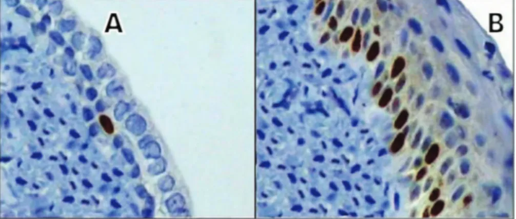
By light microscopy, cytologic examination of the vaginal epithelial cells of control group animals showed the absence of cell maturation and a predominance of basal cells, while tamoxifen-treated animals showed predominant cell maturation. Compared with control group animals, castrated female rats treated with tamoxifen showed a higher concentration of nuclei stained with anti-Ki-67 (Figure 1). The mean percentage of MIB-5-stained nuclei in the vaginal mucosa of group I (control) and group II (tamoxifen) rats was 4.04±0.96 and 26.86±2.19, respectively (po0.001) (Table 1). The boxplot illustrates the
median Ki-67 expression in the vaginal mucosal cells of animals from the control and tamoxifen groups (Figure 2).
’ DISCUSSION
In the present study, tamoxifen at a dose of 250 mg/ animal/day administered for 30 days significantly increased proliferation of the vaginal mucosa in castrated rats, as evaluated by Ki-67 protein expression. The animals received tamoxifen orally (gavage) to mimic the administration route used in women. Furthermore, according to several authors that have studied different routes of administration, the oral route led to higher serum and tissue levels of tamoxifen, thereby increasing the bioavailability of tamoxifen (18,19). Tamoxifen was diluted in propylene glycol, a neutral, colorless and odorless vehicle, allowing for the dilution and administration of predefined doses (20,21).
However, the tamoxifen dose in animal experimental studies is not easy to standardize because rats have a faster metabolism than humans. Equal-weight doses may not reproduce the serum levels and effects of tamoxifen in women (22). Dos Santos et al. (21), who studied the morpho-metry of the urethra in castrated rats given a 250mg/day tamoxifen dose for 30 days, observed that tamoxifen was capable of increasing the uterine weight and the thickness of
91
CLINICS 2016;71(2):90-93 Tamoxifen and rat vagina
the distal urethral epithelium. Thus, we used a dose of 250mg/animal/day in this study because it could mimic the effects of tamoxifen on women undergoing breast cancer treatment (22).
Friedrich et al. (13) studied the influence of tamoxifen on vaginal epithelial maturation in postmenopausal women and demonstrated an apparent increase in vaginal epithelial maturation. In another study, Friedrich et al. (14) investi-gated the effects of tamoxifen on vaginal and cervical epithelial proliferation in postmenopausal women with breast cancer and found an apparent increase not only in the incidence of metaplasia and hyperplasia of endocervical cells but also in vaginal epithelial maturation. According to Bertolissi et al. (15), tamoxifen exerts an early and persistent estrogenic effect on the vaginal epithelium during the first year of therapy in postmenopausal women with breast cancer.
Nevertheless, Berg and Dunfee (23) showed that post-menopausal women with high levels of squamous epithelial cell maturation were 15 times more likely than women in the same age group exhibiting an atrophic pattern to have coexistent endometrial adenocarcinoma because tamoxifen acts in accordance with circulating estrogen levels. Therefore, when estrogen levels are low, tamoxifen behaves as an agonist and may cause endometrial cancer. In contrast, during the premenopausal period, when estrogen levels are higher, tamoxifen appears to have an antiestrogenic action in this tissue (24). Thus, the agonist or antagonist action of SERMs in the endometrium may depend on circulating estrogen levels (25,26).
The results of the present study indicate that tamoxifen, at the dose and length of treatment used in this study, induced a significant increase in cell proliferation of the vaginal mucosa in castrated rats, as evaluated by Ki-67 protein expression. Thus, the assessment of cell proliferation in the vagina of castrated rats, used to as a mimic of postmeno-pausal women, revealed that tamoxifen displayed partially estrogenic action on the vaginal mucosa of these animals.
’ ACKNOWLEDGMENTS
We thank all the members of our research team.
’ AUTHOR CONTRIBUTIONS
Nery Aguiar AR and Aguiar YQ contributed to the data collection and to the manuscript writing. Alencar AP contributed to the statistical analysis. Conde Jr AM, Lopes-Costa PV and Tavares CB contributed to the data collection. da Silva BB conceived and planned the study and contributed to the data collection, to the manuscript writing and to conducting this study.
’ REFERENCES
1. Fernandes CE, Morita MH, Ferreira JA, da Silva EP, Wehba AS. Approach to urinary tract disorders in postmenopausal women. Rev Paul Med. 1990;108(5):230-5.
Table 1-Mean percentage of Ki-67-stanined nuclei in the vaginal mucosa of groups I (control) and II (tamoxifen).
GROUP N AVERAGE SE SD MINIMUM MEDIAN MAXIMUM
I 20 4.04 0.21 0.96 2.51 3.86 6.69
II 20 26.86* 0.49 2.19 21.77 27.38 31.23
* po0.001
Figure 2 - The median of Ki-67-stained nuclei in the vaginal mucosa of animals in the control and tamoxifen groups, as illustrated by the line in boldface.
Figure 1 -Photomicrograph of a histological section of the vaginal epithelium of castrated rats showing a greater concentration of
Ki-67-stained nuclei in the experimental group (tamoxifen)(B)compared with the control group(A)(original magnification x400).
92 Tamoxifen and rat vagina
2. de Sousa JA, Facina G, da Silva BB, Gebrim LH. Effects of low-dose tamoxifen on breast biomarkers Ki-67, estrogen and progesterone recep-tors. Int Semin Surg Oncol. 2006;3:29, http://dx.doi.org/10.1186/1477-7800-3-29.
3. Lynch BM, Neilson HK, Friedenreich CM. Physical activity and breast cancer prevention. Recent Results Cancer Res. 2011;186:13-42, http:// dx.doi.org/10.1007/978-3-642-04231-7_1.
4. Estimates of new cases and deaths from breast cancer (INCA, 2014): http://www2.inca.gov.br/wps/wcm/connect/tiposdecancer/site/home/ mama.
5. Singh MS, Francis PA, Michael M. Tamoxifen, cytochrome P450 genes and breast cancer clinical outcomes. Breast. 2011; 20 (2):111-8, http://dx.doi. org/10.1016/j.breast.2010.11.003.
6. Fisher B, Costantino JP, Wickerham DL, Redmond CK, Kavanah M, Cronin WM, et al. Tamoxifen for prevention of breast cancer: report of the National Surgical Adjuvant Breast and Bowel Project P-1 Study. J Natl Cancer Inst. 1998;90(18):1371-88, http://dx.doi.org/10.1093/jnci/ 90.18.1371.
7. Jordan VC. An overview of considerations for testing of tamoxifen as a pre-ventive breast cancer. Ann N Y Acad Sci. 1995;768:141-7, http://dx.doi.org/ 10.1111/j.1749-6632.1995.tb12117.x.
8. Vardy MD, Lindsay R, Scotti RJ, Mikhail M, Richart R, Nieves J, et al. Short-term urogenital effects of raloxifene, tamoxifen, and estrogen. Am J Obstet Gynecol. 2003;189(1):81-8, http://dx.doi.org/10.1067/mob.2003.374. 9. da SILVA BB, Lopes IM, Gebrim LH. Effects of raloxifene on normal breast
tissue from premenopausal women. Breast Cancer Res Treat 2006;95(2): 99-103, http://dx.doi.org/10.1007/s10549-005-9001-2.
10. Lara LA, Useche B, Ferriani RA, Reis RM, de Sá MF, de Freitas MM, et al. The effects of hypoestrogenism on the vaginal wall: interference with the normal sexual response. J Sex Med. 2009;6(1):30-9, http://dx.doi.org/ 10.1111/j.1743-6109.2008.01052.x.
11. Kellogg-Spadt S. Vulvovaginal atrophy. Adv Nurse Pract. 2010;18(4):31-2. 12. Kim NN, Stankovic M, Armagan A, Cushman TT, Goldstein I, Traish AM. Effects of tamoxifen on vaginal blood flow and epithelial morphology in the rat. BMC Womens Health 2006;6:14-24, http://dx.doi.org/10.1186/ 1472-6874-6-14.
13. Friedrich M, Mink D, Villena-Heinsen C, Woll-Hermann A, Wagner S, Schmidt W. The influence of tamoxifen on the maturation index of vaginal epithelium. Clin Exp Obstet Gynecol 1998;25(4):121-4.
14. Friedrich M, Mink D, Villena-Heinsen C, Woll-Hermann A, Schmidt W. Tamoxifen and proliferation of vaginal and cervical epithelium in post-menopausal women with breast cancer. Eur J Obstet Gynecol Reprod Biol 1998;80(2):221-5, http://dx.doi.org/10.1016/S0301-2115(98)00117-1.
15. Bertolissi A, Cartei G, Turrin D, Cioschi B, Rizzi V. Behaviour of vaginal epithelial maturation and sex hormone binding globulin in post-menopausal breast cancer patients during the first year of tamoxifen therapy. Cytopathology. 1998;9(4):263-70, http://dx.doi.org/10.1046/j.1365-2303. 1998.00077.x.
16. Masson P. Tumours humaines. Histologie diasgnostic et techniques.
2nded. Paris: Libraire Maloine, 1956,1061-148.
17. Kutner MH, Nachtshein CJ, Neter J, Li W. Applied linear statistical
models. 5thed. Boston: McGraw-Hill Irwin, 2005.
18. Degregorio MW, Wilbur BJ, Coronado E, Osborne CK. Serum tamoxifen concentrations in the athymic nude mouse after three methods of administration. Cancer Chemother Pharmacol. 1987;20(4):316-8, http:// dx.doi.org/10.1007/BF00262583.
19. Kisanga ER, Gjerde J, Schjott J, Mellgren G, Lien EA. Tamoxifen admin-istration and metabolism in nude mice and nude rats. J Steroid Biochem Mol Biol. 2003;84(2-3):361-7, http://dx.doi.org/10.1016/S0960-0760(03) 00051-7.
20. dos Santos AR, Lopes-Costa PV, da Silva BB. Vascular endothelial growth factor expression in the urethral epithelium of castrated adult female rats treated with tamoxifen. Gynecol Endocrinol. 2009;25(2):282-6, http:// dx.doi.org/10.1080/09513590802488420.
21. dos Santos AR, Lopes-Costa PV, de Castro JCD, Campos IC, Borges RS, Pires CG, et al. Morphometric analysis of the urethra of castrated female rats treated with tamoxifen. Maturitas 2008;59(3):275-80, http://dx.doi.org/ 10.1016/j.maturitas.2008.02.010.
22. Lien EA, Solheim E, Ueland PM. Distribution of tamoxifen and its metabolites in rat and human tissues during steady-state treatment. Cancer Res. 1991;51(18):4837-44.
23. Berg JW, Dunfee GR. Cytological presentation of endometrial carcinoma. Cancer. 1958;11(1):158-72, http://dx.doi.org/10.1002/1097-0142(195801/
02)11:1o158::AID-CNCR282011012843.0.CO;2-E.
24. Mourits MJ, De Vries EG, Willemse PH, Ten Hoor KA, Hollema H, Van der Zee AG. Tamoxifen treatment and gynecologic side effects: a review. Obstet Gynecol. 2001;97(5 Pt 2):855-66, http://dx.doi.org/ 10.1016/S0029-7844(00)01196-0.
25. Patriarca MT, Simöes RD, Smaniotto S, De Teves DC, Simöes Mde D, Evêncio-Neto et al. Morphological action of tamoxifen in the endome-trium of persistent estrous rats. Acta Obstet Gynecol Scand. 1996;75(8): 707-10, http://dx.doi.org/10.3109/00016349609065731.
26. Gielen SC, Kühne LC, Ewing PC, Blok LJ, Burger CW. Tamoxifen treat-ment for breast cancer enforces a distinct gene-expression profile on the human endometrium: an exploratory study. Endocr Relat Cancer. 2005; 12(4):1037-49, http://dx.doi.org/10.1677/erc.1.01046.
93
CLINICS 2016;71(2):90-93 Tamoxifen and rat vagina