Universidade do Algarve
Faculdade de Ciências e Tecnologia
The potential of Asparagopsis armata to control
the bacterial load associated to live feed to
improve Seabream (Sparus aurata) larvae
performance
O potencial da macroalga Aspargopsis armata no controlo da carga
bacteriana associada ao alimento vivo melhorando a performance
larvar de dourada, Sparus aurata
Sara Isabel Costa Castanho
Mestrado em Aquacultura e Pescas
ii
Universidade do Algarve
Faculdade de Ciências e Tecnologia
The potential of Asparagopsis armata to control
the bacterial load associated to live feed to
improve Seabream (Sparus aurata) larvae
performance
O potencial da macroalga Aspargopsis armata no controlo da carga
bacteriana associada ao alimento vivo melhorando a performance
larvar de dourada, Sparus aurata
Sara Isabel Costa Castanho
Mestrado em Aquacultura e Pescas
Sob a orientação de:
Laura Ribeiro, Investigadora auxiliar IPMA
Rodrigo Costa, Investigador auxiliar CCMAR, UAlg
I
The potential of Asparagopsis armata to control
the bacterial load associated to live feed to
improve Seabream (Sparus aurata) larvae
performance
O potencial da macroalga Aspargopsis armata no controlo da carga
bacteriana associada ao alimento vivo melhorando a performance
larvar de dourada, Sparus aurata
Declaração de autoria de trabalho
Declaro ser a autora deste trabalho, que é original e inédito. Autores e trabalhos consultados estão devidamente citados no texto e constam da listagem de referências incluída.
_____________________________________
Copyright Sara Isabel Costa Castanho
A Universidade do Algarve tem o direito, perpétuo e sem limites geográficos, de arquivar e publicitar este trabalho através de exemplares impressos reproduzidos em papel ou de forma digital, ou por qualquer outro meio conhecido ou que venha a ser inventado, de o divulgar através de repositórios científicos e de admitir a sua cópia e distribuição com objetivos educacionais ou de investigação, não comerciais, desde que seja dado crédito ao autor e editor.
II
Resumo
A produção em aquacultura ainda tem margem para aumentar na medida em que as taxas de sobrevivência durante a fase larvar são baixas e podem ser melhoradas. A quantidade de bactérias oportunistas introduzidas no cultivo larvar, aquando do fornecimento de alimento vivo (Brachionus spp. e Artemia sp.) é apontada como uma das causas para a elevada mortalidade nesta fase. Várias metodologias têm sido testadas para controlar a carga bacteriana associada ao alimento vivo utilizado nas maternidades de peixes marinhos. Porém, nenhuma das técnicas utilizadas é completamente satisfatória, alguns produtos usados afectam o ambiente ou produzem compostos com efeitos secundários prejudiciais. A utilização de macroalgas marinhas como alternativa a este tipo de produtos tem crescido nos últimos anos. Algumas algas são uma fonte de diversos metabolitos, com amplo espectro de actividades biológicas e diversas aplicações. De entre várias espécies de macroalgas, a alga vermelha Asparagopsis armata é uma das espécies com propriedades para inibir o crescimento de bactérias patogénicas de peixes. Esta espécie de alga está amplamente distribuída e é abundante na costa Portuguesa.
O objetivo deste trabalho foi testar a aplicação indirecta desta espécie de alga na produção larvar de dourada (Sparus aurata), analisando o seu desenvolvimento e qualidade larvar, ao reduzir a carga bacteriana associada ao alimento vivo (Brachionus spp. e Artemia sp.).
Dois produtos de A. armata, um extrato preparado em laboratório e um produto
comercial Ysaline®100 (YSA), normalmente utilizado na indústria cosmética, foram
testados no alimento vivo a diferentes concentrações, de forma a determinar o seu efeito nos rotíferos e Artemia a concentracções bactericidas. Para o ensaio larvar de dourada selecionou-se a concentração de 0.5 % de YSA para tratar o alimento vivo durante 30 min após o enriquecimento. O ensaio larvar consistiu no tratamento com YSA comparando com o grupo Controlo (em água limpa), com 4 replicados cada. O ensaio decorreu desde o início com a abertura de boca (4 dias após eclosão - DAE) até aos 35 DAE. Todos os tanques foram alimentados de forma idêntica, seguindo os protocolos estabelecidos na estação piloto de piscicultura de Olhão (EPPO) do IPMA, e a quantidade de alimento foi dividida entre 4 a 5 refeições diárias.
De forma a avaliar o desenvolvimento e qualidade larvar das douradas foram recolhidas periodicamente (aos 2, 9, 15, 20, 27 e 34 DAE), amostras para biometria
III
(comprimento total e peso seco), insuflação da bexiga gasosa, ocorrência de anomalias físicas externas e formação da barbatana caudal. A ingestão de presas foi monitorizada e amostras para análise histológica do sistema digestivo bem como para actividade enzimática foram recolhidas em pontos-chave do desenvolvimento (aos 15 DAE apenas para análise histológica e aos 20 e 34 DAE para ambas). No final do ensaio foram recolhidas amostras para realização de testes de stress, determinação da actividade de lisozima e para testes de resistência antibacteriana. Neste ponto, foram também efectuadas análises bacteriológicas às larvas e à água de produção, utilizando placas de agar com meio de cultura geral para contagem de bactérias aeróbias (TSA) e meio de cultura selectivo para contagem de Vibrionaceae (TCBS).
As larvas do tratamento YSA apresentaram maior crescimento do que o Controlo (P < 0.05), embora uma taxa de sobrevivência menor. Apesar das diferenças de tamanho as larvas apresentaram um desenvolvimento morfológico normal sem diferenças significativas entre tratamentos. Fisiologicamente, a capacidade digestiva e resposta imunológica também não foram afectadas pelo tratamento YSA à excepção da concentração de cortisol em todo o corpo da larva. Concentrações inferiores de cortisol foram observadas nas larvas do tratamento YSA em relação às larvas do grupo Controlo. Considerando os testes antibacterianos, as larvas alimentadas com as presas tratadas com o produto comercial de A. armata apresentaram alguma resistência antibacteriana enquanto as larvas do controlo não apresentaram. Na análise bacteriológica as contagens de Vibrionaceae foram significativamente inferiores no tratamento, tanto em larvas como na água de cultivo. As contagens do número de bactérias aeróbias totais foram semelhantes entre o tratamento YSA e Controlo na análise da água de cultivo e significativamente inferiores em larvas tratadas com
Ysaline®100.
O maior crescimento observado no tratamento pode estar relacionado com uma maior taxa de ingestão ou pode estar relacionado com a variação da comunidade bacteriana. Para uma maior compreensão dos resultados de sobrevivência, um maior conhecimento sobre as variações na comunidade bacteriana é necessário, bem como deve ser explorada a possibilidade de metabolitos ictiotóxicos de A. armata serem transportados até às larvas através do alimento vivo. O tratamento de alimento vivo com o produto comercial de A. armata também teve influência na concentração corporal de cortisol das larvas. A concentração de cortisol está, de uma forma geral, relacionada com respostas a situações de stress. Contudo, compreender como os mecanismos do
IV
cortisol são afectados será importante para uma melhor compreensão do seu efeito nas larvas. Os resultados da análise bacteriológica e do teste antibacteriano mostraram a capacidade das larvas do tratamento testado (YSA) em impedir ou reduzir a colonização bacteriana do intestino das larvas. Tal, pode estar relacionado com um aumento do fitness larvar ou pode estar relacionado com a possibilidade dos metabolitos de A. armata serem transportados até às larvas pelo alimento vivo.
A redução da carga bacteriana conseguida com um produto comercial de A. armata aplicado ao alimento vivo, a uma concentração de 0.5 % durante 30 min, afectou as larvas de S. aurata. Apesar de este efeito não ser completamente compreendido, a possibilidade de que o tratamento pode aumentar o fitness larvar existe. Será necessário aprofundar a investigação sobre os mecanismos em que o alimento vivo tratado com A. armata afectam as larvas de S. aurata e se resultados inferiores da sobrevivência poderão ser ultrapassados.
V
Abstract
Seaweeds are a source of bioactive compounds producing a large variety of metabolites with a broad spectrum of biological activities. This work attempted to understand the use of A. armata metabolites in larval rearing of Sparus aurata, assessing its development and larval quality, by reducing the bacterial load associated with the live feed.
Two A. armata products (a laboratory made extract and a commercial powder,
Ysaline®100 - YSA) were tested in Brachionus spp. and Artemia sp. at different
concentrations. YSA at 0.5 % was selected to be used during 30 min with live feed. The gilthead seabream larvae trial comprised one treatment where live feed was bathed with YSA vs. a Control (bathed with clear water) (each n = 4). Larval quality parameters (growth, survival, swimbladder inflation, body anomalies and caudal fin development), digestive capacity (digestive system histology and enzymatic activity), immune response (lysozyme activity and cortisol concentration) and microbiological parameters were monitored. Fish larvae from YSA exhibited higher growth rate than Control (P < 0.05). A higher food intake or a reduction of the bacterial load might justify this result. Still, lower survival rates were observed for YSA. This result might be related with variations of the bacterial community or/and to a possible ichthyotoxic effect of A. armata metabolites carried by the live feed. Treatment with YSA also influenced the whole body cortisol since lower concentrations were observed for this group. Further research on how cortisol mechanisms are affected is required to fully understand its effects. Seabream larvae from YSA exhibited lower number of bacteria, either for larvae ability to prevent or reduce the bacteria colonization, which can be related to an enhancement of larval fitness, or to the possibility that A. armata metabolites were carried to the larvae.
The reduction of the bacterial load accomplished with live feed immersed for 30 min in YSA improved S. aurata larvae fitness, although the effects are not fully understood.
VI
Index
Resumo ... II Abstract ... V Index ... VI Acronyms and abbreviations ... VIII
1. Introduction ... 1
-1.1. General overview ... 1
-1.2. Live feeds and associated bacteria ... 1
-1.3. Methods to control bacterial load... 3
-1.4. Assessment of larval quality ... 5
-1.5. Objectives ... 8
-2. Material and methods ... 9
-2.1. Biological material ... 9
-2.2. Experimental design ... 10
-2.2.1. Effects of Asparogopsis armata extracts on live feed densities and pathogenic bacteria ... 10
-2.2.2. Larval rearing trial setup ... 12
-2.2.3. Larval sampling ... 13
-2.3. Analytical methodology ... 14
-2.3.1. Fatty acid profile of live feed ... 14
-2.3.2. Survival and growth ... 15
-2.3.3. Life feed intake ... 15
-2.3.4. Morphology and development... 15
-2.3.5. Digestive system histology ... 17
-2.3.6. Digestive enzymes activities ... 18
-2.3.7. Lysozyme activity... 19
-2.3.8. Cortisol analysis from the stress test ... 20
-2.3.9. Bacteriological analysis ... 20
-2.3.10. Antibacterial test ... 21
-VII
3. Results ... 23
-3.1. Effects of Asparogopsis armata extracts on live feed densities and pathogenic bacteria ... 23
-3.1.1. A. armata extracts on live feed densities ... 23
-3.1.2. A.armata extracts on pathogenic bacteria ... 24
-3.2. Larval rearing ... 25
-3.2.1. Live feed fatty acid profile ... 25
-3.2.2. Survival and growth ... 27
-3.2.3. Swimbladder... 27
-3.2.4. Occurence of body anomalies ... 28
-3.2.5. Caudal fin formation ... 29
-3.2.6. Live feed intake ... 30
-3.2.7. Digestive system histological development ... 31
-3.2.8. Enzyme activity ... 33 -3.2.9. Lysozyme activity... 35 -3.2.10. Cortisol concentration ... 35 -3.2.11. Bacteriological growth ... 35 -3.2.12. Antibacterial activity... 36 -4. Discussion ... 37
-4.1. A. armata on live feed ... 37
-4.2. S.aurata larval development ... 38
-4.3. Larval survival and growth ... 39
-4.4. Larval digestive capacity and immune response ... 41
-5. Conclusion ... 45
-VIII
Acronyms and abbreviations
ANOVA – Analysis of Variance ARA – arachidonic acid
BAPNA – Nα-Benzoyl-DL-arginine-p-nitroanilide BSA – bovine serum albumin
CF – completed flexion stage CFU – colony forming units Ctrl – Control group
DAE – dias após eclosão DAH – days after hatching DHA – docosahexanoic acid DMSO – dimethyl sulfoxide DNA – deoxyribonucleic acid DO – dissolved oxygen DW – dried weight EF – early flexion stage
ELISA – enzyme linked immunosorbent assay EPA – eicosapentaenoic acid
EPA – environment protection agency (United States) EPPO – Estação Piloto de Piscicultura de Olhão FAME – fatty acid methyl esters
FAO – Food and Agriculture Organization (United Nations) FID – flame ionization detector
FL - flumequine FS – flexion stage
IX
HUFA – highly unsaturated fatty acids Hy – hypuralia elements
IPMA – Instituto Português do Mar e da Atmosfera LpNa – leucine-p-nitroanilide
Lv – Larvae
MH – Mueller Hinton agar
MUFA – monounsaturated fatty acids OD – optical density
OT – oxitetracicline
PBS – phosphate buffered saline pNPP – p-nitrophenylphosphate psu – practical salinity units
PUFA – polyunsaturated fatty acids SA – specific enzyme activity SFA – saturated fatty acids SFB – sodium phosphate buffer TA – total enzyme activity
TCBS – thiosulfate-citrate-bile salts-sucrose agar TSA – tryptic soy agar
TSB – tryptic soy broth UV – Ultraviolet
VNBS – Viet Nam Brine Shrimp WW – wet weight
- 1 -
1. Introduction
1.1.General overview
Aquaculture production has increased along with human population growth, and the subsequent protein demand. Fish is a nutritionally balanced source of protein and therefore its importance is increasing for human nutrition. World capture fisheries have reached a stagnant point, mainly concerning marine captures where stocks of some of the most important species are fully or over exploited (FAO, 2012). The increasing demand for marine fish implies a demand on the production of marine fish larvae. This can only be achieved with an increase in aquaculture production. In nature, only few larvae survive and become juveniles capable of integrating the stock. In aquaculture, larval survival rates are much higher but mortality rates are still significant. Thus, production efficiency has still the potential to improve in culture conditions (Planas & Cunha, 1999; Dhert et al., 2001; and Uribe et al., 2011).
The mortality observed in larvae culture is often caused by opportunistic bacteria that proliferate in the water and are mainly introduced with the live feed provided
(Olsen et al., 2000; Dhert et al., 2001; Skjermo et al., 2002; Haché & Plante, 2011). In
hatcheries hygiene procedures were implemented to minimize the infection of the larvae. The water for larvae culture is treated and disinfected before it reaches the larvae, usually by UV-irradiation or ozonation. Antibiotics can also be effectively used, but their application has diminished due to the development of antibiotic-resistant bacteria (Olafsen, 2001; Shields, 2001; Magnadottir, 2010 and Attramadal et al., 2012).
1.2.Live feeds and associated bacteria
Rotifers Brachionus spp. and brine shrimp Artemia sp. are the two most common organisms used to feed marine fish larvae at mouth opening. Rotifers were first identified as suitable for use in aquaculture by the Japanese since the 1960s. They are
- 2 -
in a wide range of salinity and temperature, with high fecundity. They can be produced at high densities and their non-selective filter-feeding characteristic allows bioencapsulation (the incorporation of substances in the live food organisms). Since rotifers are not the natural prey of fish larvae, bioencapsulation is a way to manipulate rotifers nutritional profile according to the nutritional requirements of different fish species. Artemia are a relatively primitive crustacean, also used in aquaculture due to their easiness to be produced in culture conditions and due to the possibility of
bioencapsulation. Artemia are bigger than rotifers (400 – 800 m) so the former
generally follow the latter in the diet, when larvae are bigger and have a wider mouth opening (Pousão-Ferreira, 2009; Conceição et al., 2010; Haché & Plante, 2011 and Pousão-Ferreira et al., 2012).
Several bacteria have been identified in rotifer cultures, such as Pseudomonas, Vibrio or Flavobacterium. These are known as causing agents of some diseases and can be transferred by ingestion or through the water. There are always bacteria associated to rotifer cultures, even in recirculation systems. These systems may present a more stable microbial community then the batch systems. It has been demonstrated that a stable microbial community in rotifer cultures could have a positive effect on their own production, although it may have negative impacts on the larvae. The negative impact happens even if only non-pathogenic bacteria are present, due to the risk of providing excessive bacteria (Dhert et al., 2001; Qi, et al., 2009 and Haché & Plante, 2011). The high densities and feeding demands of rotifer cultures lead to a boost of the bacterial load associated with these animals, more importantly it increases the opportunistic bacteria proliferation. This is also important in the enrichment process of both rotifers and Artemia. The high load of organic matter in the water increases the risk of providing excessive and harmful bacteria to the fish larvae (Verdonck et al., 1997; Rombaut et al., 2001; Prol-García et al., 2010; Haché & Plante, 2011 and Vladstein et al., 2013). Dehydrated cysts of Artemia are also known to have associated bacteria. Usually Artemia cysts are decapsulated with hypochlorite before cyst incubation. This process works as disinfection but does not prevent the bacteria re-colonization and
posterior boost during the enrichment procedures (Sorgeloos et al., 2001; Lee, 2003;
- 3 -
Microdiets (inert diets for fish larvae) are a possible substitution of live feeds, they are important to reduce production costs while offering the larvae high quality feed (Conceição et al., 2010). Unfortunately microdiets for marine fish larvae are still a work in progress that does not fully replace the need of live feed. Along with the efforts to develop microdiets, studies have also focused on attempting to control the bacterial load associated with the live feed (Cahu & Zambonino Infante, 2001 and Conceição et al., 2010).
1.3.Methods to control bacterial load
Apart from prophylactic and hygienic measures to control the bacterial load in marine fish larvae rearing systems, several techniques have been tested to handle the bacterial load in live preys used to feed fish larvae. Some of the bacteria associated with the live feeds can be simply washed off while rinsing or using fresh water baths in both rotifers and Artemia. But since these organisms are filter-feeders, an important part is filtered and accumulates inside them, reaching the larvae via ingestion. Different enrichment feeds or culture products can result in differences between bacteria proliferation in number and species observed. Some algae species used to feed/enrich rotifers and Artemia are known to inhibit some bacteria development, inducing an exchange of the existing bacteria in the gut of these preys, but when compared to commercial products algae have lower levels of essential fatty acids, important to some
species of marine fish larvae (Olsen et al., 2000; Ritar et al., 2004 and Castanho et al.,
2011). Ozone treatment or some disinfectant products such as formaldehyde can also be used to control bacterial proliferation in Artemia cultures, but rotifers have been described to be more sensitive than Artemia to the disinfection methods (Dhert et al., 2001). So far the rearing water is disinfected before it reaches the live feed using chemicals, UV-irradiation or ozonation. However the use of disinfectants only reduces the number of bacteria and can in some cases cause other problems (e.g. formation of by-products or toxic compounds) harmful to marine fish larval production and to the environment (Dhert et al., 2001; Conceição et al., 2010; and Haché & Plante, 2011).
- 4 -
Research on other products such pro- and prebiotics have been a major focus in recent years. Probiotics (live bacterial additives) are also being used to control the bacterial load associated with rotifers and Artemia by limiting the development of potential harmful opportunistic bacteria through competition for nutrients and space. The use of these products may also positively affect growth and survival of the culture populations of the live preys and colonize the fish larvae that eat them, improving their growth and survival rates. Bacillus spp. are commonly used as probiotics as well as yeasts. Considering that these products may affect the environment by unbalancing the microbial system, prebiotics emerged as an alternative. These products, mainly oligosaccharides, can be defined as “non-digestible (by the host) food ingredients, which beneficially affect the hosts by selectively stimulating the growth and/or the activity of specific bacteria that can improve the host health” (Dimitroglou et al., 2011, p.9). Both pro- and prebiotics can work as immunostimulants as can other products that increase the immune innate responses of the marine fish larvae until the adaptive immune response is sufficiently developed. Immunostimulants are becoming important, but they also have problems as they can cause unwanted side effects such as immunosuppression if not used correctly (Touraki et al., 1996; Vladstein, 1996; Gomez-Gil et al., 2001; Olafsen, 2001; Shields, 2001; Skjermo et al., 2002; Bricknell & Dalmo, 2005; Defoirdt et al., 2007; Conceição et al., 2010; Magnadottir, 2010; Defoirdt et al., 2011; Dimitroglou et al., 2011; Uribe et al., 2011 and Allender et al., 2012).
Some antibacterial products applied in aquaculture such as hormones, antibiotics and other chemicals, are not recommended in commercial operations. They can have residual effects and affect the human consumer with undesirable side effects, and therefore there are restrictions to their use (Citarasu, 2010). Natural alternatives such as extracts from plants, marine organisms and their by-products have been shown to be effective, low cost and environment friendly alternatives (Harikrishnan et. al., 2011). Seaweeds in particular are a prolific source of bioactive compounds with a broad spectrum of biological activities (Bansemir et al., 2006 and Manilal et al., 2009). Among several seaweed species tested in-vitro the red seaweed Asparagopsis armata was the most active species in inhibiting the growth of pathogenic bacteria of fish (Bansemir et al., 2006). A. armata is abundant in the Portuguese coast in shallow subtidal areas, occasionally down to 30 m deep. However, A. armata can also be produced in the effluents waters of fish aquaculture as a means of bioremediation
- 5 -
resulting in a sustainable source of natural bioactive compounds (Mata et al., 2010, 2013). Asparagopsis spp. antibacterial activity can be attributed to halogenated metabolites, such as halomethanes, haloacetones, haloacetates and acrylates (Kladi et al., 2004). Such compounds give these seaweeds economical value since they are valuable natural ingredients for cosmetics and medicine The cosmetic industry interest has lead to the development of a patented special technique to extract compounds for
several purposes (e.g. anti-acne treatment) (Mata, 2008). Metabolites such as
halocetones are known enzymes inhibitors and can be used in some therapies for humans and animals (Kladi et al., 2004). In this study extracts of A. armata will be used as a means to control the bacterial load associated to live feed to improve the Seabream (Sparus aurata) larvae performance and development.
1.4.Assessment of larval quality
Sparus aurata is a marine fish species reared since the 1980’s, being one of the most produced marine fish species worldwide. Larval rearing was successfully improved along the last decades, but still there is opportunity for improvement since larval survival remains low (around 20 % until approximately 30 days after hatching (DAH)) (Tandler et al., 1995; Cañavate and Fernández-Díaz, 2001; Colloca & Cerasi, 2005 and FAO, 2012).
Larval performance and development determine larvae quality. The larval quality can be hereditary or it can be related to culture conditions, and can be assessed by monitoring several aspects of their morphology and physiology. The swimbladder is the most important hydrostatic organ in pelagic fish allowing the animal to float and assisting the up and down movements, with low energy losses (Soares, 1995). Its inflation is a very important process in larvae development. As summarised by Planas & Cunha (1999) and Soares et al. (1994) abnormalities (such as non-inflation or hyperinflation) in the swimbladder can affect growth, skeletal development (hence swimming ability), or can be lethal. Non-inflation of the swimbladder in S. aurata was considered a severe problem in the 1970s and 1980s leading to approximately 90 % of farmed fry to be discarded. The problem was minimized with the adjustment of light, salinity, temperature or turbulence, and the installation of devices to clean the water
- 6 -
surface (Shields, 2001). To inflate the swimbladder larvae need to gulp atmospheric air at the surface of the water. If the water surface is not clean this may be difficult since this action is essential for a successful inflation (Planas & Cunha, 1999 and Zilberg et al., 2004). Skeleton descriptions are also often used to access larval quality. Skeleton malformations are regularly analysed using specific staining methods that highlight bones and cartilages evidencing malformations externally invisible (Koumoundouros et. al., 1997 and Faustino & Power, 1999). Malformations can occur due to several factors and complex, co-causative mechanisms. Swimbladder anomalies (Planas & Cunha,
1999 and Soares et al., 1994), nutrition (Fernández et al., 2008), temperature
(Georgakopoulou et al., 2010) or hereditary origin (Andrades et al., 1996) are some of
the important factors for skeleton malformation incidence.
An increase in larval development can also be related to an enhancement in the digestive capacity of the larvae that can also be hereditable or affected by external factors. Digestion is a physical and chemical process carried out by the oesophagus, stomach, upper and lower intestine and pyloric caeca, supported by the pancreas, gallbladder and liver. Digestive enzymes are an important part of the digestive processes along with the physical and other chemical activities that begin at ingestion and end at the excretion of feces. Enzymatic processes involve a great number of specific enzymes for the molecular breakdown of proteins, lipids and carbohydrates. Enzymes can also collaborate (directly or passively) in the transference of all types of nutrients into the enterocytes and are named according to their function or compound upon they act (e.g. lipases act on lipids) (Rust, 2002). The digestive enzymes influence what larvae are able to eat, thus they reflect their feeding habits, but what larvae eat can also influence the diversification of the digestive enzymes activity (Ribeiro et al, 2002, Caruso et al., 2009 and Gisbert et al., 2012). Algae and probiotics in the larvae diet can enhance the enzymatic activity, hence feed efficiency. Algae can trigger the digestive enzyme production or facilitate the hydrolytic functions of cell membranes (Cahu et al., 1998 and Canavate & Fernadez-Diaz, 2001), and probiotics can act through the production of supplemental digestive enzymes, metabolised by the microorganisms (Suzer et al., 2008).
- 7 -
The immune response is also an important characteristic in fish, like the previous features it can also be of congenital origin or affected by external factors. Fish immune system consists of two components a non-specific and a specific one. The non-specific component is an innate immunity, mainly of phagocytic mechanisms associated with macrophages and granular leukocytes that can attack invading microorganisms. The specific component corresponds to the immunological memory formed by humoral and cell mediated responses. The immune response can be monitored through the lysozyme activity, a small enzyme that attacks the protective cell walls of bacteria. It is present in many places with potential for bacterial growth and travels in the blood so it can be delivered to the whole body (Goodsell, 2000). This enzyme integrates the fish’s non-specific immune system and it can be found in the mucus, lymphoid tissues and serum of most fish species (Gatlin III, 2002 and Bone & Moore, 2008). Its concentration can be affected by stress or nutritional factors (Demers et al., 1997, Montero et al., 1999, Wu et al., 2007 and Costas et al., 2011).
An increase in cortisol concentration can also reflect the immune response since it activates the central nervous system to respond to stressful situations. It increases glucose and blood pressure to cope with the stress induced energy demand, and it can reduce the inflammatory/immune reaction that might lead to tissue damage (Mommsen et al., 1999). Stress response can be altered by habituation to a repeated stress situation, reproductive selection or nutrition (Tort et al., 2001 and Van Anholt et al., 2004). Cortisol is a multifaceted hormone whose concentration is one of the stress indicators most commonly measured in fish since it can be measured easily and accurately with commercial kits such enzyme linked immunosorbent assay kits (ELISA). It is always present in vertebrates, even during non-stressful occasions but tends to increase in stressful conditions.
- 8 -
1.5.Objectives
This study aimed firstly to determine the concentration of A. armata, extract (both commercial and laboratory made), to effectively reduce bacteria without harming the live prey. Secondly, to evaluate the effect of live prey (Brachionus spp. and Artemia sp.) treated with A. armata derived bioactive compounds on S. aurata larval performance, by assessing parameters related with growth, development, digestive capacity and immune response.
- 9 -
2. Material and methods
2.1. Biological material
Rotifers (Brachionus spp.) and Artemia (Artemia sp. nauplii and metanauplii) were produced at IPMA’s aquaculture research station (EPPO; Olhão, Portugal). Rotifers were produced in a batch culture system according to EPPO’s protocols. Artemia nauplii and metanauplii were respectively obtained from Viet Nam Brine Shrimp (VNBS from Golden Lotus Trading LLC, USA) and from Salt Lake Aquafeed (Catvis BV, Netherlands). All Artemia cysts were decapsulated according to the
protocol described by Pousão-Ferreira (2006) and incubated at a density of 4 cysts.mL-1
at 27 ºC, 27 psu and strong aeration. Artemia nauplii were harvested at hatching to be used while Artemia metanauplii were harvested at hatching to be enriched. Rotifers and Artemia metanauplii were enriched with the commercial product RedPepper (Bernaqua NV, Belgium) following the supplier’s indications for each.
Gilthead seabream (Sparus aurata) eggs were obtained naturally from broodstock
adapted to captivity at EPPO. Eggs were incubated at 18 0.5 °C in 200 L
cylindro-conical fibreglass tanks, at a density of 0.5 g.L-1 for 2 days. One day after
hatching (1 DAH) fish larvae were distributed across eight tanks (200 L) at a density of
100 larvae.L-1. Fish larvae were kept in a flow-through system with 20 to 40 % of water
recirculation. Water temperature was maintained at 19.2 1.23 ºC, salinity at
36 1 psu, dissolved oxygen (DO) at 7.0 1.05 mg/L and light intensity at
approximately 800 lux. Photoperiod was of 14 hours light, starting at 9 am, and 10 hours dark. Water renewal rate ranged from 20 % to 45 % per hour, draining through
- 10 -
Figure 1: Water renewal and mesh size in the outlet, for all tanks throughout the larval
rearing trial
Larvae opened their mouth at 4 DAH. Rotifers were given since mouth opening until 19 DAH, Artemia sp. nauplii were introduced at 15 DAH until 19 DAH and metanauplii were given since 20 DAH until the end of the larval rearing trial (35 DAH). Live preys were provided ad libitum considering that a minimum concentration of 5 rotifers and 0.5 to 1 Artemia nauplii per mililiter was required to balance the deficient larvae mobility in the earliest stage. The green-water technique (addition of microalgae to the rearing tanks to help maintain the water quality and the live feed nutritional profile) was applied using a mixture of Nannochloropsis oculata and Isochrysis galbana (1:1) since mouth opening until the end of the trial.
2.2. Experimental design
2.2.1. Effects of Asparogopsis armata extracts on live feed densities and pathogenic bacteria
To test the bactericide potential of the red seaweed A. armata against marine bacteria associated to the live prey commonly used in marine fish larvae production (Brachionus spp. and Artemia sp.), two products were used. A commercial powder from
the red seaweed, Ysaline®100 (Algues & Mer, France), commonly used in the cosmetic
industry, and a laboratory made extract. This extract was made from fresh seaweeds that were dried, smashed, filtered and centrifuged. Both products were used at different concentrations and for different periods of time in preliminary surveys, to select the safe
- 11 -
concentration to be used with the live preys and simultaneously the time needed to be effective as a bactericide.
Rotifers and Artemia enriched with RedPepper were tested separately using the
same protocol. After enrichment preys were harvested, washed with salt water and equally distributed between beakers of 100 mL. Preys were sampled and counted in triplicates by two operators. Sampling was made after distribution and at 30 min intervals after the addition of a product for a total of 3 h trial. During this period preys were maintained immersed in the product solution without aeration. Each concentration was tested separately with a control group (in clear water) at 5 to 10 min intervals to guarantee that the operators had time to count the live feed at the same sampling point.
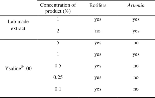
As shown in Table 1, Ysaline®100 was tested using 5 different concentrations
since the product has never been used on these organisms, whereas for fresh extract only two concentrations were tested since previous studies reported severe fish mortality when concentrations over 1 % of an aqueous solution of Asparagopsis taxiformis were used (Mata et al., 2013).
Table 1: Concentrations of the Ysaline®100 (dried powder) and the laboratory made
extract tested in rotifers and Artemia enriched with RedPepper; “yes” indicates it was
tested and “no” indicates it was not tested.
Concentration of product (%) Rotifers Artemia Lab made extract 1 yes yes 2 no yes Ysaline®100 5 yes no 1 yes yes 0.5 yes no 0.25 yes no 0.1 yes no
- 12 -
The laboratory made extract was added directly to the beakers since it was in
liquid form, whereas Ysaline®100 powder was previously diluted in salt water and then
added to the beakers. To assure that the corresponding control of each treatment had the same density of rotifers or Artemia, salt water was added to the control beakers in the same amount as the products.
Ysaline®100 (YSA) bactericide potential is well known in its target industry but
no data is available on its potential against marine bacteria commonly found in marine hatcheries. To analyse its potential in this marine environment the product was tested with a known pathological bacterium (Photobacterium damselae subsp. piscicida)
isolated from an outbreak of Senegalese Sole (Solea senegalensis).
P. damselae subsp. piscicida was cultured at room temperature in Tryptic Soy Broth medium (TSB from OXOID, USA) made with artificial seawater and used to show an exponential growth 48 h after inoculation. Concentrations of 0.01, 0.02, 0.03, 0.06, 0.125, 0.25, 0.5, 1 and 2 % were tested with the bacterium in octuplicate and compared with a control using the bacterium without YSA.
A sterile 96-well microtiter plate was filled with TSB and with overnight growing bacterial culture along with the different concentrations of YSA (except the control). Optical density (OD) readings were made at 540 nm in a Microplate reader
(SyergyTM 4-Biotek, USA) at the initial point and after 24, 48 and 120 h, following the
bacterial curve.
2.2.2. Larval rearing trial set-up
The gilthead seabream larvae trial comprised two treatments with four replicate tanks in each treatment. The trial started at mouth opening (4 DAH) and lasted until 35 DAH. All tanks were fed equally according to the protocol and the amount given per meal. Only the commercial product (YSA) was tested in this trial. YSA was selected considering two remarks. First, results from a short-term assay (until 11 DAH, Costa et al., 2013) showed no significant differences in larval survival and growth
- 13 -
against a control group. Second, advantages in using Ysaline®100 were the maintenance
of its chemical properties and availability throughout the complete larval trial.
The Ysaline®100 (YSA) treatment consisted of bathing the live preys (rotifers and
Artemia, accordingly to feeding protocol) with YSA before their provision to fish larvae, whereas for the control treatment (Ctrl) live prey were rinsed with clear salt
water. For YSA treatment live prey were bathed with 0.5 % Ysaline®100 during 30 min,
after the enrichment, and then rinsed with salt water before being fed to larvae. Live prey from the control treatment (Ctrl) were rinsed with salt water following enrichment, but were maintained in clear water for 30 min to ascertain that rotifers and Artemia from both treatments had identical nutritional value. Live preys were distributed over 4 to 5 meals along the day. Therefore, in order to maintain the nutritional value of the enriched
live preys after the 1st meal, they were stored in a cold bath of 5 1 ºC for the remaining
meals. These processes were performed daily during larval rearing.
Samples from rotifers, Artemia nauplii and metanauplii were collected before and after the 30 min bath for both treatments, for fatty acid determination. This analysis was to evaluate the impact of 30 min bath on preys’ fatty acid enrichment. These samples were rinsed with distilled water, flash frozen and freeze-dried in a Lyoquest – 50
(Telstar, Spain) freeze drier until processed for fatty acid analysis.
2.2.3. Larval sampling
To evaluate larval performance as a response to the application of a red seaweed product in the live prey, samples of 20 larvae, from each tank, for biometry (total length and dry weight) and development were collected from 2 DAH in 5 to 7 days intervals and 2 to 5 days intervals, respectively, until 34 DAH. To assess live feed intake samples (n = 15 per tank) were collected at 5, 12, 18, 23 and 30 DAH. Samples for histology and enzymatic activity (n = 15 and n = 30 respectively, per tank) were taken at 15 (only for histology), 20 and 34 DAH. Bacteriological analysis (n = 20 per tank) was performed in 34 DAH larvae and their rearing water. At the end of the trial (35 DAH) samples were also collected for stress test (n = 20 per tank), antibacterial test (n = 2 per tank), and for
- 14 -
lysozyme activity determination (n = 100 per tank). Fish larvae were sampled before food was added, exception made for the larvae collected to assess feed intake.
For enzymatic and lysozyme activity the starved larvae were collected, rinsed
with distilled water and flash-freeze. Samples were stored at -20 ºC and processed
within one month.
2.3. Analytical methodology
2.3.1. Fatty acid profile of live feed
Fatty acid methyl esters (FAME) were prepared according to Bandarra et al. (2009), using freeze-dried material and 5 mL of the acetyl chloride:methanol mixture (1:19, v/v). The transesterification was carried out at 80 ºC for 1 h. After cooling, 1 mL of water and 2 mL of n-heptane were added to the mixture, which was stirred and centrifuged at 2.150 g for 10 min. The organic phase was collected, filtered and dried over anhydrous sodium sulphate. The solvent was removed under nitrogen and the FAME dissolved in 0.1 mL of n-heptane. The analyses were performed in a Varian CP-3800 (Walnut Creek, CA, USA) gas chromatograph equipped with an auto sampler and fitted with a flame ionization detector at 250 ºC. The separation was carried out in an Omegawax (Supelco, USA) capillary column (25 m × 0.25 mm id). Temperature was programmed from 180 °C to 200 °C at 4 °C/min, holding for 10 min at 200 °C and heating to 210 °C at 4 °C/min, holding at 210 °C for 14.5 min with the injector and detector (FID) at 250 °C. FAME were identified by comparison with the retention times of known mixtures of standards (Supelco, FAME 37 and PUFA 3) and quantified using the area of the C21:0 internal standard with Varian software (USA).
- 15 -
2.3.2. Survival and growth
Larval survival was determined by counting the remaining larvae individually from all tanks at the end of the trial (35 DAH). Although dead larvae were counted every day, results were not considered to determine survival since several larvae were too degraded to be considered a reliable count.
To determine total length, larvae were measured under a Zeiss (Germany)
binocular microscope provided with an ocular micrometer (precision 0.01 mm) at
known magnification. Afterwards the same larvae were used to determine dry weight (DW), after being rinsed with distilled water, flash frozen in liquid nitrogen and stored in groups of 5 larvae at -20 Cº. Fifteen larvae were collected from each tank in a total of 60 larvae per treatment. At 2 DAH, since no treatments were in use, a total of 60 larvae were sampled from a common batch.
Frozen larvae were freeze-dried with a Lyoquest – 50 freeze drier and weighted in
a precision balance (Sartorius Pro 11, precision 0.001/0.002 mg; Sartorius AG,
Germany).
2.3.3. Life feed intake
To assess the live feed intake larvae were captured approximately 30 min after a meal and observed in a Zeiss binocular microscope to observe stomach content. This was possible because larvae were not pigmented at the surveyed sampling ages. The information was analysed according to presence or absence of food in the stomach.
2.3.4. Morphology and development
Larval morphology and development, with particular attention to swimbladder inflation, visible larval body anomalies and caudal fin formation, was assessed by collecting images of the larvae using a Canon (Japan) Power shot G5 camera linked to a Zeiss binocular microscope. These parameters are commonly monitored in hatcheries,
- 16 -
since they are good quality indicators that can be easily observed and are not costly or labour demanding.
Swimbladder and body anomalies were analysed by present or absent. Mortality at the surface of the tanks was also considered to assess fish larvae ability to inflate the swimbladder. Dead larvae on the water surface trapped on the oil film were counted daily.
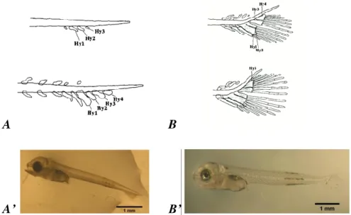
Caudal fin formation was classified according to 3 stages: an early flexion stage (EF) was considered when hypuralia elements were evident in a straight notochord (Figure 2A and A’), the flexion stage (FS) was considered when the posterior part of the notochord presented an upward curvature (Figure 2B and B’) and the last stage (CF) was considered when the flexion was concluded.
A B
A’ B’
Figure 2: Development stages of the caudal complex of S. aurata larvae. A. - early
flexion stage (EF) with the presence of hypuralia (Hy) elements; B. - flexion stage (FS) with an upward curvature of the posterior part of the notochord (in Koumoundouros et. al., 1997). A’ and B’ - examples of observed larvae at EF and FS respectively.
- 17 -
2.3.5. Digestive system histology
Larvae were fixed in 4 % buffered formaldehyde with 10 times more volume than the sample volume (10:1). Samples were kept in a fridge overnight. Afterwards they were washed, first with phosphate buffered saline (PBS) pH 7.4 (3 x 15 min) and after with tap water (15 min) and stored in 70 % ethanol at 8 ºC.
The 20 and 34 DAH larvae were decalcified before embedding in paraffin. To be decalcified the samples were rinsed in tap water (30 min for 1 h) then they were sunken in the decalcification solution (1:1 of 8 % hydrochloric acid and 8 % formic acid) during 2 h and 4 – 5 h for 20 and 34 DAH larvae respectively. After decalcification larvae were again stored in 70 % ethanol in the fridge. Afterwards all samples were dehydrated in graded ethanol before equilibration in xylene and then embedded in
paraffin wax. The paraffin was cut in 5 m thick sections and stained with
haematoxylin-eosin before observed under a light microscope.
Digital images of fish larvae sections were captured with a digital camera Nikon DS-F12 connected to a Nikon (Japan) Eclipse Ci binocular light microscope. The abdominal cavity images were examined (40 x and 100 x) and analysed using the image analysis software ImageJ 1.47t (NIH, USA) with particular attention to the liver and to the intestine. Liver and intestinal images were taken at a 400 x magnification from different sections (n = 4) of each larva (n = 3) from each tank (n = 4) of the two treatment groups sampled at different ages.
Liver was evaluated according to the hepatocyte density. Their nuclei were
counted automatically with the software, in a known area (0.01 mm2) of the photo to
assess the nutrient reserves (lipid and/or glycogen) content in the liver, assuming that a higher nuclei density always means smaller hepatocyte with lower nutrient reserves content. Intestinal morphology was evaluated according to its epithelium height as a way to assess differences in the absorption capability of the larvae.
- 18 -
2.3.6. Digestive enzymes activities
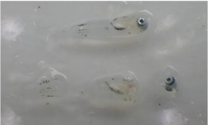
Before analysis, 34 DAH larvae were sectioned on a cold surface, to obtain the abdominal cavity as illustrated in Figure 3. Samples of the sectioned body cavity and whole body larvae (20 DAH) were then weighted and homogenised with 15 x higher volume of deionised water than their corresponding weight, using an vortexer (Ultra
Turrax T10 from IKA, Germany). The solutions were centrifuged (5000 rpm, 4 ºC)
during 5 min in a table top refrigerated centrifuge (Z383K from HERMLE Labortechnik, Germany) and afterwards the homogenates were separated from the pellets and stored at -20 ºC between analyses. Trypsin, amylase, aminopeptidase, acid phosphatase and alkaline phosphatase activity were determined.
Figure 3: 34 DAH larvae, in whole body (above) and sectioned (below) with the
abdominal cavity in the middle section.
Trypsin activity was measured at 407 nm at 25 °C using BAPNA
(Nα-Benzoyl-DL-arginine-p-nitroanilide) as substrate in Trizma-CaCl2 buffer (20 mM), pH 8.2 as
described by Holm et al. (1988). To determine amylase activity the homogenates were
combined with a starch solution (3 g.L-1 starch) as substrate (reaction tubes)
(Méthais & Bieth, 1968). The same solution was prepared without the homogenates (blank tubes). After 30 min at 37 ºC the reaction was stopped in all tubes by adding hydrochloric acid (HCl) at 1 M. The homogenate was then added to the black tubes and an iodine working solution at N/3000 was added to all tubes. The absorbance of all solutions was read at 580 nm and the results from the reaction tubes were compared to the results from the blank tubes to calculate amylase activity.
- 19 -
Aminopeptidase activity was determined using LpNa (leucine-p-nitroanilide) at 2 mM with DMSO (dimethyl sulfoxide) as substrate and Tris-HCl at 100 mM as the buffer solution combined with the homogenates (Maroux et al., 1973). Acid and alkaline phosphatase activities were determined using pNPP (p-nitrophenylphosphate)
at 5.5 mM with MgCl2.6H2O as substrate and citric acid at 0.1 M with sodium citrate at
0.1 M (acid phosphatise activity, Terra et al., 1979) or Na2CO3 at 30 mM (alkaline
phosphatise activity, Bessey et al., 1946) as the buffer solutions combined with the
homogenates. Absorbance was read at 405 for aminopeptidase and acid phosphatase
and at 407 for alkaline phosphatase, during approximately 10 min.
Absorbance readings were performed in a in a Multiskan Go apparatus
(Thermo ScientificTM, USA). Enzyme activity was calculated in micromoles of
substrate hydrolysed (mU) and expressed as specific (mU/mg protein) and total activity (mU/larva). Protein was determined according to the Bradford method (Bradford, 1976) using bovine serum albumin (BSA) as a standard.
2.3.7. Lysozyme activity
Whole body larvae were weighted and homogenised with 4 x higher volume of phosphate buffer than the corresponding weight, using a vortexer (Ultra Turrax T25
from IKA, Germany). The solutions were centrifuged (17000 rpm, 4 ºC) during 30 min
in a table top refrigerated centrifuge Z383K and afterwards the homogenates were separated from the pellets. Lysozyme activity was measured using a method based on the ability of lysozyme to lyse the bacterium Micrococcus lysodeikticus (Ellis, 1990). In
a 96-well microplate, 70 L of the samples homogenate and 200 L of the substrate
(0.5 mg.mL-1 of M. lysodeikticus suspension (Sigma, USA) in 0.05 M sodium phosphate
buffer at pH 6.2 (SFB)) were added in triplicates. The reduction in absorbance at 450 nm was measured in approximately 1 min intervals for a total of 4 min at 25 ºC
using a Multiskan Go. Lyophilized hen egg white lyzozyme (Sigma) was serially
diluted in SFB and used to develop a standard curve whose formula was used to calculate the lysozyme amount in the samples.
- 20 -
2.3.8. Cortisol analysis from the stress test
At the end of the trial, collected larvae were kept in two groups of 15 larvae (30 larvae per tank) until calm behaviour was observed. One of the groups was subjected to a stress situation of 30 s without water. Both groups were rinsed with distilled water, flash frozen and stored at -20 ºC.
The samples were unfrozen to be homogenized in 500 L of deionised water
using a vortexer (Ultra Turrax 8G from IKA, Germany). In 10 mL capacity extraction
tubes 4 mL of diethyl ether was added to the sample homogenate and stirred (10 min). Afterwards samples were centrifuged at 500 rpm at 4 ºC (5 min). To separate the aqueous phase from the formed pellet the tubes were submerged in liquid nitrogen to freeze the pellet enabling the separation of the ether to another test-tube. The pellet was submitted to a second extraction after which the test-tubes were kept in a dry bath of
37 – 40 ºC overnight. After all the ether evaporated 250 L of 0.9 % NaCl solution were
added and the extracted samples were stored at -20 ºC. Cortisol concentration was determined in vitro through immunoassay using the Cortisol Saliva ELISA kit (IBL, Germany).
2.3.9. Bacteriological analysis
Larvae and water from the larval rearing tanks were sampled at 34 DAH to assess bacterial growth. Twenty larvae were disinfected with benzalkonium chloride (0.1 %) for 30 s and then washed with sterilized saline solution (1.5 %). Afterwards larvae were homogenized with a pestle (Potter-Elehjem Tissue Homogenizer – PTFE from VWR, USA) in a final volume of 1 mL with sterile artificial sea water. This solution and the
water samples were sequentially diluted three-fold and plated. Triplicates of 100 μL of
each dilution were spread on agar plates, tryptic soy agar (TSA from Merck, USA) was
used to obtain the total number of aerobic bacteria, and
thiosulfate-citrate-bile salts-sucrose agar (TCBS from OXOID, USA) was used to
isolate and count Vibrionaceae. Concentrations of 10-1 to 10-3 and 10-2 to 10-4 were used
for TCBS and TSA respectively for the homogenized larvae. Water samples were
- 21 -
incubated at 22ºC for 7 days and CFUs (colony forming units) were counted after this period.
2.3.10. Antibacterial test
Four larvae from each tank were collected in starvation at the end of the trial to access antibacterial activity. The larvae were weighted, smashed and placed onto a disc in groups of two. The discs were placed onto Mueller Hinton agar (MH, OXOID) plates for each replicate, previously prepared with a pathogenic bacterium (Photobacterium damselae subsp. piscicida) isolated from an outbreak of Senegalese Sole (Solea senegalensis). Discs with antibiotics (Oxitetracicline and Flumequine, OXOID) were also placed in the plates to be used as a control. Plates were incubated at 22 ºC for two days and the data were treated as presence or absence of a resistance halo.
2.4. Statistical analysis
The data obtained from total length, DW, mortality at the surface of the water tank, cortisol, fatty acids, histology analysis, enzyme and lysozyme activity and bacteriological analysis were subjected to log transformation before statistical analysis. The data obtained in percentages such as preys population and survival after subject to YSA treatment, larval survival, live feed intake, swimbladder inflation, body anomalies occurrences and caudal fin formation were statistically analysed after an arcsine square root transformation.
Student’s t-test was applied to results from survival at the end of the trial and at the surface of the water tank and to the bacteriology results. A Mann-Whitney Rank Sum test was applied to Lysozyme activity results since one of the replicates was lost (no equally of variance). One Way Analysis of Variance (One Way ANOVA) was applied to fatty acids and cortisol results and Two Way Analysis of Variance (Two Way ANOVA) was applied to results from the first trial to define the adequate concentration
- 22 -
of an A. armata product to be used in live feed. This last method was also used to compare fatty acids, length and DW, live feed intake, swimbladder inflation, body anomalies occurrences, caudal fin formation, cortisol, histological analysis and enzyme activity. When appropriate both ANOVA’s were followed by an all pairwise Multiple Comparison Procedures (Holm-Sidak method).
- 23 -
3. Results
3.1.Effects of Asparogopsis armata extracts on live feed densities and pathogenic bacteria
3.1.1. A. armata extracts on live feed densities
Rotifers population was not affected by the laboratory-made A. armata extract. The percentage of active/live rotifers was of 100 % after 2 h, decreasing to
91.8 6.09 % of the initial number after 3 h. Artemia metanauplii presented 100 %
survival for the two tested concentrations of extract after 3 hours.
Artemia metanauplii also presented 100 % survival with the commercial product
(Ysaline®100). Results obtained for rotifers subjected to the different concentrations of
YSA tested are shown in Figure 4. The 5 % concentration was not included since no rotifer survival was observed after 30 min at this concentration. At 1 % concentration, the number of rotifers decreased 50 % after 30 min and no living rotifers were observed after 60 min. No significant differences were observed between the remaining concentrations during the experimental period.
Figure 4: Percentage of rotifers from the initial population number at 30, 60, 90 and
180 min submerged in a bath with the commercial product Ysaline®100 at different
concentrations. Values are presented as mean standard deviation (n = 3).
0 20 40 60 80 100 120 30 60 90 180 % t (min) 1% 0,5% 0,25% 0,1%
- 24 -
3.1.2. A. armata extracts on pathogenic bacteria
Photobacterium damselae subsp. piscicida growth was affected by YSA at concentrations higher than 0.25 % (Figure 5). Bacterial growth was inhibited during 120 h at concentrations of 2 and 1 %. YSA at 0.5 % concentration inhibited bacterial growth efficiently during 48 h. At 0.25 % YSA concentration, bacterial growth could be observed during the first 24 h of incubation, but at an inferior rate than lower concentrations. Concentrations of 0.125 % YSA and lower (not shown) presented a similar bacterial growth in comparison with the control.
Figure 5: Optical density at 540 nm (OD540) growth curves of Photobacterium
damselae subsp. piscicida growth during 120 h, using different concentrations of
Ysaline®100 (YSA). Control = no YSA. Values are presented as mean standard
deviation (n = 8). 0 0,1 0,2 0,3 0,4 0,5 0,6 0,7 0,8 0 20 40 60 80 100 120 OD540 time (h) 2% 1% 0.5% 0.25% 0.125% Control
- 25 -
3.2. Larval rearing
3.2.1. Live feed fatty acid profile
Live prey fatty acids are presented in Table 2. No significant differences were
observed for any fatty acid in rotifers, except for total 3, with a lower value in rotifers
after 30 min in clear water. In Artemia nauplii, most of the fatty acids were significantly
(P < 0.05) lower after 30 min in an YSA bath. However, the main PUFA’s
(polyunsaturated fatty acids) for fish larvae namely DHA (docosahexanoic acid), EPA (eicosapentaenoic acid) and ARA (arachidonic acid), presented no significant differences before and after the 30 min, with and without YSA for this prey. Artemia metanauplii main PUFA also presented no significant differences, but for most fatty acids significant differences were observed after 30 min in clear water and YSA bath.
- 26 -
Table 2: Fatty acids profile in mg.g-1 of dry weight of the preys (Rotifers, Artemia nauplii and Artemia metanauplii) used in the larval rearing
trial before (0) and after a 30 min period for control (30 Ctrl) and YSA (30 YSA) treatments. Values are presented as mean standard deviation.
Different superscripts indicate significant differences (P < 0.05) in the All pairwise Multiple Comparison Procedures (Holm-Sidak method)
within the same fatty acid for each live feed.
Fatty acid
Rotifers Artemia nauplii Artemia metanauplii
t = 0 t = 30 Ctrl t = 30 YSA t = 0 t = 30 Ctrl t = 30 YSA t = 0 t = 30 Ctrl t = 30 YSA
14:0 4.7 2.13 2.9 0.97 5.3 1.07 5.2 1.16a 5.4 0.78a 2.5 0.33b 6.8 0.43a 3.4 1.06b 2.8 0.57b 16:0 28.9 11.97 19.5 5.43 31.1 4.36 30.6 5.29a 34.3 2.91a 18.7 1.73b 53.1 3.38a 29.9 7.26b 26.3 4.96b 18:0 4.1 1.16 3.6 0.78 4.4 0.35 6.9 1.22a 8.1 0.61a 5.9 0.16b 12.0 0.59a 8.6 1.38b 8.6 1.19b Other SFA 3.1 1.35 2.5 0.55 3.6 0.50 19.6 2.96a 21.7 1.99a 12.2 1.13b 7.9 0.36a 5.1 0.93b 5.0 0.79b Σ SFA 40.8 16.57 28.6 .7.01 44.4 6.15 62.2 10.39a 69.5 5.62a 39.4 3.33b 79.8 4.74a 47.0 10.56b 42.7 7.46b 16:1 (n9+n7) 4.0 1.78 2.5 0.75 4.6 0.69 31.6 4.56a 35.8 4.08a 19.4 2.03b 5.7 0.44a 3.2 0.83b 3.0 0.58b 18:1 (n9+n7) 11.3 3.32 9.1 1.68 12.9 1.02 46.9 4.36a 54.1 4.52a 39.0 1.43b 39.9 2.12a 27.3 4.57b 28.2 4.22b Other MUFA 2.3 0.14 2.2 0.39 2.2 0.04 2.2 0.40 2.3 0.31 1.9 0.23 2.0 0.15 1.3 0.49 1.2 0.35 Σ MUFA 17.6 5.17 13.8 2.43 19.7 1.59 80.6 8.73a 92.2 8.17a 60.3 3.46b 47.6 2.65a 31.8 5.85b 32.4 4.44b 18:2 n 6 13.9 4.26 9.8 2.63 15.2 1.29 5.0 0.88ab 5.8 0.33a 4.1 0.18b 13.6 0.78a 9.2 1.49b 9.4 1.46b 18:3 n 3 0 0 0 2.0 0.18ab 2.4 0.23a 1.9 0.07b 43.7 2.65a 30.1 4.70b 31.0 4.92b 20:4 n 6_ARA 3.1 0.46 3.0 0.58 3.2 0.18 5.2 0.60 5.7 0.45 4.9 0.11 5.6 0.24 4.8 0.39 5.0 0.51 20:5 n 3_EPA 3.5 056 3.1 0.38 3.6 0.23 16.3 1.03 18.1 1.39 18.6 0.45 11.0 0.37 9.7 0.77 10.0 1.00 22:5 n 6 7.5 0.29 6.8 0.97 6.8 0.25 0 0 0 7.9 0.14 8.1 0.09 8.2 0.54 22:6 n 3_DHA 19.8 0.86 18.6 1.46 18.1 0.51 0 0 0.3 0.45 19.9 0.23 20.9 1.01 20.9 1.47 Other PUFA 6.0 1.72 5.2 0.65 6.2 0.63 32.3 3.80ab 36.1 3.60a 24.4 3.70b 13.4 0.84a 10.0 1.13b 10.4 1.30b Σ PUFA 53.9 5.82 46.3 4.45 53.1 1.56 60.9 5.81a 68.2 5.36a 50.5 1.72b 115.2 5.15a 92.9 7.39b 94.8 9.70b Total ómega 3 (ω3 or n3) 28.0 0.94a 25.7 1.06b 26.3 0.24ab 23.4 1.81 25.9 1.95 22.4 0.46 86.2 3.86a 69.4 5.38b 69.5 7.40b Total ómega 6 (ω6 or n6) 25.4 4.55 20.1 3.37 26.1 1.30 11.3 1.56 12.7 0.80 10.0 0.37 27.5 1.16a 22.5 1.80b 22.9 2.03b Ratio ω3/ω6 1.1 0.16 1.3 0.16 1.0 0.04 2.1 0.15 2.0 0.12 2.2 0.04 3.1 0.01a 3.1 0.02ab 3.0 0.06b Ratio DHA/EPA 5.8 1.11 6.1 0.84 5.1 0.39 - - - 1.8 0.05 2.2 0.29 2.1 0.16
- 27 -
3.2.2. Survival and growth
Larval survival at the end of the trial was significantly higher (P < 0.05) in the control treatment (Ctrl) when compared to YSA. The results obtained were
16.5 2.72 % and 8.2 1.80 % for Ctrl and YSA treatments, respectively.
Larval growth in total length and dry weight was similar for both treatments since the beginning of the trial until 15 DAH. From this age onward until the end of the trial fish larvae from YSA exhibited a higher weight and length than larvae from Ctrl (P < 0.05) (Figure 6).
Figure 6: Larval growth in total length (A) and in dry weight (B) for the different
treatments (Ctrl and YSA) along the trial period. Values are presented as
mean standard deviation (n = 45 for total length and n = 12 for dry weight).
3.2.3. Swimbladder
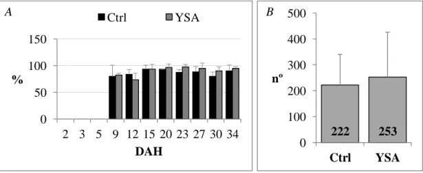
The swimblader inflation was detected since 9 DAH in more than 80 % of seabream larvae, as shown in Figure 7A. Dead larvae on the water surface of the tanks (Figure 7B) were observed between 10 and 17 DAH. These patterns and the number of
dead larvae were similar for both treatments (P > 0.05).
0 3 6 9 12 15 18 0 5 10 15 20 25 30 35 mm DAH Ctrl YSA
Polinomial (Ctrl) Polinomial (YSA)
0,0 0,5 1,0 1,5 2,0 2,5 3,0 3,5 0 5 10 15 20 25 30 35 g DAH Ctrl YSA
Exponencial (Ctrl) Exponencial (YSA)
- 28 -
Figure 7: A – Percentage of larvae with the swimbladder inflated in both treatments
tested, along the trial. B – Total number of dead larvae at the surface of the water for
each treatment throughout the trial. Values are presented as mean standard deviation
(n = 45 and n = 4 for A and B respectively).
3.2.4. Occurence of body anomalies
The incidence of visible body anomalies (Figure 8) represented 2.5 % of the total larvae analysed, with higher incidence until 12 DAH. No statistical differences were
observed between treatments (P > 0.05).
Figure 8: Examples of deformed and normal larvae from both treatments at two
different ages: A – larvae from the control treatment at 9 DAH with an abnormal larva below and B – larvae from YSA treatment at 12 DAH with an abnormal larva above.
0 50 100 150 2 3 5 9 12 15 20 23 27 30 34 % DAH Ctrl YSA 222 253 0 100 200 300 400 500 Ctrl YSA nº A B A B