Ivo Edgar Araújo Lopes
Janeiro de 2013
Development of stable lipoplexes
DODAC/MO/PEG-FOL for delivery of nucleic
acids to cells expressing folate receptor
UMinho|20 13 Iv o Edgar Ar aújo Lopes De velopment of s table lipople xes DOD A C/MO/PEG-F OL for deliver
y of nucleic acids to cells e
xpressing folate recep
tor
Ivo Edgar Araújo Lopes
Janeiro de 2013
Dissertação de Mestrado
Mestrado em Biofisica e Bionanossistemas
Development of stable lipoplexes
DODAC/MO/PEG-FOL for delivery of nucleic
acids to cells expressing folate receptor
Escola de Ciências
Trabalho realizado sob a orientação da
Professora Doutora Maria Elisabete Cunha
Dias Real Oliveira
E co-orientação da
COMPROMETE;
Universidade do Minho, ___/___/______
This thesis is dedicated to the memory of my beloved mother Maria da Conceição Dantas Araújo
v Oliveira and Dr. Andreia Gomes for the inestimable help, advice, teachings, kindness and patience that they provided me, and to have trusted and improved my capabilities as an investigator.
I extend my gratitude to my colleagues and friends João Neves, Ana Oliveira, Odete Gonçalves and Marisa Passos for their priceless help, that improved grandly my investigation, and also for their availability and sympathy, which made my work much easier and pleasant.
I also want to thank my family and friends for supporting me, during all my
academic career, and my passion for the investigation and also for being a beacon in
these extremely difficult time, especially my little brother that looks much more like a father to me, these days.
Least but not least I want to take the opportunity to go on record and thank my mother, for have worked determinedly and sacrificed herself, all of her life, to provide me and my brother a better life, for I think I have taken her for granted I didn’t thanked her enough while I had the chance.
vii versatile nonviral vectors for the delivery of nucleic acids and the discovery of RNA interference (RNAi), the development of devices to deliver RNAi molecules to specific cells as became an appealing strategy to achieve the treatment of genetic disorders.
Hence, with this work we intended to develop and characterize novel systems for therapeutic siRNA delivery based on dioctadecyldimethylammonium chloride (DODAC)/ monoolein (MO) liposomes, which previous studies developed by our group have validated as a promising system for plasmid DNA delivery, These systems demonstrated to have promising features compatible with stable and efficient encapsulation of RNA. Furthermore, DODAC/MO vesicles were coated with polyetilenoglycol (PEG) and PEG-folate grafted lipids to increase circulating half-life and obtain selectivity towards folate receptor expressing (FR+) cells (an overexpressed receptor characteristic of several cancer cells including chronic myeloid leukemia).
As a result of this project, DODAC/MO/PEG liposomes and derived RNA-containing lipoplexes showing stability in solutions of different concentrations of salt
and H3O+ and in fetal bovine serum solutions at 30 e 80% (v/v) were produced.
Additionally, it was possible to observe that the liposomes and lipoplexes coated with PEG-Folate are better internalized by FR+ cells than those with PEG only.
ix apelativos e versáteis para a entrega de ácidos nucleicos e a descoberta do RNA de interferência (RNAi), o desenvolvimento de dispositivos para entrega de moléculas de RNAi a células específicas tornou-se numa estratégia atrativa para alcançar o tratamento de doenças foro genético.
Assim, com este trabalho pretendeu-se desenvolver e caracterizar novos sistemas para a entrega de siRNA terapêutico baseados em lipossomas de cloreto de dioctadecildimetilamónio (DODAC)/monoleína (MO), que haviam já sido validados como sistemas promissores para a entraga de DNA plasmídico, em estudos anteriormente desenvolvidos pelo nosso grupo. Estes sistemas demonstraram ter características promissoras compatíveis com o encapsulamento eficiente e estável de RNA. Adicionalmente, os vesiculos de DODAC/MO foram revestidos com polietilenoglicol (PEG) e de lípidos PEG funcionalizados com folato para aumentar a tempo de circulação e obter selectividade para células expressando receptores de folato (FR +) (um receptor sobre-expresso em muitos tipos de células cancerigenas, nas quais se incluem as de leucemia mielóide crónica).
Como resultado deste projeto produziram-se lipossomas DODAC/MO/PEG e os seus respetivos lipoplexos de RNA mostrando estabilidade em soluções de diferentes
concentrações de sal e H3O+ e em soluções de soro fetal de bovino a 30 e 80% (v/v).
Além disso, foi possível observar que os lipossomas e os lipoplexos revestidos com PEG-folato internalizam melhor nas células FR +, comparativamente com os sistmas contendo apenas PEG.
xi CML - chronic myeloid leukemia
C. R. - Charge ratio
DLS - Dynamic Light Scattering
DME(M) - Dulbecco's Modified Eagle's (medium)
DNA - Deoxyribonucleic acid
DODAB - Dioctadecyldimethylammonium bromide
DODAC - Dioctadecyldimethylammonium chloride
DODAX - Dioctadecyldimethylammonium chloride or bromide
DOPE - Dioleyl-phosphatidyl-ethanolamine
DOTAP - 1,2-dioleoyl-3-trimethylammonium-propane
FBS - Fetal Bovine Serum
LDH - Lactate dehydrogenase
MO - 1-monooleoyl-rac-glycerol (monoolein)
mRNA - messenger ribonucleic acid
xii
PDI - Polydispersity
PEG (PEG (2000)-PE) - 1,2-distearoyl-sn-glycero-3-phosphoethanolamine-N-[methoxy(polyethylene glycol)-2000]
PEG-Folate (DSPE-PEG(2000)-Folate) - 1,2-distearoyl-sn-glycero-3-phosphoethanolamine-N-[folate(polyethylene glycol)-2000]
RNA - Ribonucleic acid
RNAi - Ribonucleic acid interference
Rhodamine B DHPE - Lissamine Rhodamine B 1,2-Dihexadecanoyl-sn-Glycero-3-Phosphoethanolamine Triethylammonium Salt
Index Acknowledgments ... v Abstract ... vii Resumo ... ix Abbreviations ... xi I. Introduction ... 1 1. Gene therapy ... 1
2. Cationic liposomes are promising vectors for gene therapy ... 1
3. Cellular barriers dictate transfection efficiency ... 8
4. Gene silencing through regulatory small RNA delivery as a promising therapeutic tool ... 10
5. Chronic myeloid leukemia ... 13
6. Objective ... 15
II. Materials and Methods ... 17
A. Materials ... 17
B. Methods ... 18
1. Liposomes preparation ... 18
1.1 Lipid film hydration ... 18
1.2 DODAC/MO (2:1) liposomes with 1, 2 and 5% PEG (1mM) ... 19
2. Lipoplexes preparation ... 19
3. Light interactions with matter... 20
3.1 Absorption ... 21
3.2 Fluorescence ... 23
3.3 Dynamic light scattering ... 25
3.3.1 Size ... 25 3.3.2 ζ-potential ... 26 4. Liposome stability... 28 5. Complexation assays ... 29 6. Encapsulation efficiency ... 30 7. Lipoplexes stability ... 30 8. Destabilization of lipoplexes ... 31 9. Cytotoxicity assays ... 32
9.1 LDH (Lactate dehydrogenase) assay ... 32
9.3 Liposomes toxicity ... 32
9.4 Lipoplex toxicity ... 34
10. Internalization assay ... 35
10.1 Internalization assay statistics ... 37
III. Results and Discussion ... 39
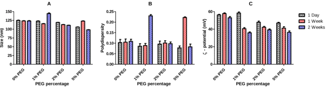
1. Characterization of liposomes DODAC/MO (2:1) at different PEG percentages ... 39
1.1 Liposomal mean sizes ... 40
1.2 ζ-potential of the liposomes ... 41
2. Cytotoxicity of liposomes DODAC/MO (2:1) at different PEG percentages in L929 cell line ... 42
3. Stability of Liposomes DODAC/MO (2:1) and DODAC/MO (2:1) 5% PEG ... 44
3.1 Effect of NaCl concentration on the mean size and ζ-potential ... 44
3.2 Stability in fetal bovine serum ... 46
3.3 Stability in different pH ... 49
4. Dynamics of RNA complexation ... 50
4.1 Dynamic Light scattering studies. ... 50
4.2 RiboGreen assay ... 53
5. RNA encapsulation efficiency of both lipoplexes at charge ratio 10 ... 55
6. Stability of Lipoplexes DODAC/MO (2:1) 5% PEG – RNA Charge ratio 10 ... 57
6.1 Stability of lipoplexes with increasing NaCl concentration and incubation time ... 57
6.2 Stability in fetal bovine serum ... 59
6.3 Stability in different pH ... 61
6.4 Destabilization with Heparin ... 62
7. Toxicity of lipoplexes DODAC/MO (2:1) 5% PEG (0,5 mM)- RNA (10 μg/mL) C. R. 10 in L929 and MDA-MB-468 cell Lines. ... 64
7.1 Toxicity of lipoplexes DODAC/MO (2:1) 5% PEG (0,5 mM)- RNA (10 μg/mL) C. R. 10 in L929 cell Line. ... 64
7.2 Toxicity of lipoplexes DODAC/MO (2:1) 5% PEG (0,5 mM)- RNA (10 μg/mL) C. R. 10 in MDA-MB-468 cell Line. ... 66
8. Internalization Assays ... 67
IV. Conclusion and future work ... 71
V. Bibliography ... 73
Appendix 1... 79
Appendix 2... 81
1 I. Introduction
1. Gene therapy
Gene therapy is the attempt to correct genetic disorders, utilizing genetic material designed to correct the anomaly. In spite of holding great promise in the treatment of hard-to-cure human diseases, the expression of therapeutic nucleic acids in cells if far from being a simple endeavor, as the human body has evolved to protect itself from environmental aggression, such as the incorporation of foreign genetic material [1].
2. Cationic liposomes are promising vectors for gene therapy
Application of naked DNA in gene therapy is rather inefficient, since the DNA molecules, large and hydrophilic due to the negatively charged phosphate groups, [2] are prone to degradation by serum nucleases [3, 4] and clearance through the mononuclear phagocyte system [4] Complexation of DNA mediated by electrostatic interactions between the negatively charged phosphate backbone of DNA and cationic molecules leads to charge neutralization and compaction of the nucleotidic fragment [2]. In the case of the application of naked RNA the molecules cannot cross the cell membrane, due to electrostatic repulsion between their negative charges and are rapidly cleared by the renal system [5] having very short half-life in blood [6].
One solution appeared in the employment of viruses, one organism that has specialized in the infection of host cells to express its own genetic material. This capability makes them obvious candidates for the use as vectors in gene therapy assays [1, 7]. Nevertheless, despite their efficient mechanisms of gene transfer, the application of viruses in gene therapy involves several drawbacks. The production of therapeutic viruses is difficult, the repeated administration leads to acute inflammatory responses and there is a risk of insertional mutagenesis associated to the integration of foreign DNA by some viruses [3]. Aditionally, the limited size of the carried transgene [8], the possibility of endogenous virus recombination and the oncogenic effects [4] impair the application of the virus in gene therapy experiments.
Nonviral delivery of nucleic acids presents an opportunity to circumvent the problems associated with viral vectors. Nonviral vectors are simple to use, can be
2
produced in a large scale and do not cause immune response [4]. They also allow specificity for the desired cells/tissues by incorporation of appropriated moieties in the vector formulation [3]. One the other hand, these vectors do not have capabilities to overcame the cellular barriers and the immune defense mechanisms as the viruses and, as a consequence, they fail to be as effective in transfecting cells as viruses. Peptides and dendrimers offer a possibility to compact DNA/RNA for systemic delivery [2] but the nonviral systems of election for the delivery of nucleic acids are the ones based in the cationic lipids and polymers [9].
Following the observation of Alec Bangham that phospholipids in aqueous
systems can form closed bilayered structures, liposomes evolved from being just an object of biophysical research to become one of the election pharmaceutical carriers for several practical applications. In the past 20 years, advances in the area resulted in the appearance of several liposomal drugs, and of many unique liposomal-based biomedical products and technologies.
Liposomes are spherical, self-assembled structures formed in aqueous solutions. They as constituted by one or several concentric lipid bilayers, with an aqueous phase in their lumen and in-between their bilayers, and can adopt the shape of multilamellar vesicles (consisting in of several concentric bilayers with sizes ranging between 500 to 5,000 nm), large unilamellar vesicles (ranging in size from 200 to 800
nmand formed by a single bilayer) and small unilamellar vesicles (around 100 nm in
diameter). Liposomes show some attractive biological properties as they are biocompatible and capable of entrapping water-soluble molecules in their internal water compartment, and water-insoluble compounds in their membrane, protecting them from the inactivating effect of external conditions and avoiding undesirable side reactions. They provide a unique opportunity to deliver drugs into cells or even to individual cellular compartments and their size, charge and surface properties can be easily tuned by adding new components to the lipid mixture before liposome preparation and by variation of the preparation methods themselves.
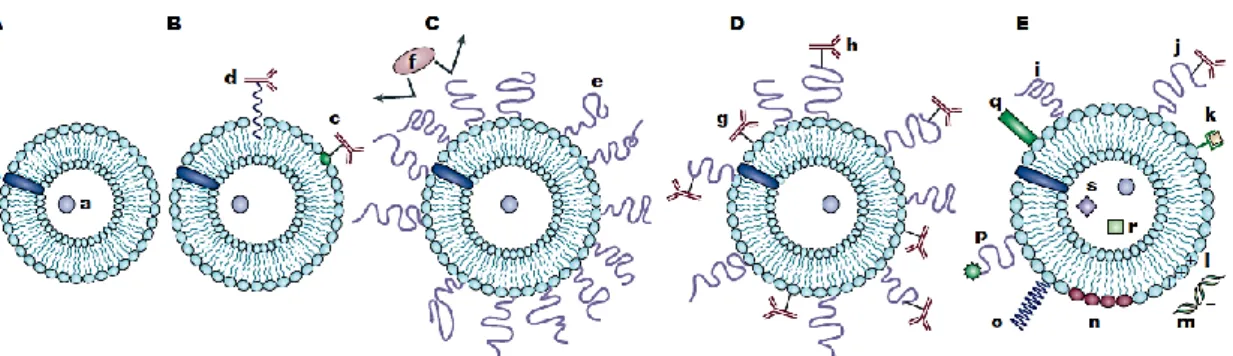
The versatility of liposomes allows them to serve for various finalities, by functionalization of their surface with various modifiers (figure 1. (A, B, C, D and E)). Modifying their surface by attachment of hydrophilic polymers, with highly flexible main chain, results in liposomes with high half-life in circulation, while the
3 incorporation of labels allows for monitoring the fate of liposomes inside an organism or cell, and for enhanced diagnostic imaging through the preparation of contrast liposomes. Liposomes with targeting functionality can be obtained by grafting specific ligands, while the attachment of either antibodies or antigens onto the surface of liposomes results in immunoliposomes. The incorporation of positively charged lipids or positively charged polymers into the liposome allows the binding of nucleic acids and the formation of systems for cell transfection [10].
Figure 1. Evolution of liposomes. A) Early phospholipid liposomes with a water soluble drug (a) entrapped into the aqueous liposome interior and a water-insoluble drug (b) incorporated into the liposomal membrane. B) Antibody-targeted immunoliposome with antibody covalently coupled (c) to reactive lipids of the membrane, or hydrophobically anchored (d) into the liposomal membrane. C) Long-circulating liposome grafted with a protective polymer (e) such as PEG, which promotes the shielding of the liposome surface from opsonizing proteins (f). D) Long-circulating immunoliposome simultaneously bearing both protective polymer and antibody, which can be attached to the liposome surface (g) or to the distal end of the grafted polymeric chain (h). E) New-generation liposome with its surface modified by attachment of: protective polymer (i) or protective polymer and targeting ligand, (e.g. antibodies) (j); diagnostic labels (k); positively charged lipids (l), allowing for the complexation with DNA/RNA, (m); stimuli-sensitive lipids (n) or stimuli-sensitive polymers (o); cell-penetrating peptides (p); and viral components (q). Additionally to a drug, liposome can be loaded with magnetic particles (r), for magnetic targeting, and/or colloidal gold or silver particles (s) for electron microscopy [10].
To promote liposomal drug delivery in specific tissues and organs, targeted liposomes with surface-attached ligands capable of recognizing and binding to markers on the cells of interest have been developed (Figure 1 B). Immunoglobulins and their fragments are widely used as targeting moieties and can be attached to liposomes, without affecting either the liposomal integrity or the antibody properties, by covalent
4
binding to the liposome surface or by hydrophobic insertion into the liposomal membrane after their functionalization in hydrophobic residues [10].
One of the disadvantages of using liposomes is their rapid elimination from blood by capture by the cells of the reticuloendothelial system. To achieve long circulation of liposomes in vivo, different methods have been suggested including the coating of the liposome surface with inert biocompatible polymers such as polyethylene gylcol (PEG) to form a protective layer over the liposome surface and
slow down liposome recognition by opsonins and subsequent clearance [11](Figure 1
C). The flexibility of the protective polymers allows a relatively small number of polymer molecules to create an impermeable layer over the liposome surface.
Research on PEGylated liposomes has been focused on the attachment of removable PEGs so that this coating detaches under the action of local pathological conditions, resulting in improved cellular capture of the liposome [10].
Clinical research showed that the anticancer agent doxorubicin has been selective delivered in PEG liposomes for the treatment of solid tumors in patients with breast-carcinoma metastases, resulting in improved survival [12-14], and impressive results were obtained for its delivery in unresectable hepatocellular carcinoma [15], cutaneous T-cell lymphoma [16] and sarcoma [17].
Experiments combining the properties of long-circulating liposomes and immunoliposomes in one preparation have been performed, by simple co-immobilization of an antibody and PEG on the surface of the liposomes, but the protective polymer can create steric hindrances for the targeting moiety [18, 19]. To improve the selectivity of PEG-coated liposomes, it is advantageous to attach the targeting ligand to a PEG spacer arm, so that the ligand is extended outside of the dense PEG layer, reducing the steric impediment and refining the binding to the target (Figure 1. (D)).
The overexpression of the folate receptors (FR) in a range of tumor cells makes tumors targeting via folate-modified liposomes an interesting approach. After studies have established the possibility of delivering macromolecules [20] and then liposomes [21] into living cells using folate receptor mediated endocytosis, which could bypass multidrug resistance, interest in folate-targeted drug delivery by liposomes grew rapidly. Daunorubicin and doxorubicin liposomes have been specifically delivered to
5 various tumor cells through folate receptor targeting and demonstrated increased cytotoxicity [10]. Recently, the application of folate-modified doxorubicin-loaded liposomes for the treatment of acute myeloid leukemia was combined with the
induction of FR using all-trans retinoic acid [22]. In gene therapy assays,
folate-targeted liposomes have been used for selective delivery of both plasmids [23] and antisense oligonucleotides [24] to tumors.
The liposome-cell interaction may be specific on non-specific (Figure 2.).
Figure 2. Liposome-cell interaction. A) Drug-loaded liposomes absorb specifically (a) or nonspecifically
(b) to the cell surface. Liposomes also fuse with the cell membrane (c), releasing their contents into the cell cytoplasm, or be destabilized, by certain cell membrane components when adsorbed on the surface, (d) releasing the carried drug, which can enter the cell via micropinocytosis. Liposome can undergo direct or transfer-protein-mediated exchange of lipids with the cell membrane (e) or enter the cell via specific or nonspecific endocytosis (f). In the case of endocytosis, a liposome is either degraded after the transformation of the endosome into lysosome (g) or the liposome can provoke endosome destabilization (h), which results in drug liberation into the cell cytoplasm. B) Liposome modified with specific viral components (a) and loaded with a drug can bound specifically to cells (b), provoke endocytosis and, by the interaction of its viral components with the inner membrane of the endosome (c), promote the drug efflux into the cell cytoplasm (d). [10]
Gene transfer mediated by liposomes have been explored since the mid 70’s when their fusogenic capabilities with the cellular membranes were demonstrated [2]. Since the first application of cationic liposomes to gene delivery in 1987 [25], these
6
became broadly utilized as nonviral vectors for gene therapy. Cationic lipid-based liposomes are easy to prepare, reasonably cheap and nonimmunogenic [10].
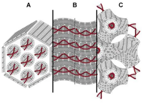
Some liposomes, with cationic lipids alone, exhibit good transfection efficiency, but the normal procedure is to incorporate helper neutral lipid, such as dioleoylphosphatidylethanolamine (DOPE) [8] and 1-monooleoyl-rac-glycerol (MO) [26], in the lipid formulation, since these non-bilayer-forming lipids facilitate the lipid mixing with the endosome membrane, a crucial step in the endosome release process [3, 8]. MO is a promising alternative to common helper lipids, as it seems to combine positive aspects of both DOPE and cholesterol: tendency to promote inverted non-lamellar structures similarly to DOPE (although different from the common inverted hexagonal structures [Figure 3. (C)]) and the fluidizing effect of cholesterol, which increases the fusogenicity of the lipoplexes [27].
Figure 3. Different types of pDNA/cationic lipid structural organizations: A) inverted non-lamellar
hexagonal structure characteristic of cationic vesicles containing DOPE at αDOPE ≥ 0.5; B) lamellar
structural characteristic of cationic vesicles containing αHelper ≤ 0.5; and C) inverted bicontinuous cubic
structure characteristic of cationic vesicles containing MO at αMO ≥ 0.5. Double-tailed surfactant with
grey-headgroup represents cationic lipid, double-tailed surfactant with white-headgroup represents DOPE and single-tailed surfactant with white-headgroup represents MO. Grey-coloured regions represent cationic lipid rich-domains and white-coloured regions represent MO or DOPE rich-domains [27].
7 Cationic liposomes are also used for the specific delivery of antisense oligonucleotides to specific tissues and papers on cationic liposome-mediated delivery of small interfering RNA have particularly compared intravenous and intraperitoneal
administration routes in adult mice[28].
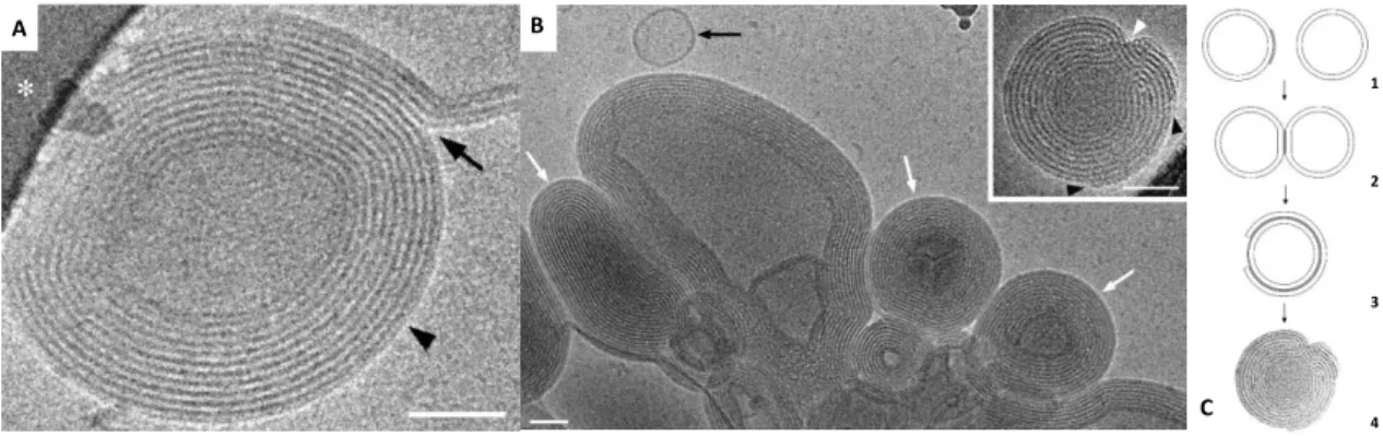
Figure 4 shows an example of cationic liposomes-nucleic acids complexes (lipoplexes) formation.
Figure 4. Formation of cationic liposomes-ODN (single-strand oligodeoxynucleotides) complexes. A and B) Cryo-TEM images showing the effect of ODN addition to cationic liposomes DOTAP/Chol (4:1). A) Observation of lipid membranes fusing (arrow) to a condensed multilamellar particle (arrowhead). B)
Lipoplexes of DOTAP with ODN, at the isoelectric point, is an aggregate of condensed multilamellar
particles (white arrows), coexisting with a few free liposomes (black arrow). In the right top corner DOTAP/Chol (1:1)-ODN lipoplex, charge ratio 1; consisting in a condensed multilamellar particle (black arrowheads pointing to incomplete outer bilayers and white arrowhead signaling a lamellar defect due to incomplete inner bilayers). Scale bars represent 50 nm. C) Diagram of lipoplex formation. (1) Initially, anionic ODN molecules (gray) may coat the cationic membrane of the liposomes. (2) Two liposomes may adsorb to each other due to ODN bridging, leading to liposome aggregation. (3) The destabilization promoted by partial charge neutralization may cause one membrane to rupture and wrap around another. (4) If local ODN concentration is sufficient more membranes may adsorb to produce a condensed multilamellar particle with intercalated ODN layers. (Adapted from [29])
Many of the finer features of these delivery systems and mechanisms remain insufficiently understood, and so the studies in this popular field have concentrated on structure, function, and structure–activity relationships, on detailed mechanisms of liposome-mediated gene delivery and on improved efficiency of transfection.
For clinical application the following quality-control assays should be applied to liposomal formulations: A B C 1 2 3 4
8
• Basic characterization of the pH, osmolarity, trapped volume, phospholipid concentration, phospholipid/lipid composition, phospholipid/lipid acyl chain composition, cholesterol concentration, active compound concentration, residual organic solvents and heavy metals, active compound/lipid ratio and proton or ion gradient before and after loading.
• Chemical stability regarding phospholipid hydrolysis, non-esterified fatty acid concentration, phospholipid acyl chain and cholesterol autoxidation and active compound degradation.
• Physical characterization of the appearance, vesicle size distribution, electrical surface potential and surface pH, ζ-potential, thermotropic behaviour, phase transition and phase separation and percentage of free drug.
• Microbiological assays of sterility and pyrogenicity (endotoxin level) [10].
3. Cellular barriers dictate transfection efficiency
Several anatomical and cellular barriers limit the overall efficiency of gene transfer by nonviral methods.
Barriers such as the epithelial, endothelial cell linings and the extracellular matrix surrounding the cells prevent direct access of macromolecules to the target cells. Professional phagocytes such as Kupffer cells in the liver and macrophages in the spleen promote an effective clearance of DNA-loaded colloidal particles administered through blood circulation. Additionally, the existing nucleases of the blood and extracellular matrix rapidly degrade free and unprotected nucleic acids following systemic administration [3].
The plasma membrane crossing is considered the most critical and limiting step for an efficient DNA/RNA transfection, as nucleic acids cannot pass through cell membrane unless their entry is facilitated by the formation transient holes [30], or through various active cellular uptake mechanisms such as endocytosis, pinocytosis, or phagocytosis [3].
Upon uptake by endocytosis, macromolecules captured within the endosomes are usually degraded as these vesicles transform into digestive lysosomes unless some escape mechanisms is triggered and the maturation process is interrupted. Two escape mechanisms have been explored. One consists in the use of membrane active or
9 fusogenic molecules, such as fusion peptides [31], or lipid components with acid-sensitive moieties and large hydrophobic portions to rupture the endosome membrane [32]. The other mechanism is based on building up osmotic pressure within the endosome to cause swelling and eventually burst of the endosomal vesicles [33].
If released from endosomes, DNA molecules in their free form or as complexes must make their way through the cytoplasmic matrix, a viscous protein solution, and the cytoskeleton network towards the nucleus where transcription takes place. Direct intracellular microinjection of naked DNA has shown that the movement by diffusion is slow and inefficient, and as a result the induced gene expression levels are very weak [34].
The nuclear envelope represents an important barrier for the entry of DNA. This double-membrane has large protein structures called nuclear pore complexes which regulate the molecular transport into and from the nucleus. In the case of the delivery of RNA, as the enzymatic machinery necessary for its action is in the cytoplasm, the nuclear envelope does not represent an obstacle for its transfection.
Finally, dissociation of DNA-carrier complexes constitutes another limitation for transfection. Cationic lipids dissociate from DNA through lipid mixing and exchange with host cell lipids, while DNA complexes formed with cationic polymers, such as PEI, remain stable after endosome escape [3].
10
4. Gene silencing through regulatory small RNA delivery as a promising therapeutic tool
The discovery of RNA interference (RNAi) [35-37] and subsequent findings of its mechanisms in mammalian cells generated excitement for its possible application for therapy of hard-to-cure diseases, including hepatitis C and human immunodeficiency virus (HIV).
This gene silencing phenomenon promoted by small pieces of double stranded RNA with sequence homology to certain genes, seems to have originated as a mechanism of defense against the insertion of foreign genetic material.
The RNAi machinery regulates the expression of genes via small hairpin RNAs, called microRNAs, processed in the cytoplasm into double stranded small RNAs [38]. MicroRNAs normally have only incomplete sequence homology to their targets and work by impeding the translation of messenger RNA (mRNA).
Figure 5 shows the RNA interference mechanism. The siRNA precursors are cleaved by Dicer (an RNAse III– like enzyme) into siRNAs (19 to 21 nucleotide double-stranded RNAs with a 2 to 3 unpaired nucleotide and characteristic 5’ phosphate and 3’ hydroxyl groups at each end). As the siRNA enter the RNA-induced silencing complex (RISC) it becomes activated and one of the siRNA strands (the guide strand) bounds to the complex. If the guide strand is homologous to an mRNA sequence, an enzyme within the RISC (Argonaute 2), cleaves the mRNA in the center of the homologous region. Since the cleaved mRNA is rapidly degraded and the guide strand of the siRNA is protected from degradation by the RISC, this mechanism enables the cleavage of many mRNAs, hence preventing the production of the protein for which the mRNA encodes [6].
11
Figure 5. RNA interference mechanism [6].
Contrarily to the initial notion that effective RNAi requires almost complete sequence homology throughout the length of the sequence, it now appears that as few as 7 contiguous complementary base pairs can direct RNAi mediated silencing [39]. RNAi is can be so specific that it has been possible to silence a mutant gene differing by
12
one nucleotide from its wild-type form [6]. For example, it has been possible to silence mutant oncogenic ras without affecting wild-type ras in vitro [40].
Inhibiting overexpressed oncogenes should block pathways that cancer cells depend on. However, in most cases it may be necessary to target pathways at several
points, or several pathways [41]. In this regard, RNAi-mediated interference of several
key oncogenes or tumor-promoting genes as inhibited the growth and survival of tumor cells [6].
Intravenous injection of immunoliposome with its surfaces functionalized with polyethyleneglycol grafted to transferrin antibodies (to aid in crossing the blood-brain barrier) and to the insulin receptor (to aid in crossing into glioma cells), have delivered DNA encoding short hairpin RNA into the brains of mice to suppress the growth of injected human glioma cells [42].
Increased resistance to apoptosis is characteristic of cancer cells. This property enables them to survive under abnormal growth stimuli and mediates their increased resistance to many chemo and radiotherapeutic agents. Hence, understanding apoptosis gives several options to induce cancer cell suicide [41].
RNA interference increases the apoptotic susceptibility of cancer cells by
inhibiting antiapoptotic genes, such as bcl-2, livin or surviving.In Hela cells, silencing of
livin increased the apoptotic rate in response to different proapoptotic stimuli [43].
Tumors do not grow more than a few mm3 in size without triggering the
angiogenic process to develop vessels and increment blood supply. This process is not fully understood, but it is most likely stimulated when hypoxia in the tumor surroundings leads to the expression of response genes, such as vascular endothelial growth factor A and placenta growth factor, which specifically stimulate the growth of endothelial cells. Still, tumors seem to easily get around the blockage of one angiogenic pathway, and clinical trials of the inhibitory drugs have underperform [41].
In spite of this, when tumor cells producing thrombospondin-1 antiangiogenic
molecule, under the control of a tetracycline promoter, were transfected with siRNA anti VEGF, and then injected into nude mice, an 86% reduction of tumor volume was observed [44].
Another tricky process to deal in cancer therapy is the development metastases development. Expression profiling of melanoma variants with low or high metastatic
13 potential, has allowed the identification of the small GTPase RhoC as a potential RNAi target to inhibit the formation of metastases [45].
Small interfering RNAs have been designed to silence BCR-ABL tyrosine kinase, an anomalous protein that results of a characteristic mutation in chronic myelogenous leukemia. Its silencing induces death of K562 leukemic cells at comparable levels to those induced by the anticancer drug STI 571 [46]. Additionally, silencing the Lyn kinase, which forms a signaling complex with BCR-ABL, induces the death of drug-resistant chronic myelogenous leukemia cells without affecting the viability of the control cells [47].
5. Chronic myeloid leukemia
Chronic myeloid leukemia occurs in approximately 25% of patients with acute lymphoblastic leukemia (ALL) and some its symptoms are weight loss, fever, and abdominal fullness but, at least in developed countries, many patients are asymptomatic until an abnormal routine blood count leads to a diagnostic work up.
The clinical sequence of CML is comprised by two stages. Most patients are diagnosed in the chronic or stable phase, which is characterized by myeloid cell growth, while cellular differentiation and function is largely maintained. After a variable length of time the disease evolves to blast crisis, which may resemble an acute leukemia of myeloid, lymphoid or undifferentiated phenotype and carries a poor prognosis.
CML was the first malignant disorder in which a reliable association with a chromosomal aberration was demonstrated. Philadelphia chromosome (Ph), originally described by Nowell and Hungerford in 1960, was originally thought to be a shortened chromosome 22 but subsequent research revealed that Ph is the result of a reciprocal translocation between chromosomes 9 and 22. This translocation was shown to fuse sequences of the ABL gene from chromosome 9 with BCR on chromosome 22, generating a chimeric BCR-ABL gene, which was the first demonstration of an oncogenic fusion gene. Subsequently, the BCR-ABL protein was shown to exhibit constitutive tyrosine kinase activity that was correlated with cellular transformation.
ABL (also referred to as ABL-1), is a tyrosine kinase involved in multiple cellular processes, including DNA repair, integrin signaling, cell cycle regulation, and signal
14
transduction from cell surface receptors. ABL knockout mice exhibit increased neonatal lethality and a number of defects, including skeletal malformations, immune
dysfunction as well as an ill-defined wasting syndrome.
The kinase activity of BCR-ABL is absolutely required for cellular transformation. The result of the enzymatic activity is a multimeric signaling complex interconnected mainly by phosphotyrosine dependent interactions, making the BCR-ABL kinase activity instrumental to its formation and maintenance. Signaling output from this complex is responsible for the activation of multiple pathways, including mitogen activated protein kinases that results in apoptosis inhibition, increased proliferation, and a perturbation of cellular adhesion to the bone-marrow stroma. There is also evidence that BCR-ABL kinase activity affects DNA repair and induces genomic instability by a variety of mechanisms. This complex signal transduction network, operated by BCR-ABL, offers a number of potential drug targets downstream of the initiating aberration.
It could be argumented that chronic phase CML would not be a significant clinical problem if it did not lead to blastic transformation. Therefore, prevention of blast crisis could be defined as the primary therapeutic goal in CML but the mechanisms responsible for disease progression in CML are not fully comprehended.
The fact that BCR-ABL kinase activity is central to its transforming potency has been the foundation for the development of imatinib mesylate for therapy of BCR-ABL positive leukemias. There is some evidence that targeting the kinase activity may have limitations as some biological effects of BCR-ABL, on migration and adhesion, are kinase independent. While these effects obviously do not prevent imatinib from inducing responses, it is possible that they contribute to the persistence of residual illness and hence, to eliminate these cells agents that target the BCR-ABL protein rather than its kinase activity would require [48].
15 6. Objective
The advent of nanotechnology along the discovery of RNA interference (RNAi), as turned the scope of nanomedicine to the development of devices to specifically delivery the pharmacological compound (drug or nucleic acids), to certain cells involved the disease processes. This approach would enable the use of lower dosages, reducing toxicity and the risk of developing resistance to treatment, as compared to other chemotherapeutic approaches.
Gene silencing by siRNA (short interfering RNA) appeared as a novel strategy for post-transcriptional gene silencing with enormous therapeutic potential. One attractive vehicle to deliver siRNA into target cells are the cationic liposomes because of the simplicity of their complexation with siRNAs, good transfection efficiency, superior pharmacokinetic properties, and low toxicity and immunogenicity [49].
The aim of this thesis was to develop and characterize novel systems for therapeutic siRNA delivery based on DODAC/MO liposomes, following previous work by our group with monoolein (MO)-based lipoplexes which have validated them as a system for plasmid DNA delivery [26, 50, 51] and demonstrated that they possess promising features compatible with stable, efficient encapsulation of RNA. Furthermore it is intended to stabilize the lipoplexes by incorporation of polyethylene glycol lipids on their membranes to increment the half-life [11] and reducing the recognition by the mononuclear phagocyte system. Finally, we intended to obtain selectivity to the folate receptor expressing (FR+) target cells (a characteristic of several cancers including chronic myeloid leukemia (CML)) [52], by coating the liposomes surface with folate grafted PEG lipids.
17 II. Materials and Methods
A. Materials
Dioctadecyldimethylammonium chloride (DODAC) was purchased from Tokyo
Kasei; 1,2-distearoyl-sn-glycero-3-phosphoethanolamine-N-[methoxy(polyethylene
glycol)-2000] ammonium salt (PEG(2000)-PE) and
1,2-distearoyl-sn-glycero-3-phosphoethanolamine-N-[folate(polyethylene glycol)-2000] ammonium salt (DSPE-PEG(2000)-Folate) from Avanti Polar Lipids; 1-monooleoyl-rac-glycerol (MO), heparin from the porcine intestinal mucosa, Dulbecco's modified eagle's medium (DME medium), Antibiotic/mycotic solution, sodium chloride (NaCl), sodium-pyruvate, Trizma Base, thiazoyl blue tetrazolium bromide (MTT), chloroform, methanol, ribonucleic acid from torula yeast and β-nicotinamide adenine dinucleotide reduced disodium salt hydrate (NADH) from Sigma-Aldrich; RiboGreen kit with TE buffer RNase
free (20x); Lissamine Rhodamine B
1,2-Dihexadecanoyl-sn-Glycero-3-Phosphoethanolamine Triethylammonium Salt (Rhodamine B DHPE) and Heat-inactivated Fetal Bovine Serum (FBS) from Invitrogen; Potassium dihydrogen
phosphate (KH2PO4•3H2O) from Panreac; Dipotassium hydrogen phosphate trihydrate
(K2HPO4) and ethanol absolute from Merck; RNase Zap from Ambion; L929,
18
B. Methods
1. Liposomes preparation
1.1 Lipid film hydration
5 mL of DODAC, MO and Polyethylene glycol (DSPE-PEG 2000) solutions were prepared at a 20 mM concentration, by diluting correct masses of the first two lipids in ethanol and the latter in chloroform, respectively. As for the preparation of the PEG-Folate 1 mL lipid solution, to obtain the concentration of 0,31mM, the lipid was diluted in a mixture of methanol: chloroform (9:1). These solutions were conserved at 4˚C.
Appropriated volumes of the desired lipid solutions, as to obtain the wanted lipid ratios and concentration of liposomes after hydration, were mixed with a fair volume of ethanol in a round bottom flask. Then the mixture is evaporated in a rotatory evaporator VV Micro Rotary Evaporator (Heidolf), under vacuum and at 30˚C, for 10 minutes. This promotes the formation of a homogenous lipid film in the flask. Afterwards the film was hydrated, during 10 minutes and at 30˚C, with an appropriated volume of ultrapure water, as to obtain the desired concentration. At this point, the liposomes are formed but they are multilamellar and very heterogeneous in size. This can be attested by the turbidity of the resulting solution. To overcome these issues, and obtain unilamellar and homogeneous liposomes, they are subjected 5 extrusion cycles. They are compressed with air, through a filter with a pore size of 400 nm in the first cycle, and then four times more, through a filter of 100 nm of pore (Track-Etched Membranes (Nuclepore)), in a pre-heated extruder (Lipex Extruder (Northern Lipids)) at 55˚C, to augment their fluidity and facilitate the extrusion. The final solution is much clearer.
19 1.2 DODAC/MO (2:1) liposomes with 1, 2 and 5% PEG (1mM)
1mM liposomes DODAC/MO (2:1) with different percentages of PEG (1, 2 and 5%) were prepared by hydration of the lipid film with ultrapure water, according to previously described method (1.1).
These liposomes were subject to DLS analysis in order to determine their hydrodynamic diameter (in Polystyrene (10x10x45 mm) cells (Sarstedt)) and ζ-potential (in folded capillary cells (Malvern)) in a Zetasizer nano ZS (Malvern).
2. Lipoplexes preparation
To prepare lipoplexes in the determined charge ratios, the proper volume of the solution of the intended liposomes (1 mM), diluted to 0,5 mM, was added to the 100 μL of a RNA solution at 10 μg/mL. The mixture was then subjected to vortex stirring, allowed to rest for an hour and then diluted to a final RNA concentration of 1 μg/mL.
The RNA solution was prepared, at the chosen concentration, by diluting an adequate mass of RNA in a 20 mM phosphate buffer (pH 6,8, 150 mM of NaCl). This
Buffer was prepared by mixing 51 mL of a 0,2 M aqueous solution of KH2PO4 with 49
mL of a 0,2 M aqueous solution of K2HPO4, diluting an appropriated mass of NaCl and
adding ultrapure water, to obtain a final volume of 200 mL.
⁄
Equation 1. Charge ratio with np the number of moles of positive charge n(DODAC) for liposomes
without PEG or PEG-Folate and n(DODAC) – n(PEG) for liposomes with the PEG moieties) and nN the
number of moles of negative charge (n(phosphates) considering the RNA molecules are composed of 22 nucleotides)
20
3. Light interactions with matter
As all the experiments performed on this work are based on light interactions with matter, an introduction on the subject is adequate.
Einstein demonstrated that light exists as bundles of wave energy called photons, with energies corresponding to the frequency of their waves in the bundle. The higher the frequency, the greater the energy carried by that bundle [53].
⁄
Equation 2. The wave frequency (ν) relation with wavelength (λ), where c it’s the speed of light in the
vacuum.
Figure 6. The electromagnetic wave with its electric and magnetic fields. Adapted from [54]
While propagating through space, light behavior is better described as a wave, when interacting with matter it behaves as a particle (photon), in which its energy (E) is given by:
Equation 3. Photon’s energy relation with its frequency, with h being the Planck constant (6.626×10⁻³⁴ J/s) and ν the frequency of the electromagnetic wave (s-1).
21 The electromagnetic radiation is sorted in a spectrum by the frequency of its waves.
Figure 7. The electromagnetic spectrum with the classification of the types of electromagnetic radiation.
Adapted from [54]
3.1 Absorption
When light goes through an absorbing medium, if the energy of the photons matches that required to promote an energetic level transition of the electrons from a ground state to a particular excited state, they are absorbed. The absorption proprieties of some compounds can be utilized to quantify them, by measuring the intensity of a light beam, of a specific wavelength, after and before its interaction with
the sample. Comparing the intensity of the incident light (I0) with the intensity of the
transmitted light (I(λ)) it is possible to infer about the absorption capability of the medium.
Equation 4. Light absorption comparing the intensity of the incident light (I0) with the intensity of the
22
The light, of a determined wavelength, that passes unaltered through the
medium is designated by transmittance (Tλ) andis given by:
⁄
Equation 5. Transmittance (Tλ) of the medium.
and it is directly related to the absorbance through:
Equation 6. Relationship between absorbance (Aλ) and transmittance(Tλ) .
As the concentration is related to the absorbance coefficient (αλ) through:
Equation 7. Relationship between the concentration (C) and the absorbance coefficient (αλ), where ελ is
the molar absorption coefficient.
Considering these previous relations, Beer – Lambert law can be attained [55]:
Equation 8. Absorption of the medium in function of the concentration of absorbent species.
This law only applies for monochromatic radiation. For each wavelength radiation, there’s a corresponding absorption coefficient. If there is more than one absorbent species the absorption becomes the sum of their absorbances.
Regarding this work, the absorbance measurements were performed in a SpectraMax Plus 384 absorbance Plate Reader (Molecular Devices).
23 3.2 Fluorescence
After the electronic excitation promoted by the absorption of electromagnetic radiation within the molecules, processes of relaxation take place so that the electrons can return to the fundamental state and the molecule to stabilize.
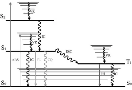
Figure 8. Jabłoński diagram displaying the formation of a molecular excited singlet state trough
absorption (ABS), and its decay processes, including fluorescence (FL), phosphorescence (PH), internal conversion (IC), intersystem crossing (ISC), vibrational relaxation (VR) and collisional quenching (CQ) [56].
The relaxation processes can be radiative or non-radiative. The latter include: i) vibrational relaxation, where the surplus of energy is dissipated (in the form of heat) though collisions between the excited molecules and the solvent molecules; ii) internal conversion, where transfer of the excited electrons happens between states of equal multiplicity; iii) Intersystem crossing, where transfer of the excited electrons arises between states of different multiplicity; [57] and iv) collisional quenching, where transfer of the excited electrons ensues to a quencher species trough collision.
The radiative decay processes are the phosphorescence and fluorescence. While phosphorescence occurs through transitions from states of different multiplicity (from a triplet to a singlet state), with spin-forbidden moves, in fluorescence, the transition is spin-allowed and always occurs from the first excited singlet to the fundamental state [58].
24
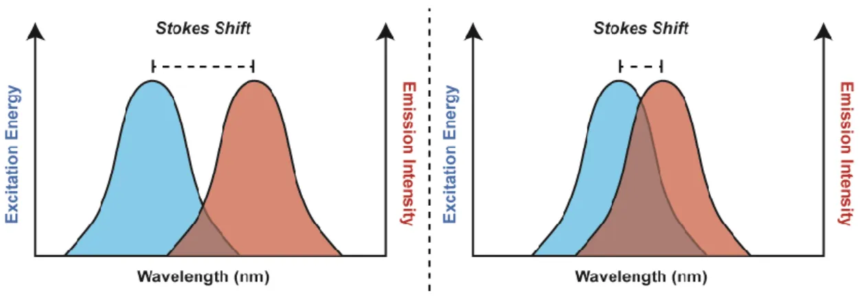
In the fluorescence decay process the fluorescence emission maximum is
observed at a longer wavelength than the absorption maximum.This energy difference
between the absorption maximum and the emission maximum is designated as the Stokes shift and it’s a result of the loss of vibrational energy in the excited state before emission of the photon [59]
Figure 9. Representation of the Stokes shift exhibiting the energy difference between the absorption
maximum and the emission maximum. Adapted from [59].
Fluorescence can be characterized by several parameters, of which some of
the most important are the fluorescence intensity at a given wavelength (IF), the
fluorescence spectrum (emission intensity variation with the emission wavelength), the fluorescence quantum yield (φF), the fluorescence lifetime (tF) and polarization
[56].
The fluorescence measurements were performed in a Jobin Yvon Fluorolog-3
(Horiba) spectrofluorimeter in disposable Polystyrene (10x10x45 mm) cells (Sarstedt)) and in a Fluoroskan ACEN FL Microplate Fluorometer/ Luminometer (Thermo scientific).
25 3.3 Dynamic light scattering
3.3.1 Size
Particles, emulsions and molecules in suspension undergo Brownian motion [60] (random movement). This motion is induced by the collision with solvent molecules that are moving due to their thermal energy or, in other words, by the spontaneous diffusion towards a homogeneous distribution in a liquid. This diffusion (D) is compared to a well understood reference process: the free diffusion of a single particle in a liquid, distant from other particles or a barrier, for which the diffusion coefficient (D) is given by:
⁄
Equation 9. Einstein’s equation with kB being the Boltzmann's constant (1.38 × 10⁻²³ m2.kg.s-2.K-1), T the absolute temperature and f the friction coefficient of the particle .
In the simplest case, a spherical particle with in a Newtonian fluid (fluid with a proportional viscosity to the flow velocity), the friction coefficient for translational motion is given by:
Equation 10. Stokes friction factor, where η is the viscosity and r the radius of the particle.
The combination of these equations results in:
⁄
Equation 11. Stokes-Einstein equation, for diffusion of spherical particles through liquid with low
26
The intensity of the scattered light fluctuates with time at a rate that is dependent upon the size of the particles: the smaller the particles are, the more rapidly they move. Analysis of these intensity fluctuations yields the velocity of the Brownian motion and hence the particle size distribution using the Stokes-Einstein equation.
Other parameters produced by dynamic light scattering are the Z-average size and the polydispersity (PDI). The Z-average parameter accounts for the mean hydrodynamic radius of the particles and is obtained of the scattered light fluctuations through:
Equation 12. Polynomial fit to the log of the of the scattered light fluctuation in time
The value of b is known as the z-average diffusion coefficient and is converted to a size using the dispersant viscosity and some instrumental constants. The analysis of the intensity of scattered light only gives two values, a mean value for the size, and a width
parameter known as the Polydispersity. If the distribution is very broad, orrephrasing,
if the polydispersity is too high (superior to 0,5) the z-average value is unreliable and the distribution analysis should be used [61].
3.3.2 ζ-potential
The charge at the particles surface affects the distribution of ions in the surrounding interfacial region. As a result, an increased concentration of counter ions (ions of opposite charge to that of the particle) gathers close to the surface, and an electrical double layer forms around each particle. The closer layer to the particle surface is called the Stern layer, and their ions are strongly bound. Contiguously to the Stern layer there’s a diffuse layer, with its ions less firmly attached, where exists a theoretical boundary within which the ions and particles form a stable entity. When the particle moves, ions within the boundary move with it, but the ions beyond it do not. This boundary is called the surface of hydrodynamic shear or slipping plane. The potential that exists at this boundary is known as the ζ-potential [61].
27 When a charged particle in a gas or liquid is under the influence of a uniform electric field, it will be accelerated until it reaches a constant drift velocity according to:
Equation 13. Drift velocity (v) equation where E is the magnitude of the applied electric field and μ the
mobility of the particle.
Reformulating, the electrical mobility of the particle is defined as the ratio of the drift velocity to the magnitude of the electric field:
⁄
Equation 14. Rearrangement of the equation X.
The electrophoretic mobility of a spherical colloidal particle carrying low zeta (ζ) potential moving in an electrolyte solution is given by:
⁄
Equation 15. Henry’s equation, where εr is the relative permittivity, ε0 is the is the permittivity of a
vacuum,f(kr) is the Henry’s function and η is the viscosity of the electrolyte solution [62].
Henry’s function (f(kr)) is a function used in the theoretical treatment of the
electrophoretic mobility that accounts on the radius of the macromolecule (r) and its
reciprocal ion-atmosphere radius (κ). When the limit of krtends to infinity f(kr) tends
to 1 and the equation becomes:
⁄
28
When the limit of kr tends 0 f(kr) tends to 2/3 and the takes the form of:
⁄
Equation 17. Hückel’s equation [62].
Hence, measuring the electrophoretic mobility through the analysis of the intensity fluctuations of scattered light, it is possible to convert the signal into ζ-potential, through the presented theoretical considerations.
The Dynamic Light Scattering (DLS) measurements were performed in Zetasizer
Nano ZS (Malvern) placing 1 mL of the intended solution in a disposable Polystyrene
(10x10x45 mm) cells (Sarstedt)) for the determination of the hydrodynamic diameter and 800 μL in folded capillary cells (Malvern) for the determination of the ζ-potential. The measurement was made with appropriated SOPs at 25ºC.
4. Liposome stability
Liposomes DODAC/MO (2:1) and DODAC/MO (2:1) 5% PEG, were prepared at a concentration of 1 mM by hydration of the lipid film with ultrapure water, in agreement to the method described in 1.1.
Afterwards, the liposomes were diluted to a final lipid concentration of 100 μM, in appropriated volumes of saline, serum, aqueous solutions at pH 3 and 5 and ultrapure water (to obtain the conditions pH≈6 and 0 mM NaCl).
The saline solutions were prepared in ultrapure water, at such concentrations that after the addition of the liposomes the final concentrations of NaCl were 5, 10, 50, 100 and 150 mM. The same was done for the serum solutions, which were diluted, in ultrapure water, as to be at 30 and 80% (v/v) after the addition of liposomes.
The acidified aliquots of ultrapure water (pH 3 and 5) were obtained by titration with HCl solutions (0,5 mM and 10 μM) and for pH 5 solution, the final pH was adjusted with the addition of NaOH solutions (0,5 mM and 10 μM).
29 These preparations were then subject to dynamic light scattering studies to determine the salt and pH effect on the size and ζ-potential and the serum effect on the size of the lipid particles in a Zetasizer nano ZS (Malvern).
5. Complexation assays
Liposomes DODAC/MO (2:1) and DODAC/MO (2:1) 5% PEG (1mM), prepared by hydration of the lipid film with ultrapure water as described in 1.1, were diluted to 0,5 mM, with ultrapure water and used to prepare lipoplexes at charge ratios 0,5; 1; 2; 4; 6; 8; 10; 15; 20; 25 and 30. This lipoplexes were obtained according to the method described in section 2.
These lipoplexes were subjected to dynamic light scattering analysis, to determine their hydrodynamic diameter (in Polystyrene (10x10x45 mm) cells (Sarstedt)) and ζ-potential (in folded capillary cells (Malvern)) in a Zetasizer nano ZS (Malvern), and to RiboGreen assay, to follow the complexation of the RNA with the liposomes as RiboGreen is a RNA intercalating probe that fluoresces when in the RNA midst. The RiboGreen assay was conducted, considering manufacturer specifications, by plating 100 μL of lipoplexes in a 96 well plate and ultrapure water (for blank) and adding 100 μL of RiboGreen (200x). Also, a calibration curve was made, according to the manufacture, to assure linearity between the RiboGreen fluorescence and the RNA concentration. Briefly, 100 μL of RNA solutions, in phosphate buffer, of 0,04; 0,2; 1 and 2 μg/mL concentrations with 100 μL of RiboGreen (200x) were plated to obtain the final RNA concentrations of 0,02; 0,1; 0,5 and 1 μg/mL.
The mixtures were allowed to incubate during 5 minutes and then fluorescence was measured in a Fluoroskan ACEN FL Microplate Fluorometer and Luminometer (Thermo scientific), using the excitation/emission filter pair of 485/538 nm.
RiboGreen solution was prepared by 200x dilution of the RiboGreen stock solution (750 mM) in 10 mM Tris-HCl, 1 mM EDTA, pH 7,5 (TE) buffer, diluted 20 fold, from the stock solution with ultrapure water RNase free.
This procedure was carried on ice and the utilized micropipettes and gloves were cleaned with RNAse zap cleansing solution and RNAse free tips were used.
30
6. Encapsulation efficiency
To determine the encapsulation efficiency of the lipoplexes at charge ratio 10, prepared with Liposomes DODAC/MO (2:1) and DODAC/MO (2:1) 5% PEG as described
before, were centrifuged, 15 minutes, at 1000 G in 50 KDa amicons (Millipore)
(Centrifuge 5804 R (Eppendorf)). These falcon-like tubes have a filter unit, which allows the separation of the liposome – RNA complexes of the free RNA. While the lipoplexes are retained in the filter unit, the free RNA molecules are small enough to pass through the filter.
After centrifugation, 100 μL of the free RNA fractions and of a RNA solution of 1 μg/mL (corresponding to the initial concentration of RNA after their dilution to the final volume) were plated, in a 96 well plate, along with 100 μL of RNA solutions, in
phosphate buffer, of 0,04; 0,2; 1 and 2 μg/mL concentrations, to obtaining a
calibration curve . Then 100 μL of RiboGreen (200 x), prepared in the same fashion as described previously, were added to each well and the mixture was allowed to incubate for 5 minutes. Also a blank, consisting in a mixture of 100 μL of ultrapure water and 100 μL of RiboGreen (200 x), was made. Finally the fluorescence was measured in Fluoroskan ACEN FL Microplate Fluorometer and Luminometer (Thermo scientific), using the excitation/emission filter pair of 485/538 nm.
7. Lipoplexes stability
Once again lipoplexes DODAC/MO (2:1) 5% PEG – RNA at C. R. 10 were prepared as described previously and diluted to final RNA concentration of 1 μg/mL, in appropriated volumes of saline, serum, aqueous solutions at pH 3 and 5 and ultrapure water (to obtain the conditions pH≈6 and 0 mM NaCl).
As in the case of the liposome stability studies, the saline solutions were prepared, in ultrapure water, at such concentrations that, after the addition of the lipoplexes the final concentrations of NaCl were 5, 10, 50, 100 and 150 mM. The same was done for the serum solutions, which were diluted, in ultrapure water, as to be at 30 and 80% (v/v) after the addition of lipoplexes.
The acidified aliquots of ultrapure water (pH 3 and 5) were obtained by titration with HCl solutions (0,5 mM and 10 μM) and for pH 5 solution, the final pH was adjusted with the addition of NaOH solutions (0,5 mM and 10 μM).
31 These preparations were then subject of dynamic light scattering studies to determine the salt and pH effect on the size and ζ-potential and the serum effect on the size of the lipid particles in a Zetasizer nano ZS (Malvern).
8. Destabilization of lipoplexes
Lipoplexes DODAC/MO (2:1) and DODAC/MO (2:1) 5% PEG – RNA C. R. 10 were prepared as described in 2, with a difference in the in the dilution to obtain the final volume. In this case lipoplexes C. R. 10 were diluted by adding the appropriate volumes of heparin from the porcine intestinal mucosa (0,121 mM) (a polysaccharide with a high negative charge density), as to form 5; 2,5 and 0,5 charge ratios. The final volume of 1000 μL was completed with the addition of ultrapure water and the solutions were subjected to vortex agitation.
As for the previous experiments with RiboGreen, 100 μL of each solution and of a RNA solution of 1 μg/mL (corresponding to the initial concentration of RNA after their dilution to the final volume) were plated, in a 96 well plate, along with 100 μL of RNA solutions, in phosphate buffer, of 0,04; 0,2; 1 and 2 μg/mL concentrations, to obtaining a calibration curve . Then 100 μL of RiboGreen (200 x), prepared in the same fashion as described previously, were added to each well and the mixture was allowed to incubate for 5 minutes. Also a blanks, consisting in a mixture of 100 μL of ultrapure water and of a solution of heparin with the same concentration used to obtain the charge ratio 0,5 with 100 μL of RiboGreen (200 x), were made. Finally the fluorescence was measured in Fluoroskan ACEN FL Microplate Fluorometer and Luminometer (Thermo scientific), using the excitation/emission filter pair of 485/538 nm.
32
9. Cytotoxicity assays
9.1 LDH (Lactate dehydrogenase) assay
This technique allows an indirect measurement of cell viability, since LDH is released to the extracellular medium by cells with compromised membranes. LDH catalyzes the interconversion of pyruvate and lactate, with NADH oxidation
(Nicotinamide adenine dinucleotide) or NAD+ reduction acting as reaction catalyst.
The LDH enzymatic kinetic can be monitored by measuring the absorbance variation at 340 nm, due to the oxidation of the NADH. As the quantities of NADH and pyruvate are a constant in the experimental reaction, the LDH quantity in the extracellular medium
will determine the slope of the enzymatic kinetic curve.
9.2 MTT assay
This method involves the conversion of MTT (3,(4,5-dimethylthiazol-2-yl)-2,5-diphenyl tetrazolium bromide) into formazan crystals. When cells are viable, this tetrazolium salt is metabolized by mitochondrial enzymes, forming insoluble colored crystals that absorb light at a wavelength of 570 nm. If the cellular metabolism somehow becomes impaired, formation of formazan crystal will be blocked and, as consequence, the absorption of light will be inferior. Thus, measuring the absorption of the formed crystals, allows the estimation of cell viability [63]
9.3 Liposomes toxicity
Liposomes DODAC/MO (2:1) and DODAC/MO (2:1) with 1, 2 and 5% of PEG were prepared, at 3 mM lipid concentration, by hydration of lipid film with ultrapure water, as described in 1.1.
L929 cells (cell line of adherent fibroblasts of subcutaneous connective tissue of Mus musculus (mouse)) were cultured in DME medium supplemented with 10% (v/v) of heated inactivated FBS and 1% (v/v) of antibiotic/antimycotic solution and in an
atmosphere of 5% CO2 and 37˚C.
When the cells reached the exponential phase of growth they were seeded, at a ratio of 30000 cells per well, in 24 well plates and left overnight, for adhesion, in an
33 In the following day, the prepared liposomes were sterilized by filtration through a membrane filter with a pore diameter of 200 nm (Filtropur S 0,2 (Sarstedt)), and diluted in DME medium to a 100 μg/mL concentration. These solutions were then used to prepare the 25 and 5 μg/mL concentrations by preforming the dilutions in appropriated volumes of DME medium. The cells’ medium was aspirated and they were incubated, in duplicates, with 500 μL of each liposome formulation at 5, 25, 100 μg/mL concentrations. As control conditions were included cell viability control (adding 500 mL of fresh DME medium), cell death control (DMSO diluted at 30% (v/v) in DME medium) and water control (approximately the same volume of water used to dilute the liposomes to a concentration of 100 μg/mL in DME medium). The work with the cells was conducted in asepsis within the confinement of a laminar flux chamber Bio II A (Telstar).
After 48 and 72h of incubation of the L929 cell line, extracellular LDH and MTT toxicity assays were conducted using the cell medium and the adherent cells respectively. The supernatant medium of each well was transferred to eppendorfs and
centrifuged for one minute at 13000 rpm in a Centrifuge 5418 R (Eppendorf) to
sediment cells or cellular debris in suspension. After centrifugation, 40 μL of the supernatant medium of each condition were placed in a 96 well plate. 250 μL of a 0,31 mM NADH solution were added, followed by 10 μL of 8,96 mM pyruvate solution in phosphate buffer (0,05M, pH 7,4). The measurement of the LDH enzymatic activity was made immediately after the addiction of pyruvate, at 30˚C, in a SpectraMax Plus 384 absorbance Plate Reader (Molecular Devices) using the appropriated software (SOFT Max Pro) protocol.
For the MTT assay, 500 μL of a MTT (10x) (Stock solution of 5 mg/mL) in DME
medium solution were added to the adherent cells in the wells and left to incubate for
two hours in an humidified incubator at 37˚C, with 5% CO2. The supernatant medium
was then carefully aspirated and 500 μL of a DMSO/Ethanol [1:1 (v/v)] solution was added to each well. The plate was shaken to dissolve the formazan crystals, after which 150 μL of each well, as well as 150 μL of DMSO/Ethanol [1:1 (v/v)] (as blank), were transferred into a reading 96 well plate. The measurement was made in a SpectraMax Plus 384 absorbance Plate Reader (Molecular Devices) using the appropriated software (SOFT Max Pro) protocol.

![Figure 6. The electromagnetic wave with its electric and magnetic fields. Adapted from [54]](https://thumb-eu.123doks.com/thumbv2/123dok_br/17780794.838079/35.892.271.616.489.800/figure-electromagnetic-wave-electric-magnetic-fields-adapted.webp)