iv | António José Almeida Rebelo | Universidade do Minho
António José Almeida Rebelo
Sleep stage classification based on
cardiorespiratory signals
Dissertation of Master’s Degree
Master’s Degree in Informatics Engineering
Work done over the orientation of Paulo Novais and Pedro Fonseca Departamento de Informática
António José Almeida Rebelo | Universidade do Minho | iii
The reproduction of this thesis integral only for research purposes by written declaration of the person concerned, that such commits.
Acknowledgments
I would like to thank Pedro Fonseca for giving me the opportunity to come to the Neth-erlands and to work within Philips Research allowing me to live a new experience and to grow and build up professionally and personally. I am also thankful for the patience, ad-vice and guidance given throughout the year.
To professor Paulo Novais for the support, availability and guidance given along this year.
To my friends and colleagues from Eindhoven and Portugal for the friendship, support and the leisure activities.
To my family for all the support and care given throughout my whole university years. Finally, to Ana, who helped me in all moments giving me companionship and support.
Title
António José Almeida Rebelo | Universidade do Minho | v
Resumo
O sono está ligado a uma quantidade bastante considerável de patologias que têm impacto direto na maioria das atividades diárias tais como a aprendizagem, memorização ou pro-dutividade. Assim, reduzir as consequências pessoais e os custos associados com os dis-túrbios do sono tornou-se num dos maiores desafios das últimas décadas. Patologias como distúrbios respiratórios do sono, sonolência, síndrome de pernas inquietas ou distúrbios do sono relacionados com o ritmo circadiano são bastante prevalentes, produzindo gran-des distúrbios no dia-a-dia dos pacientes. Para o diagnóstico e tratamento gran-deste tipo de patologias, a capacidade de avaliar o padrão de sono do paciente por períodos de tempo mais alargados poderá ser necessária. A necessidade de avaliação de um determinado medicamento ou a monitorização da qualidade do sono do paciente ao longo do tempo são bons exemplos. O teste clínico PSG, atualmente padrão para a avaliação do sono, é um método caro e complexo, disponíveis apenas em hospitais especializados e equipados com um laboratório do sono e profissionais qualificados. Para além de nem sempre estar disponível, PSG é considerado um procedimento muito penoso devido aos diversos elé-trodos em contacto com o corpo e cabeça, que causam desconforto e possivelmente um padrão de sono anormal. Para além destes incómodos, os pacientes têm ainda que dormir num laboratório, sendo continuamente observados ao longo da noite. PSG é, portanto, uma técnica cara, geralmente limitada a uma ou duas noites num laboratório do sono. Métodos como actigraphy, que utilizam sensores semelhantes a relógios de pulso para medir os movimentos corporais dos pacientes, podem dar informações úteis sobre os pa-drões de sono dos indivíduos durante períodos de tempo mais alargados sem perturbar significativamente os hábitos normais de sono dos pacientes. No entanto, este método tem várias limitações, uma vez que apenas avalia movimentos corporais, o que é insuficiente para informações relativas à arquitetura do sono dos pacientes.
Para ultrapassar as limitações dos métodos acima descritos, seria relevante a criação de um novo procedimento capaz de complementar os já existentes. Um sistema de monito-rização do sono baseado em informação cardiorrespiratória poderá fornecer mais infor-mação sobre a arquitetura do sono, de forma não intrusiva e durante períodos de tempo alargados, no conforto e privacidade da residência dos pacientes. Esta informação poderia
ser utilizada para o rastreio de doenças, acompanhamento e monitorização de tratamentos ou mesmo complementar o PSG para o diagnóstico de algumas doenças do sono.
O sistema apresentado neste trabalho aborda parte desta hipótese, classificando automa-ticamente várias fases do sono usando apenas informação cardiorrespiratória. Embora os dados utilizados para este estudo, tenham sido adquiridos através do uso de sensores de contacto, no futuro, esta informação poderá ser obtida através da utilização de métodos não intrusivos, que já se encontram disponíveis comercialmente. Esta hipótese é bastante interessante porque consegue fornecer mais informação aos profissionais do sono, sem interferir com o dia-a-dia do paciente.
António José Almeida Rebelo | Universidade do Minho | vii
Abstract
Sleep pathologies have a direct negative impact into most of daily activities such has learning, memorization or productivity. Decreasing the personal burden and the societal cost associated with sleep disturbances has become one of the major challenges in the last decades. Pathologies like sleep disordered breathing, insomnia, restless leg syndrome or circadian rhythm sleep disorders are fairly prevalent, heavily disturbing the life of af-fected subjects. For the diagnosis and treatment of these disorders, the ability to assess a patient’s sleep pattern over longer periods of time may be required. The need of evalua-tion of a certain medicaevalua-tion or the monitoring of the sleep quality of the patient over time can be named as good examples. The polysomonographic (PSG) clinical test, current gold standard for sleep assessment, is an expensive and complex method only available in specialized hospitals equipped with a sleep lab and qualified professionals. Not always available, PSG is considered a very stressful procedure because of the various electrodes attached to the body and head, which cause discomfort and potentially disrupt the usual sleep patterns. Furthermore people need to sleep in an unfamiliar environment while be-ing observed throughout the entire night. PSG is therefore an expensive technique usually limited to one or two nights in a sleep laboratory. Methods like actigraphy, which measure body movements, can give useful insight about the sleeping patterns of the subjects during longer periods of time without significantly disrupting the normal sleeping habits of a person. However this method has several limitations as it only assesses the movements of the patients and therefore provides little insight about the subjects’ sleep architecture. In order to address the shortcomings of the existing techniques, the introduction of a new system, easy and cheap to deploy and use, capable of complementing the existent ap-proaches is relevant. A sleep monitoring system based on cardiorespiratory data may be able to provide bigger insight of the sleep architecture, while having the potential to be unobtrusive and able to monitor sleep during longer periods of time, in the comfort and privacy of the subject’s own room. Furthermore it can potentially enable the screening of diseases, follow-up on treatments, or even complementing PSG for diagnosis of some sleep disorders.
The system presented in this work addresses part of this system by automatically classi-fying multiple sleep stages using cardiorespiratory information. Although the data used for this study was acquired with contact sensors, in the future, this information might be obtained through the use of non-obtrusive methods that are already commercially availa-ble. This possibility is interesting as it provides bigger insight of the subject sleeping patterns and architecture for the sleep professional, without interfering with the daily life of the patient.
António José Almeida Rebelo | Universidade do Minho | ix
Contents
Acknowledgments ... iv Title ... iv Resumo ... v Abstract ... vii Contents ... ixList of figures... xiii
List of tables ... xvi
Glossary ... xvii
Introduction ... 1
Ambient assisted living ... 1
AAL for health, rehabilitation and care ... 2
Sleep monitoring ... 3
Scope of the dissertation ... 4
Objectives ... 5
Research methodology ... 7
Structure of the document ... 7
Sleep monitoring ... 9
History of sleep medicine ... 9
2.1.1 Early theories ... 9
2.1.2 REM sleep ... 10
2.1.3 Sleep clinics and sleep disorders ... 10
Normal human sleep ... 11
2.2.1 Sleep architecture... 13
Sleep disorders ... 15
2.3.1 Insomnia ... 16
2.3.2 Sleep disordered breathing ... 17
State-of-the-art in home sleep monitoring ... 17
2.4.1 Actigraphy ... 17
2.4.2 Unattended portable monitors in the diagnosis of OSA ... 18
2.4.3 Ambulatory polysomnography ... 20
Unobtrusive monitoring in bed ... 20
2.5.1 Ballistocardiography ... 21
2.5.2 Doppler radar ... 21
2.5.3 Video ... 22
Sleep stage classification using unobtrusive modalities ... 23
2.6.1 Sleep and wake Discrimination ... 23
2.6.2 Sleep staging using cardiorespiratory signals ... 23
2.6.3 Sleep staging based on signals acquired through bed sensor ... 24
2.6.4 Analysis and Comparison ... 25
Summary ... 27
Datasets ... 28
Boston Healthy dataset... 28
Boston Insomniacs dataset ... 28
Eindhoven dataset ... 29
SIESTA dataset... 29
Invalid data handling... 30
Subjects selection... 30
António José Almeida Rebelo | Universidade do Minho | xi
Establishing ground truth ... 33
Cardiorespiratory features ... 33
3.8.1 ECG data ... 34
3.8.2 Respiratory inductance plethysmography ... 35
Summary ... 37
Feature Analysis ... 38
Class separability measures ... 38
4.1.1 Absolute standardized mean distance ... 40
4.1.2 One way analysis of variance F-statistic ... 41
Unprocessed features ... 41 Feature normalization ... 44 4.3.1 Motivation ... 44 4.3.2 Normalization Techniques ... 46 4.3.3 Combining techniques ... 49 4.3.4 Normalization results ... 50 Feature transformation ... 53 4.4.1 Motivation ... 54 4.4.2 Techniques ... 54 4.4.3 Combining techniques ... 59
4.4.4 Optimizing best transformations for each class ... 60
4.4.5 Transformations results ... 61
Normalizations and transformations on subjects with insomnia ... 62
Classifier ... 65
Bayesian decision theory... 65
Bayesian linear discriminant ... 67
From binary classification to multiple class ... 69
Feature selection ... 70
Temporal stage changes ... 72
Temporal a Priori probabilities calculation ... 76
Stage change probability and Markov Chain ... 76
Combination with Linear Discrimination formula and output ... 77
Post processing and establishing final classification ... 78
Validation ... 79
Conclusions ... 80
Results and Discussion ... 81
Cohens’s kappa coefficient ... 81
Results for with the original data ... 82
Results for with the normalized data ... 82
Results for with the transformed data ... 83
Comparisons with literature results ... 84
Conclusions ... 88
Bibliography ... 91
António José Almeida Rebelo | Universidade do Minho | xiii
List of figures
Figure 1.System overview ... 6
Figure 2. Hypnogram of a healthy adult ... 13
Figure 3. Age related trends for stage 1, stage 2, slow wave sleep (SWS), REM sleep, wake after sleep onset (WASO) and sleep onset latency (minutes) [21] ... 14
Figure 4. Actiwatch Spectrum, Philips Electronics ... 18
Figure 5. Stardust II, Philips Electronics ... 19
Figure 6. Alice PDx, Philips Electronics ... 20
Figure 7. Under-Mattress Bed Sensor, Emfit ... 21
Figure 8. An example set up of SleepMinder, Biamcamed [33] ... 22
Figure 9. Total epochs spent in each stage from lights out, for Boston Insomniacs dataset ... 29
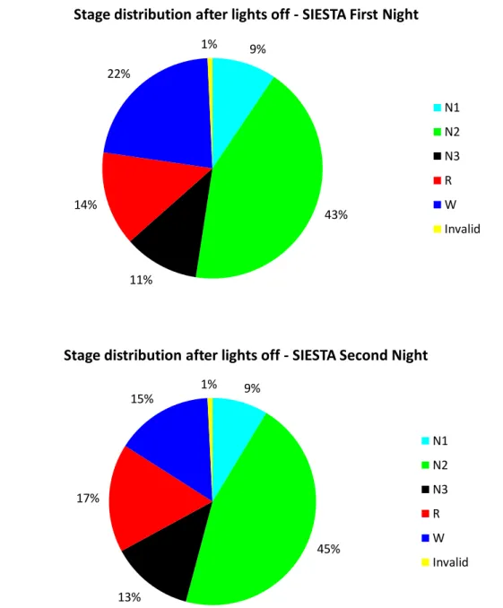
Figure 10. Comparison of sleep stages distribution of first and second night ... 32
Figure 11. Example of an ECG recording ... 34
Figure 12. Schematic representation of a typical ECG tracing ... 34
Figure 13. Example of feature 27, mean RR interval, for a given subject ... 35
Figure 14. Pro-Tech ezRIP, Philips Electronics ... 36
Figure 15. Example of feature 13, respiration frequency, for a given subject ... 36
Figure 16. Example of feature 169 for a given subject, a feature with good discrimination power for deep sleep and REM. ... 39
Figure 17. Example of feature 13 for a given subject, a feature with low discrimination power for all classes ... 40
Figure 19. Histograms relating the ASMD with the number of features per class ... 43 Figure 20. Mean and pooled ASMD for deep sleep versus the remaining classes, for each feature. ... 45 Figure 21. Mean and pooled ASMD for REM versus the remaining classes, for each feature. ... 46 Figure 22. An example of feature values of feature 169 before quantile normalization 49 Figure 23. An example of feature values of feature 169 after quantile normalization wit h normal distribution as reference ... 49 Figure 24. Normalization techniques search graph ... 50 Figure 25. Pooled ASMD of all normalized features for four classes ... 52 Figure 26. Comparison between Original (O) and Normalized (N) feature set with histograms indicating the number of feature for different ASMDs ... 53 Figure 27. Example feature values for feature 13 for a given subject ... 56 Figure 28. Example feature values for feature 13, for the same subject, after the application of the std method ... 56 Figure 29. Contour lines of the ASMD of feature 13 for Deep sleep, varying the size of the median filter window and the std window... 57 Figure 30. Contour lines of the ASMD of feature 27 for Deep sleep, varying the size of the median filter window and the std window... 58 Figure 31. Transformations search graph ... 60 Figure 32. Comparison between Original (O), Normalized (N) and Post-processed (P) dataset with histograms relating the ASMD with the number of features per class ... 62
António José Almeida Rebelo | Universidade do Minho | xv Figure 33. Comparison between Original (O), Normalized (N) and Post-processed (P) dataset with histograms relating the ASMD with the number of features per class for
subjects with insomnia ... 63
Figure 34. Example of two regions R1 and R2 formed by the bayesian classifier on probability density functions with equal prevalences for both classes ... 66
Figure 35. Filter and wrapper feature selectors [72] ... 70
Figure 36. Sub Matrix of Temporal Stage Changes being processed ... 75
Figure 37. Temporal a Priori Probabilities for all classes ... 76
Figure 38. Averaged kappa coefficient of agreement for different weight values of prior probability and threshold values for the confidence points... 79
Figure 39. Diagram of the Classification procedure ... 80
List of tables
Table 1. Results with the original data ... 82
Table 2. Results with the normalized data ... 83
Table 3. Results with the transformed data ... 84
Table 4. Comparison with results from literature ... 86
Table 5. The impact of age in the classification results ... 86
António José Almeida Rebelo | Universidade do Minho | xvii
Glossary
AAL Ambient Assisted Living AI
ICT NREM REM
Artificial Intelligence
Information and Communication Technologies Non Rapid Eye Movement
Rapid Eye Movement
PSG Polysomnography EEG Electroencephalogram ECG Electrocardiogram EOG Electrooculogram EMG Electromyogram AASM SWS
American Academy of Sleep Medicine Slow Wave Sleep
WASO Wake After Sleep Onset OSA Obstructive Sleep Apnea SDB Sleep Disorder Breathing
OSAS Obstructive Sleep Apnea Syndrome ASMD Absolute Standardize Mean Difference PDF Probability Density Function
António José Almeida Rebelo | Universidade do Minho | 1
Introduction
Modern lifestyles introduced a wide variety of factors capable of perturbing the ordinary habits of the population. Stress, overworking and a fast paced lifestyle made sleep depri-vation and fatigue a common problem and hence the source of many accidents, depres-sions, and health related problems. Although sleep is increasingly recognized as im-portant to public health, it is essential to realize that sleep deprivation is very often due to undiagnosed sleep disorders. After a disturbed night of sleep, a person might not feel restored and refreshed and be sleepy during the day, but be totally unaware that is sleep-deprived or has a sleep disorder. On the other hand existing diagnosis are expensive, ob-trusive, limited to one or two night, and require a patient to sleep in an unfamiliar envi-ronment. The solution to this problem may reside in new technologies in the area of am-bient assisted living that may provide new solutions for a better and cheaper care provid-ing.
Ambient assisted living
The continuous increase of older population in Europe and worldwide, associated with increased costs of healthcare favor the deployment of AAL intelligent systems for a bet-ter, healthier and safer life in the preferred living environment. AAL comprises concepts, products and services that interlink and improve new technologies and the social envi-ronment, with a focus on older people.
AAL relies greatly on Ambient Intelligence (AmI) to provide seamless and unobtrusive interaction in the human environment, thus radically moving away from more traditional assistive technologies towards universal access.
Ambient Intelligence is a relatively new field of Artificial Intelligence, in which comput-ers interaction is made in a more natural way since it’s made using friendly interfaces such as gestures. The underlying goal of Ambient Intelligence is to involve a wide variety of different technologies, hiding their presence from users or soothingly integrate them within the surrounding context as augmented physical objects, rather than technological
gadgets. This methodology makes ICT accessible and usable for the largest possible pop-ulation and takes into account the requirements of older users.
The AAL domains range from the AAL for home and mobile support, including AAL for health, rehabilitation and care, personal and home safety and security, AAL in the com-munity, addressing social inclusion, entertainment and mobility and lastly AAL at work, addressing the needs of older people in the workplace.
AAL for health, rehabilitation and care
In this work we will be focusing on AAL for health, rehabilitation and care, specifically home care monitoring systems, which are intelligent technologies capable of monitoring home inhabitants in their daily activities, and thus preventing health and security risks or alerting family members or healthcare providers when specific situations occur. Current efforts in this context address fall detection and prevention, detection of helplessness, as well as the long term vital signals monitoring.
The monitoring of the vital signs during sleep plays an important role in the quality of life of an individual and therefore in the AAL context. The available knowledge has es-tablished that sleep serves an important function, as evidence by the rebound of sleep loss and the developmental, functional, and metabolic impairments produced by sleep depri-vation. Persons experiencing sleep insufficiency are more likely to suffer from chronic diseases such as hypertension, diabetes, depression, and obesity, as well as from cancer, increased mortality, and reduced quality of life and productivity[1].
The analysis of a survey taken in the United States in 2009, regarding perceived insuffi-cient rest or sleep, and on sleep behavior, determined that among 74,571 adult respond-ents in 12 states, 35.3% reported less than 7 hours of sleep during a typical 24-hour period, 48.0% reported snoring, 37.9% reported unintentionally falling asleep during the day at least once in the preceding month, and 4.7% reported nodding off or falling asleep while driving at least once in the preceding month [2].
Although sleep is increasingly recognized as important to public health, it is important to realize that sleep deprivation is very often due to undiagnosed sleep disorders. After a typical night's sleep, a person might not feel restored and refreshed and be sleepy during
António José Almeida Rebelo | Universidade do Minho | 3 the day, but be totally unaware that is sleep-deprived or has a sleep disorder. On the other hand existent treatments are expensive, obtrusive, and long-lasting requiring a patient to sleep in a sleep clinic which is a hassle to the patient. These facts favor alternative solu-tions following the AAL approach, capable of monitoring sleep at home, in an unobtru-sive way during extended periods of time.
Sleep monitoring
Sleep is a natural, periodic and easily reversible state characterized by reduced or absent consciousness and sensory activity as well as inactivity of nearly all voluntary muscles. It’s a state of extreme rest observed in most animals. Sleep scientists remain in the delicate position of not knowing why we sleep, however it is accepted that the function of sleep is likely multidimensional and differential depending on the organism’s stage of devel-opment [3].
Sleep disorders interfere with the normal sleeping pattern of a patient, and sometimes are serious enough to interfere with normal physical, mental and emotional functioning. In-adequate or non-restorative sleep can markedly impair a patient’s quality of life [4]. Sleep disordered breathing, insomnia, restless leg syndrome or circadian rhythm disor-ders may need the examination of the patient’s sleeping patterns over extended periods of time for a correct diagnosis and treatment. The current gold standard for sleep assessment is the clinical procedure known as polysomnography (PSG). This procedure comprises a com-prehensive recording of the bio-physiological changes that occur during sleep, monitoring the brain (EEG), eye movements (EOG), muscle activity or skeletal muscle activation (EMG), heart rhythm (ECG) and breathing functions. Due to the number of functions monitored in a standard PSG, the number of attached electrodes and the overall complex-ity of the method, PSG is an expensive method, only available in specialized hospitals equipped with a sleep lab and qualified professionals. Moreover, carrying out a PSG test forces the patient to spend the night in a sleeping lab which cause discomfort and poten-tially an unusual sleeping pattern. The “first night effect” is good example of a phenom-enon that may alter the usual sleeping pattern of the patient [5–7].
To overcome some these problems, PSG is sometimes complemented by actigraphy. This technique is a non-invasive method capable of distinguishing rest from activity cycles. Actigraphy sensors are generally watch-shaped devices, which can be worn for several weeks on the wrist of the non-dominant arm. An actigraphy sensor, usually equipped with accelerometers, continually records the gross movements of the body. Actigraphy has been indicated by the American Academy of Sleep Medicine (AASM) as a suitable method to assist in the evaluation of patients with circadian disorders and sleep-wake disturbances, and also to assess response to therapy of circadian disorders and insomnia [8][9]. Although having some advantages over the PSG test, actigraphy is only capable of differentiating between rest – wake, which is insufficient for the diagnosis of certain diseases.
As a form of support for sleep assessment techniques, a sleep diary is usually used, in which patients are asked to report their perceived sleep quality. The patients use subjec-tive measures that may turn out to be hard to interpret. Moreover different people have different opinions about the quality of their sleep when experiencing a night with similar sleep quality. As a consequence data obtained from PSG and data derived from a sleep diary often do not coincide very well. An alternative system with objective measures, capable of supporting long-term and unobtrusive sleep monitoring at home is therefore extremely interesting.
This work has the aim of developing a system for the analysis of cardiorespiratory signals and automatically classify sleeping stages. Through this method, sleep parameters can be objectively analyzed in an unobtrusive manner during long periods of time.
Scope of the dissertation
Systems currently implementing multiple sleep stage classification based on cardiorespir-atory signals are considered to have great potential. This potential comes from the varia-tions in features related to cardiac or respiratory activities, induced by different sleeping stages [10][11]. The use of unobtrusive techniques to retrieve data as well as the use of hardware, which is simple and cheap to deploy and use, instead of specific and expensive reveals interesting and conforms to the standards of AAL. Moreover this technique is promising as it offers objective measures to assess sleep staging.
António José Almeida Rebelo | Universidade do Minho | 5 This investigation took as a starting point the work already developed in the Sleep Mon-itoring project at Philips Personal Health Solutions department [3][4].
Objectives
The goal of this research is to provide a method capable of automatically classify multiple sleep stages based on cardiorespiratory data. This method, when integrated in a system capable of monitoring and capturing cardiorespiratory data from a patient, should be able to monitor the patient’s sleeping patterns over long periods of time, in the comfort of his house. Although the available data for this study was collected with contact sensors, in the future, these sensors can be substituted with non-obtrusive devices that are already commercially available. This method would be a very good complement to other methods of sleep assessment and help in the diagnose, treatment and monitoring of patients with sleep disturbances.
In this work, the main objective is to further improve the existent technologies and meth-ods for the creation of a multiple sleep stage classifier based on cardiorespiratory data. To address the objectives proposed, the following tasks will be performed:
Research on sleeping patterns and sleep architecture of a normal healthy adult. o The aim is to provide new designs to acquire new information for the
clas-sification process.
Improve data quality and reduce between-subject variability.
o Research normalization techniques capable of reducing the differences in the features belonging to different subjects.
o Search for transformations of the features, such as statistical transfor-mations, capable of providing new information for the classification pro-cedure.
Use an adequate classifier set-up in order to successfully extend the existent work for the multiple class situation
o Research and apply feature selection algorithms.
o Define and set up an appropriate classifier, capable of good discrimination between multiple classes.
o Create methods capable of using probabilistic techniques, to further im-prove classification techniques.
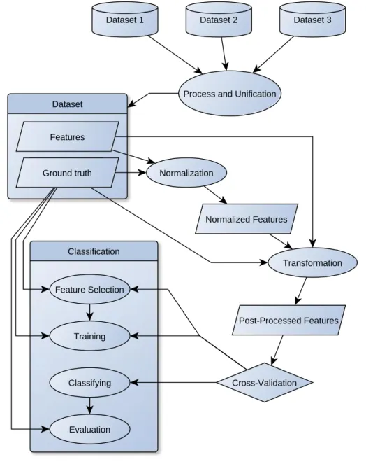
As a way to have an idea of the overall system, as well as the way the proposed objectives and subsystems are going to be organized, Figure 1, shows the archi-tecture of the system that will be constructed.
Figure 1.System overview
Dataset
Ground truth Normalization Features
Normalized Features
Post-Processed Features Transformation
Dataset 1 Dataset 2 Dataset 3
Process and Unification
Classification Feature Selection Training Classifying Evaluation Cross-Validation Ground truth
António José Almeida Rebelo | Universidade do Minho | 7 Research methodology
This work was developed according to an Action-Research methodology. This method is suitable for solving a problem that seeks to obtain information leading to its resolution through an iterative and recurrent process. This method starts with the identification of the problem so that several hypotheses can be formulated on which development work will be based. Afterwards, the gathered information will be recompiled, structured and analyzed, in order to continuously develop a proposal to solve the identified problem. As an end result, one can make conclusions based on the outcomes obtained during the re-search.
Using this research model, six complementary steps are defined to achieve the planned objectives:
Specification of the problem and its characteristics;
Incremental update and review of the state of the art;
Idealization of new methods and iterative development of the proposed model;
Experimentation and implementation of the prototype;
Results analysis and conclusions;
Diffusion of knowledge with the scientific community.
Structure of the document
This document is organized as follows: Section 1 starts with the description the main techniques for sleep assessment focused on the insufficiencies and problems associated with them. After defining the main problem, the scope of the project, followed by the objectives and research methodology are introduced.
Section 2 will begin with a brief introduction to sleep medicine, describing the essential aspects of normal human sleep and some sleep disorders. Some studies related to sleep
assessment are reviewed. Studies that are in the same scope of the research being devel-oped are presented, examined and compared.
In section 3 the datasets used in this work are described. A review and a comparison of the current data with sleep literature is presented as well. The sleep architecture of the subjects of this study will be further analyzed, culminating in the selection of the subjects with a normal healthy sleep architecture.
Section 4 will cover the techniques used to improve the quality of the data and its dis-criminating power. These techniques will be divided in two distinct procedures, namely normalization and transformation. Normalization aims at reducing between-subject vari-ability while transformation aims at maximizing the number of features with a high dis-criminative power. The results of the application of this techniques are discussed in this section as well.
Section 5 will describe the classifier, the feature selection algorithm and the probabilistic post-processing step used to improve the classification results. This step aims at capturing the non-stationary temporal characteristics of sleep.
The final results are presented, discussed and compared with literature in section 6. Finally, section 7 will review the work performed and draw the final conclusions of the present work.
António José Almeida Rebelo | Universidade do Minho | 9
Sleep monitoring
History of sleep medicine
It is difficult to determine or even produce an estimated point in time when interest in sleep first occurred. Insomnia was reported in ancient Egyptian texts and it is thought that opium was used as the first hypnotic medication [5]. Despite early curiosity, the scientific interest in sleep has emerged over the past century and the field of sleep medicine itself has existed for only about five decades. Sleep medicine is devoted to the diagnosis and therapy of sleep disturbances and disorders.
2.1.1 Early theories
In the late nineteenth and early twentieth century, a variety of theories were formed re-garding the nature of sleep. A theory gained popularity around the end of the nineteenth century, hypothesizing that toxins were developed during wakefulness and were gradu-ally eliminated during sleep. Legendre and Pieron injected serum from sleep deprived dogs into awake dogs and observed that they became fatigued. The term ‘hypnotoxin’ was introduced to describe this endogenous sleep factor, which promoted sleep [6]. The development of the electroencephalogram (EEG) in 1929 by the German Psychiatrist, Hans Berger allowed the examination of brain activity during sleep [7]. This measuring technique recorded the electrical activity of the human brain, allowing for a continuously and quantitatively measure of the neural activity of the sleeper without disturbing it. In-vestigation in the following years established the characteristics of the EEG during sleep. High amplitude, slow waves, and spindles were found to be typical during sleep while wakefulness was characterized by fast and lower amplitude waves and alpha rhythms [8]. Using the EEG it became clear that the brain was not idle and that it actually followed a synchronized pattern of neuronal activity.
Sleep stages were first categorized and described in 1937 by Alfred Lee Loomis and his team, who distinguished the different electroencephalography features of sleep into five levels representing the spectrum from wakefulness to deep sleep [9].
2.1.2 REM sleep
The phenomenon of rapid-eye movement (REM) sleep and its association with dreams was discovered by Eugene Aserinsky, Nathaniel Kleitman and William C. Dement in 1952 at the University of Chicago [10]. The creation of a method which could measure the eye mobility, called electrooculography (EOG), made possible the analysis of the eye mobility across the night. They found that during certain periods in the night, there was a substantial increase in the speed of the movement of the eyes. These periods were there-fore called rapid eye movement (REM) sleep [11]. During their investigation, recurring variations were observed, corresponding to cyclical occurrences of REM sleep at intervals of 90 to 100 minutes, with intervals tending to be longer toward the end of night. The total sleep time spent in REM sleep was estimated to be between 20 and 25% of the whole sleep time. The association between REM sleep and dreaming was also established [12]. Meanwhile in 1960, Michel Jouvet demonstrated through electromyographic (EMG) re-cordings that activity and muscle tone are completely suppressed during REM. Presently it is established that muscle atonia is a fundamental characteristic of REM sleep [13]. Because of these discoveries sleep was therefore reclassified into REM and four non-REM (Nnon-REM) stages.
2.1.3 Sleep clinics and sleep disorders
With the discovery of REM sleep, sleep research relying on all-night sleep recordings became standard and was the precursor of sleep medicine and particularly of the clinical test, polysomnography.
The first sleep disorders center was established as a narcolepsy clinic at Stanford Univer-sity in 1964, evolving into evolved into a full-service sleep disorders clinic by 1970. The sleep center was directed by a sleep specialist, and had the ability to perform polysomnog-raphy and multiple sleep latency tests [14].
In 1965, sleep apnea was discovered independently by Gastaut, Tassinari and Duron in France and Jung and Kuhlo in Germany [15][16].
António José Almeida Rebelo | Universidade do Minho | 11 The staging criteria were standardized in 1968 by Allan Rechtschaffen and Anthony Kales in the "R&K sleep scoring manual" [17].
At Stanford University in 1972, respiratory and cardiac measurements became a standard of the all-night diagnostic test. In 1974, Dr. Jerome Holland, a member of the Stanford group named this test ”polysomnography” [18].
Presently people are much more aware of the significance of sleep in their lives as well as the consequences of sleep disorders. However the percentage of undiagnosed sleep disorders is still extremely high.
Normal human sleep
Healthy sleep is characterized by a consistent and cyclic process in which phases of deep sleep alternate with lighter sleep. Normal human sleep is composed by two major phases of sleep called REM and NREM (Non Rapid Eye movement). These major phases alter-nate cyclically across the night. In 2007, the former Rechtschaffen and Kales scoring system [28], which was comprised by Wake, Rem and four individual stages of Non-Rem sleep, was revised by the AASM, which resulted in several changes. Arousals and respir-atory, cardiac, and movement events were added and stages III and IV were combined into a single stage, stage N3 [28]. Using the AASM definition, NREM sleep is further divided in N1, N2 and N3, increasing depth towards N3 which is sometimes also called slow-wave sleep. When comparing ASSM and Rechtschaffen and Kales scoring, it is ac-ceptable to compare N1 to stage I, N2 with stage II and N3 with stages III and IV. Presently the current gold standard for sleep assessment is the polysomnography clinical test. As mentioned before, this is a very reliable manual scoring performed by sleep pro-fessionals who visually inspect the recordings of brain activity, eye movements and mus-cle activity in order to determine, for each period of the night, in which sleep stage the subject is. Sleep stages are consequently used to plot a so-called hypnogram, as illustrated in Figure 1. If sleep diseases are suspected, sleep professionals might use additional sen-sor modalities such as using respiratory flow and respiratory effort to score apnea events, or use video recordings to diagnose REM behavior disorder.
According the AASM guidelines, the night is divided into 30-second epochs which are manually classified as Wake, N1, N2, N3 or REM.
Stage N1 is common in the beginning of the sleeping period, accompanied with slow eye movement and a decline in the tonic activity when compared with the wake state. It is normally a transition phase between wake and sleep. It can also emerge briefly during transitions from sleep to wake or after brief body movements. People are normally una-ware of this stage and when aroused they often believe they were still wake. The EEG is characterized by relatively low voltage slow activity in the theta range (4 - 7Hz). N1 is distinguishable from relaxed wake, with closed eyes, since wake during wake higher fre-quency alpha waves (8 - 13Hz) are generated by the brain [17].
Stage N2, is marked by the appearance of EEG spindles (fast activity in the 7-14 Hz range lasting at least half a second) and K-Complexes, which consist of high-voltage waves with a negative sharp component followed by a positive component. Cardiac and respir-atory frequencies are usually slower than during the wake state. Conscious awareness completely vanishes [3].
Stage N3, is the deep sleep stage where slow-wave sleep (SWS) occurs. This stage is scored if delta waves, large amplitude figures with a frequency range of 0.5 - 2Hz, occupy
at least 20% of the thirty second epoch in the EEG. These very pronounced waves are much “slower”, have a lower frequency, than those characteristic of N1 and N2, explain-ing why this stage is also called slow-wave sleep. Deep sleep is characterized by lower variations in respiratory and cardiac activity when compared against the other sleep stages. Sleepwalking, sleep-talking or other parasomnias are typically encountered in this stage [29].
REM sleep represents an active form of sleep and is characterized by low amplitude and high frequency cerebral activity, very similar to wake, associated with muscle atonia and rapid eye movements. Both cardiac and respiratory activity are highly variable during REM sleep. Most dreams occur in this phase of sleep. REM sleep can be further divided in tonic and phasic components. Tonic REM sleep is associated with near paralysis of most muscular groups. Only the diaphragm, the cardiac muscle, and some sphincters at
António José Almeida Rebelo | Universidade do Minho | 13 the top and at the bottom of the gastrointestinal tract remain active during REM sleep. Transient swings in blood pressure, heart rate changes, and irregular respiration are asso-ciated with tonic REM sleep as well. Phasic REM sleep is characterized by irregular epi-sodes of EMG activity and rapid eye movements [17].
2.2.1 Sleep architecture
In a healthy adult, sleeping on a normal schedule, sleeps follows a certain architecture beginning with NREM, progressing towards N3, before arriving at the first episode of REM sleep approximately 80 to 100 minutes after sleep onset. Afterward NREM sleep alternates with REM sleep with a periodicity of approximately 90 minutes. Across the night the length of the episodes of deep sleep and REM sleep vary. In the first cycles of sleep, N3 episodes are longer than REM sleep episodes. As the nigh progresses, N3 epi-sodes start to be smaller or even absent and REM sleep epiepi-sodes become longer. In young adults, N1 sleep constitutes about 5-10% of the night. The largest amount of sleep time, 50-60%, is spent in stage N2. Stage N3 constitutes about 10-20% of the total sleeping time while REM sleep 20-25% [19].
Figure 2. Hypnogram of a healthy adult
Figure 2 illustrates an actual hypnogram of a single night of healthy subject, monitored in a sleep laboratory. Hypnograms were developed as an easy tool to present the sleep architecture during a period of sleep. They allow the easy visualization of the sleep stages, which in turn allow certain parameters which might be indicative of sleep disorders to be
100 200 300 400 500 600 700 800 N3 N2 N1 REM Wake Epoch
easily identified, for example, high sleep fragmentation or low sleep efficiency, too short REM sleep onset, long sleep onset, low percentage of deep sleep among others. Each epoch correspond to 30 seconds starting from the moment the lights were turned off. In this image it is possible to see the NREM - REM cycles as well as the changes in the duration of these two stages throughout the night.
2.2.2 Changes in sleep with age
In the absence of disorders, the most significant factor affecting total sleep time and sleep stages is age. Newborns have so-called passive and active sleep stages, which are the precursors of NREM and REM sleep respectively. Newborn infants, during the first months of life, sleep 17-18 hours a day, and spend 50% of their sleep time in active sleep. The duration of the passive-active sleep cycle is also shorter in the newborn, when com-paring with NREM-REM cycle in adults, at about 50-60 minutes [20]. Also, SWS is max-imal in young children and decreases markedly with age. REM sleep and sleep efficiency decrease with age as well.
Figure 3. Age related trends for stage 1, stage 2, slow wave sleep (SWS), REM sleep, wake after sleep onset (WASO) and sleep onset latency (minutes) [21]
António José Almeida Rebelo | Universidade do Minho | 15 In Figure 3 it is possible to observe how sleeping time is divided according to the age of the subjects. Sleep onset latency is the length of time that it takes to accomplish the tran-sition from wake to sleep. The changes in sleep onset latency are not very significant, and remain fairly constant as subject’s age. WASO is the amount of time spent wake after sleep has been initiated and before final awakening. It is a good metric for measuring the subject’s difficulty to stay asleep, a common occurrence in sleep disorders such as insom-nia but also a normal occurrence in elderly subjects, since it increases with age.
Sleep efficiency is the ratio of time spent asleep (total sleep time) to the amount of time spent in bed from light out to lights on. Since WASO increases with age, sleep efficiency consequently decreases.
Sleep disorders
The prevalence of sleep disorders in the general population is considerably high. In fact hundreds of millions of people over the world suffer from sleep disturbances [2]. Sleep disorders can be the cause of impaired academic or occupational performance, accidents at work or while driving and disturbances of mood and social adjustment. Somnolence and the predisposition to fall asleep during the performance of dangerous tasks, is recog-nized as an important problem in our society. Furthermore, sleep disorders may lead to or aggravate serious medical, neurologic and psychiatric problems. There are some com-mon complaints regarding disturbances in sleep. Patients usually mention problems like insomnia, excessive daytime sleepiness and abnormal movements, behaviors or sensa-tions during sleep or nocturnal awakenings.
Ever since sleep disorders were first accepted, their classification has been of particular interest to clinicians. The Diagnostic Classification of Sleep and Arousal Disorders [22], published in 1979, was the first significant classification and organized sleep disorders in categories which influenced the current classification systems. In order to standardize the sleep disorders and create a systematic approach for their diagnose, the International Clas-sification of Sleep Disorders (ICSD) was created in 1990 and in 2005 it was updated and named ICSD-2 [23]. The sleep disorders were organized into eight distinct categories:
1. Insomnias.
2. Sleep related breathing disorders.
3. Hypersomnias of central origin not due to a circadian rhythm sleep disorder, sleep related breathing disorder or other cause of disturbed nocturnal sleep.
4. Circadian Rhythm Sleep Disorders. 5. Parasomnias.
6. Sleep Related Movement Disorders.
7. Isolated Symptoms, Apparent Normal Variants, and Unresolved Issues. 8. Other Sleep Disorders.
This classification has been widely accepted and used by sleep professionals and has al-lowed better international communication and cooperation in sleep disorder research. 2.3.1 Insomnia
Insomnia is by far the most common form of sleep disturbances [24]. Usually insomnia is defined as the symptom of difficulty falling asleep or remaining asleep and occasionally as the inability to obtain restorative sleep. Insomnia has a very peculiar characteristic as it is both a symptom and a disorder. It can be secondary to another disorder or an inde-pendent disorder. Thus, insomnia may be classified into primary or secondary, with both forms leading to sleep - wake disturbances although the secondary form arises as a func-tion of psychiatric condifunc-tions, medical diseases or substance abuse [25]. Insomnia is usu-ally linked with a very active life, high levels of stress, age, shift working, and psychiatric and medical disorders. It is also more prevalent in women. The clinical assessment of insomnia is usually based on a clinical interview with a patient, often supplemented by questionnaires, psychological screening tests, sleep diaries, and interviews with the bed partner. The use of actigraphy in addition to the questionnaires is a common practice before PSG [26]. An efficient assessment and management of insomnia must address psychological and behavioral factors such as poor sleep routines with irregular sleep-wake schedules, hyperarousability, and wrong attitudes and beliefs about sleep. Several treatments are applied in order to address primary insomnia. Examples include sleep
re- António José Almeida Rebelo | Universidade do Minho | 17 striction, stimulus control, cognitive theraphy. Relaxation training and sleep hygiene ed-ucation are used as well as a combination of those methods, which is referred together as cognitive behavior therapy.
2.3.2 Sleep disordered breathing
These types of sleep disorders are characterized by an irregular and unnatural respiration during sleep. Among this disorders, sleep apnea is by far the most common and well-known. It is associated Sleep apnea can be distinguished between central apnea and ob-structive sleep apnea. Central apnea disorders are characterized by diminished or absent respiratory effort intermittently or cyclically as a result of central nervous system dys-function. Obstructive sleep apnea disorders are formed by an obstruction in the airway resulting in an increased breathing effort and inadequate ventilation. In adults, obstructive sleep apnea is characterized by repetitive episodes of breathing cessation (apneas) or par-tial upper airway obstruction (hypopneas). The typical consequences are snoring and sleep disruption leading to secondary insomnia or excessive daytime sleepiness [28]. It is also associated with cardiovascular morbidity and hypertension. Although significant prevalent in preschool children and middle age adults, in many countries, up to 90% of the affected subjects remain undiagnosed due to the lack of resources [27–29].
State-of-the-art in home sleep monitoring
As presented in the previous sections, although sleep disturbances have a significant neg-ative impact on health, Laboratory-based polysomnography (PSG), the gold standard for sleep monitoring, is impractical for long-term and home use. Hence, alternative devices have been developed for home sleep assessment and diagnose of sleep disorders. This section reviews the literature, providing an overview of available projects and commer-cially available products for sleep monitoring outside the laboratory or the sleep clinic. 2.4.1 Actigraphy
Actigraphy is a non-invasive method that is able to monitor human wake and sleep activ-ity cycles. An actigraph is generally a watch-shaped device, as seen in Figure 4, worn by the patient on the wrist of the non-dominant hand, in order to measure gross motor activ-ity. It continually records the movements it undergoes over extended periods of time,
while allowing the patients to proceed with their normal daily routine and to sleep in their natural environment. Depending on the type of device, the data can be later transferred to a computer or mobile device and further analyzed. The recorded movements, when par-titioned by thirty second epochs, lead to the widely known feature called activity counts, which is a set of numbers expressing the total number of movements within each epoch. Based on the acquired data, scoring algorithms are used to identify sleep and wake states from the activity counts. This information allows the objective assessment of several sleep parameters like time in bed, total sleep time, sleep efficiency or wake after sleep onset. Actigraphy is suitable to provide sleep and wake information that can supplement PSG tests or be used to pre-screen some patients. Furthermore it can track longitudinal sleep information that may be missed by a one-night PSG study.
Actigraphy has been indicated by the AASM as a suitable method to assist in the evalua-tion of patients with circadian rhythm disorders, such as advanced sleep phase syndrome, delayed sleep phase syndrome or shift work disorder, and also to assess response to ther-apy of circadian disorders and insomnia [26]. The AASM’s Standards of Practice Com-mittee (SPC) also provided recommendations for the use of actigraphy in clinical practice.
Figure 4. Actiwatch Spectrum, Philips Electronics
2.4.2 Unattended portable monitors in the diagnosis of OSA
As a related project regarding sleep monitoring and diagnosis of obstructive sleep apnea, Collop et al. published a review, including an overview [30], and clinical guidelines [31]
António José Almeida Rebelo | Universidade do Minho | 19 on portable monitoring methods specifically for the diagnosis of OSA. As a result of the work performed, the authors concluded that portable monitoring may be used as an alter-native to polysomnography (PSG) for the diagnosis of OSA in patients with a high prob-ability of moderate to severe OSA. For patients with significant comorbid medical con-ditions, portable monitoring is not appropriate for the diagnosis of OSA, as it may signif-icantly degrade the accuracy of the system. This method may be indicated for the diag-nosis of OSA in patients for whom in-laboratory PSG is not possible because of immo-bility, safety, or critical illness. The portable monitoring must record, at least, airflow, respiratory effort, and blood oxygenation. The airflow, effort, and oximetric biosensors should be similar to the ones conventionally used for in-laboratory PSG.
A good example of an unattended portable monitor is the Philips Stardust II. This system records the body position as well as the airflow, gathered by the nasal cannula, in Figure 5, and/or the oral thermistor. The pulse oximetry, gathered by the SpO2 sensor which
corresponds to the grey sensor in the finger in Figure 5. Pulmonary ventilation is meas-ured by respiratory inductance plethysmography which consists of two sinusoid wire coils installed on a flexible band, one placed around the rib cage under the armpits, visible in Figure 5, another placed around the abdomen at the level of the umbilicus (belly button).
Figure 5. Stardust II, Philips Electronics
Although these methods allow measurements to be performed at home, which greatly improves the comfort of the patient who is being monitored, the collected data must be
analyzed by a sleep clinician, and the diagnosis of obstructive sleep apnea (OSA) must be performed only in conjunction with a comprehensive sleep evaluation. Furthermore, the application of the sensors must be performed by an experienced sleep technician or the patients must be educated in sensor application.
2.4.3 Ambulatory polysomnography
Sleep in a laboratory may not be representative of the typical sleep of a subject suspected of a sleep disorder. One of the main reasons is related to the unfamiliarity or intimidation with the environment [32]. The labour-saving and cost-saving benefits of home record-ings as well as the increased comfort, privacy, and convenience are the main advantages of ambulatory polysomnography.
There are a lot of products capable of performing ambulatory polysomnography. Philips Alice PDx, visible in Figure 6, is a good example, which is a multi-purpose device capable using different sensors depending on the study being performed. Examples of such sen-sors are the ones used for OSA monitoring as well as others like ECG, EEG and EOG.
Figure 6. Alice PDx, Philips Electronics Unobtrusive monitoring in bed
Ambulatory PSG systems greatly reduce the problems related to sleep monitoring in an unfamiliar environment, potentially diminishing effects such as the “first night effect”. However, these types of approaches use obtrusive methods with several contact sensors and wires, which decrease the level of comfort of the patient. Furthermore, these systems are still costly to use and not easy to deploy, generally limiting their application to one or two nights. Although some modalities such as airflow or neural activity cannot be readily replaced by unobtrusive counterparts, other modalities such as cardiac and respiratory
António José Almeida Rebelo | Universidade do Minho | 21 activity, and body movements can be monitored using already available commercial sys-tems.
2.5.1 Ballistocardiography
Ballistocardiography is a technique for producing a representation of repetitive motions of the human body, induced by the heartbeat, occurring due to acceleration of blood as it is ejected and carried through the great vessels. Ballistocardiography obtains mass move-ments of the body, caused by the heart contraction, giving information regarding the over-all performance of the circulatory system. Through this technique the mechanical move-ment of the heart can be captured by unobtrusive methods from the surface of the body. An example of a device relying on this technology is the EMFIT’s bed foil sensor in Figure 7. Through this type of devices it is possible to unobtrusively acquire cardiac in-formation as well as movement and respiration inin-formation.
Figure 7. Under-Mattress Bed Sensor, Emfit
2.5.2 Doppler radar
The Doppler radar, uses the Doppler Effect, to acquire information regarding the velocity of the objects at a distance. The Doppler Effect is the change in frequency of a wave, an observer experiences, when moving relatively to its source. The radar beams a microwave signal against the target, waits for the reflection and analysis the frequency of the returned signal, which has been altered due to the target’s motion. Doppler radars have a wide
variety of applications including aviation, satellites, meteorology, police speed guns and healthcare.
In the context of sleep monitoring, unobtrusive sleep monitoring devices use the Doppler Effect to measure respiration signals. The emitted microwave signals hit the patient’s moving chest wall and are modulated in amplitude and phase. The frequency of the mov-ing chest wall can be calculated through the reflected signal.
An example of a device using this principle for unobtrusive respiration monitoring during sleep is SleepMinder from BiancaMed. This device is capable of unobtrusively monitor respiration during sleep and detecting sleep-disordered breathing events.
Figure 8. An example set up of SleepMinder, Biamcamed [33]
2.5.3 Video
Different methods have been proposed that rely on video recording for monitoring sleep in an unobtrusive way. For instance Duffy et al.[34] proposed a methodology to access the respiration information of the patient based on an optical chest-wall measurement system. The system used a low powered helium-neon laser to illuminate the chest wall of the patient.
António José Almeida Rebelo | Universidade do Minho | 23 In 2010, Kuo et al.[35] sugested a method to monitor gross body movements and body positions as well as respiration activity of a sleeping patient. In their study they used near-infrared camera with a near-near-infrared lighting source.
Sleep stage classification using unobtrusive modalities
The systems described in the previous section have the potential to use non-obtrusive techniques for data collection, which makes then valid options for comfortable, non-ob-trusive sleep monitoring over extended periods of time.
In order to compare their performance, the studies presented in this section are described in terms of Cohen’s Kappa coefficient. This is a statistical measure of inter-rater agree-ment for qualitative items. Although there is no consensus about what constitutes a good or a bad performance, Landis and Koch [36], characterized values less than zero as indi-cation of no agreement, 0 to 0.20 as slight agreement, 0.21 to 0.40 as fair agreement, 0.41 to 0.60 as moderate agreement, 0.61 to 0.80 as substantial agreement, and 0.81 to 1 as almost perfect agreement. The metric is further described in section 6.1.
2.6.1 Sleep and wake Discrimination
The study described in this document had used the work of Devot et al. [4] developed within Philips Research as the starting point. It used a Linear Discriminant (LD) classifier trained to classify sleep and wake using actigraphy, cardiac and respiratory features, on a dataset comprised of 35 middle-aged subjects (9 healthy, 27 insomniacs, 16 males and 20 females) and reported an overall Cohen’s kappa coefficient of agreement of 0.62 (overall accuracy of 86.7%) , 0.7 for healthy subjects, 0.61 for insomniacs.
This system has been further extended with additional features, such as described by Xi et al. [3]. Using only actigraphy and features derived from respiratory effort extracted from a dataset comprised of nine healthy subjects (eight females) with a mean age of 32 ± 13, it reports a κ of 0.69 (overall accuracy of 95.4%).
2.6.2 Sleep staging using cardiorespiratory signals
The work reported by Redmond et al. [37] is a method for the discrimination of Wake, Non-REM and REM stages, using ECG and respiratory effort signals. In previous work
[38], Redmond et al. executed a similar study, aimed at performing sleep stage classifi-cation on subjects being assessed for Obstructive Sleep Apnea. The newly developed study, aimed at examining the effectiveness of the same system when applied to normal healthy subjects.
The dataset used for this work was composed by 31 male subjects with a mean age of 42±7 years.
This study tested and compared the performance of linear and quadratic discriminant clas-sifiers. Temporal varying priori probability information was introduced in the classifica-tion results to further increase the classificaclassifica-tion results. The best performance was ob-tained by linear discriminant classifier model using temporal varying priori probability. For the 3 class system an agreement of κ = 0.45 was achieved. When considering only a two class system, Sleep and wake, the agreement would increase to κ = 0.57.
2.6.3 Sleep staging based on signals acquired through bed sensor
The study performed by Kortelainen et al. [39], had the peculiar characteristic of acquir-ing the signals usacquir-ing non-obtrusive sensors. It was based on previous work developed in the same scope but with signals acquired through contact sensors[40]. Emfit sensor foils, such as that illustrated in the previous section in Figure 7, were used and placed under the bed mattress, from which the heart-beat interval (HBI) and body movements were ob-tained.
The dataset used for this work was composed by 18 recordings from shift-work subjects. Nine females, aged between 20 and 54 years, participated in this study with two record-ings each.
A time-variant autoregressive model was used for the extraction of the relevant features, and the classification was performed with a hidden Markov model (HMM). The classifi-cation results for the 3 classes Wake, NREM and REM achieved κ of 0.44 ±0.19 and an accuracy of 79 ±10% using only three HBI features and one movement feature.
António José Almeida Rebelo | Universidade do Minho | 25 2.6.4 Analysis and Comparison
All the studies presented above reported very promising results, which shows why this area of research is interesting and continues to progress towards a reliable unobtrusive sleep staging system. However, since obtaining cardiorespiratory and movement data in an unobtrusive manner and using this data for automatic sleep stage classification, is not a trivial challenge, there is room for improvements.
In terms of data used, Redmond et al.[37] had the bigger dataset, has he had the bigger number of subjects. Their study had a total of 31 subjects, however all subjects were men of the same age group, suggesting that performance might degrade when using the same system in datasets with female subjects, or with subjects of other age groups. Kortelainen et al.[39] used a total of 18 recordings from healthy subjects, however the subjects were all female and only nine subjects participated in the study (with two nights each). Since the sleep architecture of a subject is usually similar across different nights, classification results when training and testing with data from the same subject, even though they be-long to different nights, might be biased. Long et al.[3] used only nine healthy sleeping subjects. The dataset to be used in this research project will consist of more subjects. Furthermore it will be interesting to observe the results obtained with a bigger number of subjects and with a classification process using more features. Moreover it is possible that during the research project more features are created and added or the method or the cre-ation method of the features is modified.
For the maximization of the discrimination power, these studies used various methods like detrended fluctuation analysis or time-dependent auto regressive models. Such meth-ods aim to retrieve information from the features taking the time factor into consideration. Furthermore Redmond et al.[37], suggested that the use of time varying prior probabilities would result in improved results. Time plays therefore an important role in the sleeping architecture and should not be forgotten when developing the classifier.
In terms of the classification process, two of these studies chose a Linear Discriminant and another used a Hidden Markov model. Hidden Markov Models and Bayesian Linear Discriminants are widely known classification algorithms, each with some advantages
and disadvantages. A hidden Markov model (HMM) is a statistical Markov model in which the system being modelled is assumed to be a Markov process with unobserved (hidden) states [41]. Bayesian Classifiers are based on the idea of assigning unknown patterns to the most probable class within a known set of classes. Each pattern is charac-terized by a feature vector [42].
Although sleep follows a certain sequence of states throughout the night, Hidden Markov Models may not be appropriate for this specific classification process. The complexity of the model and wrong assumptions regarding the process may reduce the classification performance. Assumptions such as the Markov assumption which requires that the next state is dependent only upon the current state, and the stationary process assumption which requires that state transition probabilities are independent of the actual time at which the transitions takes place, may cause the classification results to drop. Besides the probability of a certain sleep stage may not only depend on the previous epoch, but on the sequence of previous epochs. Moreover the probabilities of stage change vary across time, in fact, before the study of Kortelainen et al.[39] and in an attempt to capture the time varying stage probabilities, Redmond et al.[37], proposed a method that uses time-varying a priori probabilities, accomplishing positive results.
Linear discriminants are very simple classifiers where probabilities can be added or al-tered in order to achieve the best representation of the problem at hand [41]. Although time-varying a priori probabilities played an important role in the task of capturing the time varying probabilities of sleep stages, this method can be further explored.
Classification results are very similar for the studies performed by Kortelainen et al. [39] and Redmond et al. [37]. The study performed by Long et al.[3] only analyzed the classi-fication for Wake and Sleep which makes the comparison against the systems classifying Wake, Non-REM and REM stages difficult since systems have different problem com-plexities.
One important detail is the high standard deviation in the classification results of the sub-jects reported in the literature. The very high standard deviation suggest that these results are being affected by large between-subject variability. Between-subjects variability,
António José Almeida Rebelo | Universidade do Minho | 27 when all data is pooled, greatly decreases the discrimination power comparing against the average discrimination power of the same feature for the each subject. Normalizing the feature values across each subject this between-subject variability can be reduced, possi-bly improving the classification results.
Summary
This section began with an introduction to sleep medicine, with information regarding the normal human sleep and sleep disorders. Some studies, methods and devices related to sleep assessment were reviewed.
This section finished with an examination of some techniques, technologies and studies within the same scope of this project. The different studies were compared, taking into consideration the datasets used, the methods applied for the classification and the classi-fication results.
Datasets
For the investigation of the automatic classification of sleep stages based on cardiorespir-atory data and evaluation of developed algorithms, annotated data is needed. Since the goal of this research work is to use supervised learning to classify data into sleep stages, the annotated sleep stages and corresponding cardiorespiratory measurements are needed. The annotated sleep stages were annotated by a sleep professional, in 30-second epochs following the AASM or R&K guidelines.
In this work four different datasets are considered, namely the Boston healthy, The Bos-ton insomniacs, Eindhoven and SIESTA.
Boston Healthy dataset
The Boston Healthy dataset comprises 10 healthy subjects (8 females) with an average age of 31 ± 12 years. Sleep stages were scored by an expert according to the AASM guidelines.
Boston Insomniacs dataset
The Boston Insomniacs dataset comprises 27 subjects (13 females) diagnosed with in-somnia, with an average age of 46 ± 14 years.
Sleep stages were scored by an expert according to the AASM guidelines.
Looking at Figure 9, high amount of epochs score as wake is observable. Sleep efficiency is therefore very low on insomniacs subjects. Moreover the number of epochs scored as deep sleep is much lower than in normal healthy subjects considering the same age of the subjects. As expected, a person who suffers from insomnia, has a predominance in N2 and N1, with difficulty to fall asleep followed by several awakenings which explains the high amount of N1 and wake epochs.
António José Almeida Rebelo | Universidade do Minho | 29 Figure 9. Total epochs spent in each stage from lights out, for Boston Insomniacs dataset
Eindhoven dataset
The Eindhoven dataset comprises 12 subjects (6 females) with an average age of 29 ± 5 years.
Sleep stages were scored by an expert according to the AASM guidelines.
SIESTA dataset
SIESTA was a project funded by the European Commission which involved several Eu-ropean partners. The aim of the project was to research the nocturnal human sleep as well as to develop and evaluate new methods of sleep analysis [43].
The SIESTA dataset is the largest dataset in this study. It has a total of 292 subjects, amongst which subjects diagnosed with general anxiety disorder, depressive disorder, sleep apnea syndrome, restless legs or Parkinson.
They have an average age of 52 years ± 17. This dataset is composed by 126 females and 166 males.
Sleep stages were scored by a consensus of at least two experts according to the R&K guidelines. 8% 47% 6% 13% 26%
Sleep stage distribution after lights off - Boston Insomniacs N1 N2 N3 R W Invalid

![Figure 3. Age related trends for stage 1, stage 2, slow wave sleep (SWS), REM sleep, wake after sleep onset (WASO) and sleep onset latency (minutes) [21]](https://thumb-eu.123doks.com/thumbv2/123dok_br/17758584.835321/31.892.131.688.649.1030/figure-related-trends-stage-stage-sleep-latency-minutes.webp)

![Figure 8. An example set up of SleepMinder, Biamcamed [33]](https://thumb-eu.123doks.com/thumbv2/123dok_br/17758584.835321/39.892.179.741.614.878/figure-example-set-sleepminder-biamcamed.webp)