RESUM O RESUM ORESUM O
RESUM ORESUM O.- [M ycopl asm a su i sM ycopl asm a su i sM ycopl asm a su i sM ycopl asm a su i sM ycopl asm a su i s em su ín o s n at u r al m en t e em su ín o s n at u r al m en t e em su ín o s n at u r al m en t e em su ín o s n at u r al m en t e em su ín o s n at u r al m en t e infect ados: um est udo ult ra-est rut ural e mor
infect ados: um est udo ult ra-est rut ural e morinfect ados: um est udo ult ra-est rut ural e mor
infect ados: um est udo ult ra-est rut ural e morinfect ados: um est udo ult ra-est rut ural e mor fomét rico.fomét rico.fomét rico.fomét rico.fomét rico.] A eperitrozoonose suína é uma doença hemotrópica causada por Eperit rozoon suis, atualmente denominado Mycoplasma suis, uma bactéria extracelular que, aparentemente, adere à mem-brana dos erit rócit os suínos, induzindo sua deformação e lesionando-os. O presente trabalho busca estabelecer os as-pectos estruturais e ultra-estruturais, pouco conhecidos, deste microorganismo. O estudo ultra-estrutural revelou a presen-ça de estruturas correspondentes a túbulos disseminados no soma bacteriano. Obser vou-se também uma separação variá-vel ent re a membrana do microorganismo e a parede do eritrócito. O estudo morfométrico e a localização de M . sui s pode permitir especulação sobre seu mecanismo de ação.
TERMOS DE INDEXAÇÃO: Eperitrozoonose, M ycoplasma sui s, ultra-estrutura, morfometria, patogenia.
INTRODUCTION INTRODUCTION INTRODUCTION INTRODUCTION INTRODUCTION
Eper ythrozoonosis is a haemotrophic disease in swine caused by a bact erium formerly classified as t he ricket t sial agent Eper yt hrozoon suis (Hoelzle et al. 2003). The disease is clinically characterized by anemia, jaundice and a variety of associated syndromes (Hsu 1986, Henderson et al. 1997, Machuca et al. 1999).
Eper yt hrozoon suis, now allocated to the genus M ycoplasma, is an extracellular bacterial organism that attaches to and causes deformit y and damage t o porcine red blood cells (Hoelzle et al. 2003). The accepted actual name of the agent is M ycoplasma sui s (Neimark et al. 2002). It has a restrictive cellular membrane, lacki ng organelles and nucleus. It s cyt oplasm cont ains as much DNA as RNA (Gwalt ney 1995, Gresham 1996). M ycoplasma sui s can be identified by optic microscopy in Romanowsky stained blood smears (Gwaltney 1995). It may be round, oval or ring-shaped with diameters ranging from 0.8 µm to 2.5 µm or even larger (Kreier & Ristic 1984, Gwaltney 1995).
Although the pathogenesis of the disease is not completely known, it is considered that once M .sui s comes into contact with the er ythrocyte, it uses the plasmatic glucose for its own metabolism. On the other hand, upon damaging the cellular m em br ane of t he r ed bl ood cel l , i t w oul d i nduce t he
M ycoplasma sui s
M ycoplasma sui s
M ycoplasma sui s
M ycoplasma sui s
M ycoplasma sui s
in nat urally infect ed pigs: an ult rast ruct ural and
in nat urally infect ed pigs: an ult rast ruct ural and
in nat urally infect ed pigs: an ult rast ruct ural and
in nat urally infect ed pigs: an ult rast ruct ural and
in nat urally infect ed pigs: an ult rast ruct ural and
morphomet ric st udy
morphomet ric st udy
morphomet ric st udy
morphomet ric st udy
morphomet ric st udy
11111Enrique L. Portiansky2*, María A. Quiroga2, Mariana A. Machuca2
and Carlos J. Perfumo2
ABSTRACT ABSTRACTABSTRACT
ABSTRACTABSTRACT.- Portiansky E.L., Quiroga M.A., Machuca M.A. & Perfumo C.J. 2004. M ycopl asm aM ycopl asm aM ycopl asm aM ycopl asm aM ycopl asm a sui s
sui ssui s
sui ssui s in nat urally infect ed pigs: an ult rast ruct ural and morphomet ric st udy in nat urally infect ed pigs: an ult rast ruct ural and morphomet ric st udy in nat urally infect ed pigs: an ult rast ruct ural and morphomet ric st udy in nat urally infect ed pigs: an ult rast ruct ural and morphomet ric st udy in nat urally infect ed pigs: an ult rast ruct ural and morphomet ric st udy... Pesquisa Veterinária Brasi lei ra 24(1):1-5. Inst it ut o de Pat ología, Faculdad de Ciencias Vet erinarias, Universidad Nacional de La Plata, Calle 60 y 118, C.C. 296, (1900) La Plata, Buenos Aires, Argentina. E-mail: elport i@ fcv.unlp.edu.ar
Swine eper ythrozoonosis is a haemotrophic disease caused by Eper yt hrozoon sui s, actually called M ycoplasma sui s, an extracellular bacterial organism that apparently adheres to pig erythrocyte membrane, inducing its deformation and damage. Since little is known about the ultrastructural and morphometrical aspects of this microorganism, the present work aimed t o deal wit h t hese issues. The ult rast ruct ural st udy revealed t he presence of st ruct ures corresponding to tubules disseminated throughout the soma of M . sui s. A variable separation between the microorganism membrane and that of the erythrocyte was also obser ved. The structural and positional attitude of M . sui s could allow speculation about its mechanism of act ion.
INDEX TERMS: Eperythrozoonosis, M ycoplasma sui s, ultrastructure, morphometr y, pathogenesis.
1Received on August 27, 2003.
Accepted for publication on September 12, 2003.
2Instituto de Patología, Faculdad de Ciencias Veterinarias, Universidad
Na-cional de La Plata, Calle 60 y 118, C.C. 296, (1900) La Plata, Buenos Aires, Argentina.
cells and their removal by way of the phagocyte-mononuclear system (Gwaltney 1995, Gresham 1996).
Since little is known about the structural (morphometric) and ultrastructural aspects of this microorganism (Pospichil & Hoffmann 1982), the goal of the present work was to obtain more details regarding its morphology that could explain its pat hogenesis.
MA MA MA MA
MATERIALS AND METHODSTERIALS AND METHODSTERIALS AND METHODSTERIALS AND METHODSTERIALS AND METHODS
Blood samples were obtained from 27 naturally infected feeder male and female pigs, from a semi-intensive establishment located in the town of Junín, Buenos Aires Province, Argentina. The samples were obt ai ned by veni punct ur e of t he ant er i or cava vei n, usi ng et hyl endi am i not et r acet i c aci d (EDTA) as ant i coagul ant , i n a concentration of 0.1 mg/ml of blood. The samples thus obtained were centrifuged at 800 rpm, for 5 min. Later, the cells were washed 3 times with PBS, for 5 min each time. The washed samples were fixed in 2% glutaraldehyde solution. After 30 min, the pellet was broken into small pieces, which were left in contact with the fixer for another 30 min. The fragments were later washed 3 times with PBS and post -fixed wit h osmium t et roxide, for 30 min. Aft er dehydration of the samples, they were infiltrated and embedded with epoxi resins (Quetol 812, Nisshin EM Co. Ltd, Tokyo, Japan). Blood smears were stained with May-Grünwald-Giemsa and Wright st ains.
Semi -t hi n 1 mm t hi ck cut s were obt ai ned usi ng an ult ra-microtome. The samples were stained with methylene blue and a “ Multiple Stain” solution (Polysciences Inc., Warrington, USA) and observed with an optic microscope. Ultra-thin cuts were stained with uranyl acet at e/lead cit rat e and obser ved wit h a t ransmission electronic microscope JEM-1200 (JEOL Co. Ltd, Tokyo, Japan).
Images of blood smears and semi-thin cuts were captured from a microscope (Olympus BX50 system microscope, Tokyo, Japan) with a microscopic magnificat ion of x100, t hrough an analogical RGB video-camera (Sony DXC-151A CCD, Tokyo, Japan) and digitized with a fr am e gr abber (Fl ashpoi nt 128, Int egr al Technol ogi es Inc., Indianapolis, IN, USA) connect ed t o a comput er. Images were pr ocessed usi ng t he Im age-Pr o Pl us v4.50 sof t w ar e (M edi a Cybernetics, Silver Spring, MA, USA), with a depth of pixel of 24 bits, RGB and TIFF format. The resolution of the microscopic images was of 640x480 pixels with a spatial calibration yield of 0.13 µm/pixel. The phot ographs obt ained wit h t he elect ron microscope were digit ized t hrough a full-page scanner (Microt ek Scanmaker E6, Rot t erdam, Holland), wit h a resolut ion of 300 ppp. In order t o separat e t he desired object from t he rest of t he t issue, color segmentation, based on the optical density of the object, was issued. Once separ at ed fr om t he r est of t he i m age, t he obj ect w as quantitatively characterized based on its area (in µm2), major and
minor axis (t o obt ain t he aspect of t he object ), maximum and minimum diameter, perimeter and roundness, all of them measured in lineal µm. Table 1 describes t he morphomet ric paramet ers evaluated.
RESUL RESUL RESUL RESUL RESULTSTSTSTSTS
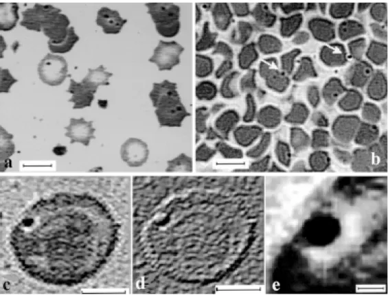
The blood smears revealed the presence of M ycoplasma sui s in close contact with the red blood cells (Fig. 1a). The stained semi-thin cuts showed a regular quantity of spherical elements appearing alone or in groups of 2 or 3, stuck to the membrane of the er ythrocyte or free among the red blood cells (Fig. 1b).
Fig.1c shows a magnificat ion of M .sui s in a red blood cell membrane. Fig.1d shows a 3D-like image obt ained by a filtering process, in which a depression of the erythrocyte membrane surrounding M . sui s can be obser ved. A higher magnificat ion of t he microorganism and t he depression it produces on t he red blood cell surface membrane can be obser ved in Fig.1e.
The ultra-structural studies revealed some morphological details of the M . sui s. The organisms were observed as round or oval structures, limited by a membrane (Fig.2a). Neither cellular organelles nor nuclear elements were obser ved. In some cases, widespread electron-dense granules were identi-fied in t he cyt oplasm. Some microorganisms evidenced st ruct ures similar t o microt ubules. Alt hough M . sui s was present on the plasma membrane of the er ythrocyte, in most cases a separation between both structures was observed. No cellular membrane damage was detected in the red blood cells, although some irregularities in the shape of the infected er ythrocyte were observed on many occasions (Fig.2b).
Table 2 shows the morphometrical values obtained from the light and electron microscopy studies. These values are the average of at least 30 images obtained from each analyzed sample. It was obser ved that there is coincidence between the major and minor axes and the maximum and minimum di amet ers, respect i vely. The elect ron mi croscopi c dat a demonstrate that a distance of 37 to 64 nm separates those parasites that are not touching the er ythrocytes from these cells. The real and calculated data for the inner tubule-like
sui s sui ssui s
sui ssui s charact erist ics charact erist ics charact erist ics charact erist ics charact erist ics
Parameter Description Area Aspect Major Axis Minor Axis Max. Diameter Min. Diameter Max. Distance Min. Distance Perimeter Roundness
Reports the area of each object
Reports the ratio between the major axis and the minor axis of the ellipse equivalent to the object (i.e., an ellipse with the same area, first and second degree moments), as determined by Major Axis/Minor Axis
Reports the length of the main axis of the ellipse equivalent to the object (i.e., an ellipse with the same area, first and second degree moments)
Reports the length of the minor axis of the ellipse equivalent to the object (i.e., an ellipse with the same area, first and second degree moments)
Reports the length of the longest line that can be drawn to pass through the centroid position and join two points on each object’s perimeter
Reports the length of the shortest line that can be drawn to pass through the centroid position and join two points on each object’s perimeter
Reports the maximal separation between M ycoplasma sui s
and the red blood cell membrane
Reports the minimal separation between M . sui s and the red blood cell membrane
Reports the length of the outline of each object
Reports the roundness of each object, as determined by the following formula: (perimeter2) / (4p * area). Circular
structures of M .sui s (Fig. 3a,b), maintain certain proportion. The calculat ed values were obt ained from measurement s
taken after a filtration process on the images from the electron microscope. The filtration process preser ved just the desired obj ect s for m easur i ng at t he expense of som e i m age modifications. Since certain proportion exists, a correction index can be established for the filtered images. The area fraction of the real and calculated values of the tubules is shown in Table 3. In both cases the values are quite similar.
DISCUS DISCUS DISCUS DISCUS DISCUSSIONSIONSIONSIONSION
Swine eper yt hrozoonosis is a cont agious disease charac-terized by a marked paleness of the mucous surfaces, presence of blood of water y aspect, delay of growth and increase of Fig. 1. Blood smears (a) and semi-thin cuts (b) showing spherical elements corresponding to M ycoplasma
sui s in close contact with the red blood cells (100x). Arrows point to the microorganism attached to two er ythrocytes; Magnification (2x) of an erythrocyte appearing in (a) infected with M . sui s (c); The same cell as in (c) with a 3D-like aspect obtained by filtering process; Magnification (4x) of M . sui s (a) and the depression it produces on the er ythrocyte surface membrane. Bars represent either 5 µm (a, b); 1 µm (c, d) or 0.25 µm (e).
Fig. 2. Elect ron microscopic phot ographs showing a spherical st ruct ure corresponding t o M ycoplasma sui s (a). Widespread electron-dense granules were identified in the cytoplasm that resemble microtubules. M . sui s could also be obser ved as an oval st ruct ure (b). Alt hough no cellular membrane damage was det ect ed some i rregulari t i es i n t he shape of t he i nfect ed erythrocyte could be obser ved. In neither case is M . suis touching the red blood cell it infects. Bars represent 400 nm.
mortality in feeder pigs, with a death toll that ranges between 3.1% and 18.7%, monthly. M ycoplasma sui s can be detected by light microscopy in Romanowsk y st ained blood smears (Gresham 1996). In our work, round elements were obser ved ranging from 0.25 to 0.40 µm, stuck to the membrane of the er yt hrocyt e in coincidence wit h t hose described by ot her authors (Hsu 1986, Solignac et al. 1996). In the 1µm semi-thin cuts similar structures were obser ved but with smaller dimensions. This could be attributed to the stretching of the cells in the smears.
The ult rast ruct ural st udy revealed some det ails of t he morphology of M . sui s. Alt hough an apparent cont act was obser ved bet ween t he microorganism and t he er yt hrocyt e in t he semi-t hin cut s, t he ult rast ruct ural st udies revealed a separat ion ranging from 37 t o 64 nm bet ween t he red blood cell membrane and M . sui s. The closeness of t he st ruct ures persist ed even aft er t he blood samples had been washed, last ing few free element s among t he cells. Most of t he infect ed er yt hrocyt es showed variat ions in t he cont our of t heir membrane at t he point of cont act wit h t he microor-ganism, but did not show any damage. These obser vat ions coincide wit h t hose described by Pospichil & Hoffmann (1982).
According t o t he morphomet rical values obt ained, a concordance bet ween t he major and minor axes and t he maximum and minimum diameters, respectively, was observed.
This fact endorses the roundness equal to 1. This means that M. suis has a spherical form although the ultra-structural study revealed t hat M . sui s present s an oval shape on cert ain occasions.
As described for other microorganisms, the microtubules obser ved in the cytoplasm of some M . sui s could be a part of it s cyt oskelet on (Ghadially 1988). On t he ot her hand, and keeping in mind t hat t his microorganism uses t he plasma gl ucose for i t s ow n m et abol i sm (Gw al t ney 1995), t he microtubules could demarcate channels through which the transport of necessar y products would take place. This allows us t o speculat e t hat M . sui s does not need t he physical atmosphere of the red blood cells to live and reproduce, but si m pl y i t s nut r i ent s, and t hat t he dest r uct i on of t he erythrocyte by exhaustion of its reserves would not necessarily signify the destruction of the agent, which would simply move in search of its next target cell.
The descr i bed m or phom et r i c det ai l s const i t ut e an int erest ing fact for t hemselves, as t hey had never been depict ed for M . sui s. The use of an aut omat ed image analyzer allows object ive appreciat ion, repet it ion of t he measure-ment s and simple operat ion. Dat a relat ed t o M . sui s can t hus be analyzed st art ing from blood smears as well as from semi-t hin cusemi-t s. This allows a good diagnosis asemi-t a relasemi-t ive speed. The fine det ail, represent ed by t he presence of microt ubules and t he separat ion of M . sui s from t he er yt hrocyt e mem-brane, adds t o t he precision of t he diagnosis and leaves an open door t o t he analysis of a possible pat hogenesis of t he infect ion.
There are bact eria t hat int eract wit h t heir t arget cells t hrough a het erot ypic union of sur face glycoprot eins t o further internalize them by attaching to their cytoskeleton (Cossart & Bierne 2001). Although no surface proteins have been ident ified in t he bact eria yet , it could be speculat ed that M .sui s might interact with the erythrocyte in a similar fashion but without entering into the cell.
In conclusion, the use of the optic and electron microscope T
T T T
Table 3. Area fract ion of inner t ubules-likable 3. Area fract ion of inner t ubules-likable 3. Area fract ion of inner t ubules-likable 3. Area fract ion of inner t ubules-likable 3. Area fract ion of inner t ubules-like st ruct ures ofe st ruct ures ofe st ruct ures ofe st ruct ures ofe st ruct ures of M ycoplasma sui s
M ycoplasma sui sM ycoplasma sui s M ycoplasma sui s M ycoplasma sui s
Total Total tubule areaa M . sui s area Area
number of fraction
tubules
Real values 44± 11 25437.09± 1392.05 117911.60± 21758.02 21.57% Calculated 35± 5 23406.58± 846.00 117911.60± 21758.02 19.85% values
aArea values are expressed in nm2.
Measurementsa M . sui s soma (LM values) Tubules-like structures (EM Values)
Blood smears Semi-thin cuts Grid Real values Calculated values
Area 0.33± 0.07 0.18± 0.06 0.17± 0.09 221.47± 126.55 304.08± 169.20 Major axis 0.65± 0.06 0.57± 0.11 0.49± 0.27 20.94± 8.00 23.14± 7.79 Minor axis 0.57± 0.08 0.39± 0.09 0.44± 0.21 12.95± 4.73 16.11± 4.60 Aspect 1.14± 0.31 1.49± 0.38 1.11± 0.24 1.79± 0.68 1.46± 0.30 Max. Diameter 0.66± 0.04 0.57± 0.11 0.49± 0.29 20.31± 7.04 22.78± 7.32 Min. Diameter 0.57± 0.08 0.39± 0.09 0.45± 0.26 12.76± 4.63 15.51± 3.66 Avrg. Diameter 0.61± 0.06 0.48± 0.09 0.47± 0.28 16.55± 5.37 19.17± 4.82 Perimeter 1.23± 0.19 1.07± 0.35 1.47± 0.86 42.58± 21.61 51.71± 24.23 Roundness 1.00± 0.00 1.00± 0.00 1.01± 0.05 1.03± 0.10 1.02± 0.13
Max. Distance 64
Min. Distance 37
aArea values are expressed either in µm2 (M . sui s soma) or nm2 (tubules). All other values are expressed either in
µm (M . sui s soma) or nm (tubules). Tubules values represent each tubule characteristic. Maximal and minimal
distance between M . sui s and a red blood cell membrane are expressed in nm. Values represent the average of at
in combination with an image analyzer would allow not only the diagnosis of an infection, as in eper ythrozoonosis, but also the analysis of possible mechanisms of action.
Acknowledgement s.- Acknowledgement s.- Acknowledgement s.-
Acknowledgement s.- Acknowledgement s.- The authors wish to thank Silvina Viviana Macnie for her skillful technical assistance in the proof reading of the manuscript. The technical assistance of Mr. Silvio Igal of the Ser vicio Central de Microscopía Electrónica, Facultad de Ciencias Veterinarias, Universidad Nacional de La Plata, is acknowledged. This study was partially supported by a grant from t he Consejo Nacional de Ciencia y Tecnología (CONICET) and from t he Secretaría de Ciencia y Técnica, Universidad Nacional de La Plata. ELP is a Research Career Member of CONICET.
REFERENCES REFERENCESREFERENCES REFERENCESREFERENCES
Cossart P. & Bierne H. 2001. The use of host cell machinery in the pathogenesis of Li st eri a monocyt ogenes. Curr. Opin. Immunol. 13:96–103.
Ghadially F.N. 1988. Microt ubules, p. 937-952. In: Ghadially F.N. (ed.) Ultrastructural Pathology of the Cell and Matrix. Vol. 2. 3rd ed. Butter-worths, London.
Gresham A.C. 1996. Eper ythrozoon infection in pigs. Pig J. 37:20-26. Gwaltney S.M. 1995. Eper yt hrozoon sui s infections in pigs: clinical syndromes
and diagnosis. J. Swine Health Prod. 3:25-27.
Henderson J.P., O´ hagan J., Hawe S.M. & Pratt M.C.H. 1997. Anaemia and low viability in piglets infected with Eper yt hrozoon sui s. Vet. Rec. 140:144-146.
Hoelzle L.E., Adelt D., Hoelzle K., Heinritzi K. & Wittenbrink M.M. 2003. Development of a diagnostic PCR assay based on novel DNA sequences for the detection of M ycoplasma sui s (Eper yt hrozoon sui s) in porcine blood. Vet. Microbiol. 93:185-196.
Hsu F.S. 1986. Pat hol ogi cal feat ur es of nat ur al l y occur r i ng por ci ne eperythrozoonosis, p. 372. In: Proc. 9th Int. Pig Veterinary Society Congress, Barcelona, Spain.
Kreier J.P. & Ristic M. 1984. Eper ythrozoon, p. 726-729. In: Kreig N.R. & Holt J.G. (ed.) Bergey’s Manual of Systematic Bacteriology. Vol. 1. 1st ed. Williams & Wilkins, Baltimore.
Machuca M., Quiroga M.A., Armocida A.D., Arauz S., Idiart J.R., Stornelli M.A. & Perfumo C.J. 1999. Eperitrozoonosis porcina: descripción de un brote en la provincia de Buenos Aires. Revta Med. Vet. 80:470-474.
Neimark H., Johansson K.E., Rikihisa Y. & Tully J.G. 2002. Revision of haemotrophic Mycoplasma species names. Int. J. Syst. Evol. Microbiol. 52:683.
Pospichil A. & Hoffmann R. 1982. Eper yt hrozoon sui s in naturally infected pigs: a light and electron microscopic study. Vet. Pathol. 19:651-657. Solignac T., Nicolas Y., Fourchon P. & Blaisot S. 1996. Eper ythrozoonosis in