Int J Anat Res 2014, 2(4):684-88. ISSN 2321-4287
Original Article
ARTHRI TI S OF THE SU BTALAR JOIN T ASSOCI ATED W ITH
SUSTENTACULUM TALI FACET CONFIGURATION
K.S. Nemade
* 1, M .M . M eshram
2, A. P. Kasote
3, N. Y. Kamdi
4.
ABSTRACT
Address for Correspondence: Dr. K. S. Nemade, Flat no. 301, Swami Samart h vihar-3, plot no. 88, Ekmat Nagar, Nagpur 440034, M aharasht ra, India. M obile No.: +919623619311.
E-M ail: knkirt i84@gmail.com
Access this Article online
Quick Response code Web site:
* 1 Assistant Professor, Department of Anat omy, Government M edical College, Nagpur, M aharasht ra,
India.
2 Professor & Head, Depart ment of Anat omy, Government M edical College, Nagpur, M aharasht ra,
India.
3 Associate Professor, Depart ment of Anat omy, Government M edical College, Nagpur, M aharasht ra,
India.
4 Associate Professor, Depart ment of Anat omy, Government M edical College, Nagpur, M aharasht ra,
India.
Variat ion in t he art icular facet of t he sust ent iculum tali have been described by m any aut hors. M ost researchers view t hese differ ences in f acet conf igurat ion as anat om ical variat ions of no funct ional significance. Bruckner (1987), for t he first t im e argued t hat t hese facet configurat ions affect joint st abilit y.
The purpose of t his st udy w as t o det erm ine t he t alar facet configurat ion of calcanei in India, m easure t he angle bet w een t he ant er ior and m iddle facet planes of t hese calcanei, and assess t he relat ion bet w een t he above param et ers and t he degenerat ive changes in t he subt alar joint s. St udy w as conduct ed in 220 calcanei of unknow n age & sex. The facet patt erns observed w ere fused ant erior and m iddle facet s (Type I), t hree separat e facet s (Type II), absence of t he ant erior facet (Type III) and t hree m erged facet s (Type IV).
Ost eoart hr it ic changes st udied are lipping, ebur nat ion on visual inspect ion and subchondral sclerosi s on r adiogr aphs.
Present st udy reveals t hat t he t alar facet configur at ion of calcanei and t he angle bet w een t he ant erior and m iddle f acet s inf luence t he st abilit y of t he subt alar joint s and developm ent of ost eoar t hrit is.
KEY W ORDS: Talar facet , Calcanei, Subtalar art hrit is, Forefoot pain.
INTRODUCTION
Int J Anat Res 2014, Vol 2(4):684-88. ISSN 2321- 4287 DOI: 10.16965/ ijar.2014.525
Received: 21 Jul 2014
Peer Review : 21 Jul 2014 Published (O):30 Nov 2014 Accept ed: 10 Nov 2014 Published (P):31 Dec 2014 Internat ional Journal of Anat omy and Research
ISSN 2321-4287 w w w.ijmhr.org/ ijar.ht m
DOI: 10.16965/ ijar.2014.525
Evolut ion of bipedalism has result ed in many changes in human foot . One of t he most impor-t animpor-t changes is developmenimpor-t of arches of fooimpor-t . Among t hese arches, medial longit udinal arch is most important arch. Subt alar joint is t he main joint of t his arch. This joint involves t hree fecet s
on calcaneum— anterior, middle and posterior. Out of t hese, ant erior and middle facet s are lo-cat ed on sust ent iculum t ali and post erior facet is locat ed on dorsal surface.
calcanei and are not development al responses t o physical act ivit ies [5]. M ost of t he research-ers view t hese differences in facet configura-t ion as anaconfigura-t omical variaconfigura-t ions of no funcconfigura-t ional significance. But , Bruckner argued t hat t hese facet configurat ions affect joint st abilit y & re-sponsible for early art hrit ic changes in some t ype [6] w hich is also support ed by some ot her au-t hors [7].
Subt alar inst abilit y is a major podiat ric problem for foot & ankle surgeons. In children, it can lead t o severe flat foot w it h grow ing pain & quick fat igue w hile w alking & running. It can lead t o many orthopedic problems affect ing ankle, knee, hip & low er back & have clinical present at ions like anterior or posterior tibial tendonit is, planter fasciit is, forefoot pain et c. [8,9].
Hence t he present st udy has been carried out t o st udy variat ion in sust ent iculum t ali facet configurat ions in vidarbha and t o evaluat e t he proposed relat ionship bet w een subt alar joint morphology and t he frequency of ost eoart hrit ic changes in sust ent iculum t ali facet configura-t ions i.e. subconfigura-talar joinconfigura-t sconfigura-tabiliconfigura-t y.
M ATERIALS AND M ETHODS
Total 220 calcanei of unknow n age and sex w ere st udied in present st udy. These w ere classified according t o sust ent icular t alar facet int o t hree groups.
Type I- Sustent iculum tali w it h single facet and It had t w o subt ypes.
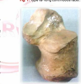
Type Ia- Sustent iculum tali w it h long cont inuous facet (Figure 1)
Type Ib- Transit ional figure 8 forms w it h fused anterior & medial facet s (Figure 2)
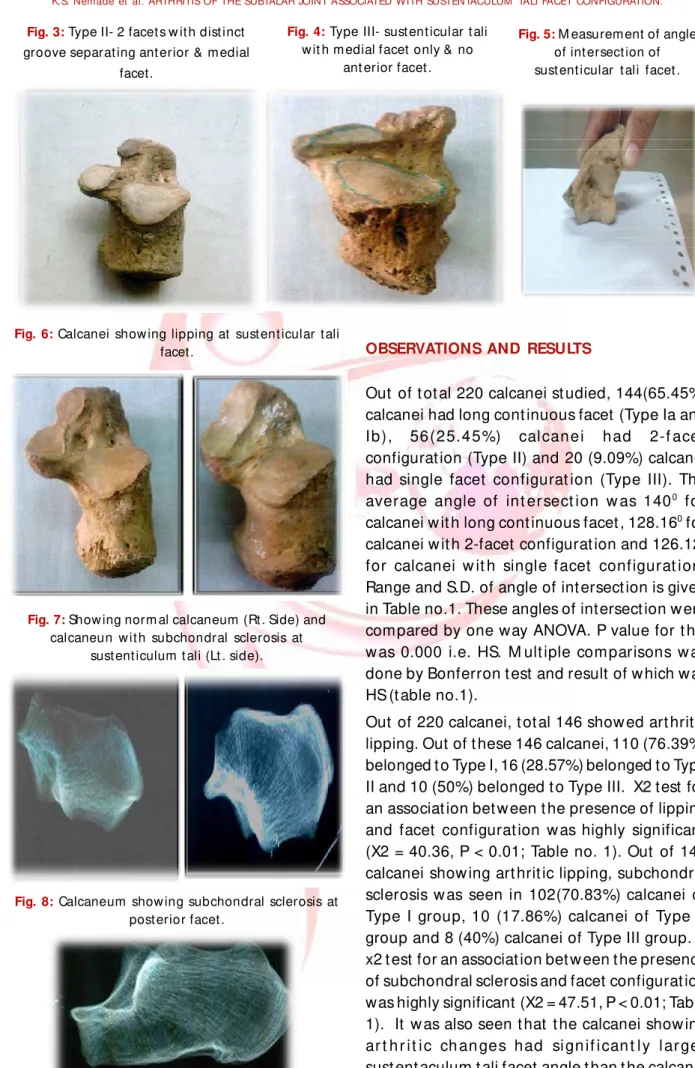
Type II- 2 facet s w it h dist inct groove separat ing anterior & medial facet s (Figure 3)
Type III- Sustent icular tali w it h medial facet only & no anterior facet (Figure 4)
The configurat ion in w hich all t alocalcaneal facet fused in one single facet w as not observed
Aft er classifying calcanei, angle of sust ent icular t alar f acet w as det er m ined. For t his, The calcaneus w as placed on it s medial side on a piece of paper so t hat t he medial border of t he sust ent aculum t ali facet s cont act ed t he paper and t he planes of t he facet s w ere perpendicular
t o t he surface of t he paper The cont our of t he f acet s w as t r aced w it h a pencil. The angle m easu red w i t h a p ro t r act o r. Rep eat ed observat ions w ere made on t he same series of bones indicat ed t hat t he average error of t his measurement is about 20 (Figure 5) [7].
Visual inspect ion of art icular surface w as made t o find art hrit ic changes consist ing periart icular rem odeling & eburnat ion. Calcanei show ing l i pp i n g w er e sub j ect ed t o r ad i ol o gical examinat ion t o find subchondral sclerosis.
Art hrit is w as scored as present if any of t hese o sseo us chan ges w as f ou n d o n t h e sust ent aculum t ali facet s, post erior t alocal-caneal facet or tarsal canal. (Figure 6 and 7).
Fig. 1: Type Ia- long cont inuous facet .
Int J Anat Res 2014, 2(4):684-88. ISSN 2321-4287
Fig. 3: Type II- 2 facet s w it h dist inct groove separat ing ant erior & m edial
facet.
Fig. 5: M easurem ent of angle of int ersect ion of sust ent icular t ali facet .
Fig. 8: Calcaneum show ing subchondral sclerosis at post erior facet .
Fig. 4: Type III- sust ent icular t ali w it h m edial facet only & no
ant erior facet .
Fig. 6: Calcanei show ing lipping at sust ent icular t ali facet.
Fig. 7: Show ing norm al calcaneum (Rt . Side) and calcaneun w it h subchondral sclerosis at
sust ent iculum t ali (Lt . side).
OBSERVATIONS AND RESULTS
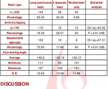
Out of t ot al 220 calcanei st udied, 144(65.45%) calcanei had long cont inuous facet (Type Ia and Ib), 56(25.45%) cal canei had 2-f acet configurat ion (Type II) and 20 (9.09%) calcanei had single facet configurat ion (Type III). The average angle of int er sect ion w as 1400 for
calcanei w it h long cont inuous facet , 128.160 for
calcanei w it h 2-facet configurat ion and 126.120
for calcanei w it h single facet configurat ion. Range and S.D. of angle of int ersect ion is given in Table no.1. These angles of intersect ion w ere compared by one way ANOVA. P value for t his w as 0.000 i.e. HS. M ult iple comparisons w as done by Bonferron t est and result of w hich w as HS (t able no.1).
Table 1: Tot al num ber and percentages of facet t ypes, art hrit ic lipping, degr ees of int ersect ing angles.
Facet t ype Long cont inuous
facet
2 separate face t
M edial facet only
St at ist ical analysis
n = 220 144 56 44
Percentage 65.45 25.45 9.09
Art hrit ic lipping
n= 146 110 16 10 Chi sq.=40.36
Psercent age 76.39 28.57 50 P < 0.01 (HS)
Subchondral
sclerosis 102 10 8 Chi sq.=47.51
Percentage 70.83 17.86 40 P < 0.01 (HS)
Int ersect ing angle
Average 140.6 128.16 126.12
M inim um 111 89 101
M aximum 167 145 151
S. D. 10.62 12.59 11.86
DISCUSSION
Acco r d in g t o t he p resent st u d y, Ty pe I configurat ion is most common configurat ion in In dian s. Ar t h r it ic chan ges li ke li ppi ng and subchondral sclerosis are significant ly less in calcanei belonging t o Type II Group t han ot hers. Thus result s of present st udy indicat es t hat subt alar joint st abilit y depends on sust ent icular t ali facet morphology and is consist ent w it h Buckner ’s hypot hesis that joint s w ith t he 2-facet configurat ion are comparat ively more st able [6]. How ever, Buckner has included calcanei having medial facet only configurat ion in Type I Group and not st udied t hem as a separat e group[6]. Result s of present st udy are also consistent wit h t he findings of Francine-Drayer Verhagen and M adhavi et .al [7,10].
In 2-separat e facet configurat ion, t alus and calcaneum art iculat e at t w o different locat ions. Articular surfaces are separat ed by a groove and t his result in t he format ion of V shaped art icular surface w it h an average angle of 128.160
. This l im i t s m edi al r o t at i o n o f t al ar h ead an d minimizes t he st rain on it during heel st rike. In cont rast t o t his, in Type I group, int ersect ing angle is obt use w hich put s more st rain on t alar head during m edial rot at ion. Event ually, talar head exert s cont inuous and excessive pressure o n sp r i n g l i gam en t w h ich co n n ect s sust ent iculum t ali w it h navicular leading t o ligament laxit y in t his configurat ion. Laxit y of l i gam ent s an d m u scles is t ho u ght t o b e r espo n si b le f o r m ob il e o r u nst ab l e f eet [ 4,11,18].
St abilit y of t he subt alar joint also depends on
t he height of t he longit udinal arch, w hich is det ermined by t he inclinat ion of t he subt alar joint axis [14,16]. A high arch represent s a more st able st ruct ure and is commonly referred t o as a ‘ r i gid ’ o r cavu s f oot [ 6,12,16] . Br u ckn er m easured inclinat ions of subt alar joint s, and found t hat joint s w it h t he 2-facet configurat ion have a higher subt alar joint axis t han t he ot her configurat ions [6]. This can be explained by t he analysis of int ersect ing angle of t hese facet s. Relat ive t o t he rounded, cont inuous facet , t he 2-facet configurat ion has an ant erior facet t hat is slant ed upw ards and raises t he t ot al subt alar joint axis. On t he ot her hand, t he cont inuous facet is horizont ally inclined, w hich result s in a low er arch and a less st able foot [4,6,18].
In Type III calcanei, int ersect ing angle and arthritic changes are midway bet ween other t wo groups. Here, as t here is only medial facet , t alar head is inadequat ely support ed w hich allow s excessive ant erior and inferior rot at ion of t he t alus during w eight bearing. This result s in a v algu s p o sit i o n o f t h e cal can eus and a dow nward t ilt of t he t alar head [4,16]. The vastly increased pressure on t he ant erior subt alar joint capsule causes ligament ous laxit y [17]. CT scans also show t hat t he planus foot (hypermobile or flat foot ) has no ant erior sust ent aculum t ali facet [19]. These are t he possible explanat ion of t hese changes in t his group.
CONCLUSION
Acco r d in g t o t he p resent st u d y, Ty pe I susent iculum t ali facet configurat ion is m ost com m only observed am ong Indians. Present st udy support s t he view t hat certain morphologi-cal variat ions of t he sust ent iculum t ali predis-pose people t o t he development of art hrit ic changes in t he subt alar joint . Present st udy also concludes t hat people w it h t he long cont inuous facet (t ype I) & medial facet only(t ype III) con-figurat ion have great er risk for subt alar joint inst abilit y t han individual w it h t he 2-f acet configurat ion(t ype II).
Conflicts of Interests: None
REFERENCES
Int J Anat Res 2014, 2(4):684-88. ISSN 2321-4287
How to cite this article
:
K.S. Nemade, M .M . M eshram, A. P. Kasote, N. Y. Kamdi. ARTHRITIS OF THE SUBTALAR JOINT ASSOCIATED WITH SUSTENTACULUM TALI FACET CONFIGURATION. Int J Anat Res 2014;2(4): 684-688. DOI: 10.16965/ ijar.2014.525
[2] . EL-EIshi. Variat ions in t he talar ar t icular facet s in Egypt ian calcanei. Act a Anat om ica.1974; 89: 134-138.
[3] . Gupt a S.C., Gupt a C.D., Arora A.K. Pat t ern of t alar ar t i cu lar f acet s in In di an cal can ei . Jo ur nal o f Anat om y 1977; 124: 651-655.
[4] . Kapandji I.A. The Physiology of t he Joint s, vol. 2, Low er Lim b Churchill Livingst one; New York 1970. [5] . Bunning PSC, Bar net t C.H. A com parison of adult f o et al t al o cal can eal ar t i cu l at i o n s. Jo u r n al o f Anat omy1965; 99: 71-76. Bruckner J. Variat ions in t he hum an subt alar joint . Journal of Ort hopaedic and Spor t s Physical Therapy 1987; 8: 489-494. [6] . Bruckner J. Variat ions in t he hum an subtalar joint .
Journal of Ort hopaedic and Sports Physical Therapy 1987; 8: 489-494.
[7] . Francine drayer -verhagen. Art hrit is of t he subt alar jo int associat ed w it h sust ent acu lum t ali f acet configurat ion. J. Anat 1993; 183: 631-634. [8]. John E. et al. Sprains- residual instabilit y of subtalar,
Lisfranc joint and t urf t oe. Clin. Sport s M edicine 2004; 23: 97-121.
[9]. Low ell Weil. Em phasizing careful assessm ent of t he et iol ogy and u nder lying cont r ibu t ing f act or t o subt alar joint inst abi lit y. Podiat r y t oday 2007; 20:5.
[10]. M adhavi et . al. Sout h Indian calcaneal t alar facet conf igur at ions and ost eoar t hrit ic changes. Clin Anat 2008 ;21(6):581-6.
[11]. Harr ingt on I.J. Biom echanics of joint injuries. In Biom echanics of M usculoskelet al Injur y E. Gozna; W illiam s & Wilkins1982: 31-69.
[12]. Glancy J. Ort hot ic control of ground react ion forces during running (a prelim inary repor t ). Or t hot ics and Prost het ics 1984; 38:12-40.
[13]. Laidlaw P.P. The os calcis. Journal of Anat omy 1905; 39: 161-177.
[ 14 ] . M an n R. A. Ov e r vi e w o f f o o t an d an kl e biom echanics. In Disor ders of t he Foot and Ankle: M edical and Surgical M anagem ent E. H. W ickland Jr; Philadelphia W. B. Saunders 1991: 385-408. [15]. Norkin C.C. Joint St ruct ure and Funct ion F. A. Davis;
Philadelphia 1983.
[16]. Perry J. Anat om y and biom echanics of t he hindfoot . Clinical Ort hopaedics and Relat ed Resear ch 1983; 177: 9-16.
[17]. Rose G.K. Pes planus. In Disorder s of t he Foot and Ankle: M edical and Surgical M anagem ent E. H. W ickland Jr ; Phi ladelphia W. B. Saunders 1991: 892-919.
[ 18] . Sam i lson R.L., Di llin W. Cavus, cavovar us and calcaneocavus. Clinical Or t hopaedics and Relat ed Research 1983; 177: 125-132.