R E S E A R C H
Open Access
Preparation and neutralization efficacy of
IgY antibodies raised against
Deinagkistrodon acutus
venom
Jinhua Liu
1, Qiyi He
1, Wenwen Wang
1, Bin Zhou
2, Bo Li
1, Yingfeng Zhang
1, Cong Luo
1, Diancheng Chen
1,
Jia Tang
1and Xiaodong Yu
1*Abstract
Background:The five-paced pit viper (Deinagkistrodon acutus), endemic to China and northern Vietnam, is responsible for most snakebites in the Chinese territory. Antivenom produced from horses is the main treatment for snakebites, but it may cause numerous clinical side effects and have other disadvantages involved in their production such as the welfare of animals. The present study was conducted aiming to develop an alternative antibody (IgY) from the egg yolk of leghorn chickens immunized with snake venom.
Methods:IgY from the egg yolk of white leghorn chickens previously immunized intramuscularly withD. acutus
venom was extracted by water, precipitated by ammonium sulfate and purified by affinity chromatographic system. IgY was identified by SDS-PAGE, ELISA and Western blot. Finally, IgY neutralization assays to test its efficacy against
hemorrhagic, edema-forming and myotoxic activities ofD. acutusvenom were conducted on mice.
Results:For the first time, IgY antibodies againstD. acutusvenom were raised successfully in egg yolk of chickens injected withD. acutusvenom multiple times. By three steps, including caprylic acid extraction, ammonium sulfate precipitation and affinity chromatography, IgY antibodies were isolated and purified from egg yolk, which exhibited a single protein band on SDS-PAGE and two bands (about 65 kDa and 35 kDa, respectively) under reducing conditions, and presented a high titer (1:40,000) tested by ELISA. Immunoblot analysis confirmed that these IgY
were polyclonal antibodies since they bound to components ofD. acutusvenom. Furthermore, immunodiffusion
assay showed that anti-D. acutusvenom IgY cross-reacted with the venoms ofTrimeresurus albolabrisand
D. saxatilisEmelianov, but did not react to the venoms ofBungarus multicinctusandNaja atra. In the
neutralizing lethal assay, the median effective dose of anti-D. acutusvenom IgY was 14.14 mg/kg of mouse
body weight under the challenge dose (3 LD50ofD. acutusvenom). In neutralizing the hemorrhagic, edema-forming and myotoxic activities ofD. acutusvenom, IgY showed the characteristic dose-dependent neutralization effects against all these toxic activities ofD. acutusvenom.
Conclusion:Anti-D. acutusvenom IgY antibodies with high purity and titer were for the first time raised successfully in egg yolk of chickens immunized withD. acutusvenom. They were effective in neutralizing the lethal effects, and the hemorrhagic, edema-forming and myotoxic acitivities ofD. acutusvenom. IgY could be an effective source to develop a treatment against snake bites in humans or animals in the future.
Keywords:IgY antibody, Egg yolk, Snake venom, Snakebite,Deinagkistrodon acutus, Venom neutralization
* Correspondence:yxd@cqnu.edu.cn
1Animal Toxin Group, Chongqing Key Laboratory of Animal Biology, Chongqing Engineering Research Center of Bioactive Substances, Engineering Research Center of Active Substances and Biotechnology, Ministry of Education, College of Life Science, Chongqing 401331, China Full list of author information is available at the end of the article
Background
Snake envenomation is an important public health issue in the world. There are more than 420 species of ven-omous snakes, which are widely distributed throughout the globe (including oceans), except for a few islands, frozen environments and high-altitude regions. These animals provokes 5.4 million bites, about 2.5 million of envenomation cases and over 0.125 million of deaths annually. As is well known, the highest burden of snakebites is found in South Asia, Southeast Asia and sub-Saharan Africa [1–4]. In addition, the burden of snakebites is heavy in China, where there are over 50 species of venomous snakes. One species, the five-paced pit viper (Deinagkistrodon acutus), is responsible for most snakebites in the country. It is a large sized and highly venomous snake, found from the southern provinces of China (including Taiwan) to northern Vietnam [5, 6].
The intravenous administration of antivenom is the mainstay treatment for envenomated victims. The con-ventional antivenom is generally produced from the blood of the large animals, frequently horses or sheep, immunized with the venom of a single snake or mixed venoms obtained from several animals to eliminate the intraspecific diversification [7]. Nevertheless, the serum used to treat snakebite victims may cause various side effects, such as anaphylactic shock, pyrogen reaction and serum sickness, which are mainly caused by the nonspe-cific proteins found in commercially available antive-noms [8–11]. Moreover, frequent immunization and bleeding during antivenom production may cause great distress to the employed animals. In addition, the pro-duction of sufficient pure serum antibodies against snake venom may require more investment and time, which may be conflicting with interests of serum manufac-turers, especially in developing countries. Thus, it is ne-cessary to find an alternative way of antibody creation, which would be safer, cheaper, short-cycle and non-invasive.
In recent years, eggs of chicken immunized with gens have been recognized as a good source of IgY anti-bodies [12–14]. So far, many research results about snake antivenom IgY have demonstrated that IgY in eggs of chicken is an excellent alternative to the conventional antivenom produced in large mammals [15, 16]. The main advantages of IgY include:
collecting eggs instead of bleeding animal meets the animal welfare requirement;
the cost of keeping hens is lower than that of raising horses or sheep;
IgY antibodies do not cross-react with Fc receptors, thereby reducing the cause of false positive results in immunological assays;
IgY, like mammalian IgG, is a reasonable stable protein over time;
the yield of IgY is large and its production may be readily scalable [17–20].
In this study, hens were hyperimmunized with low doses ofD. acutusvenom to produce specific antibodies. IgY was extracted from egg yolk and further purified by affinity chromatography. Subsequently, we assessed and examined the purity, the binding specificity, the titer and the neutralization efficiency of IgY. All the results will provide a basis for developing IgY into a clinical agent in the future.
Methods
Reagent and supplies
Freund’s complete adjuvant (FCA), Freund’s incomplete adjuvant (FIA), rabbit anti-IgY peroxidase conjugate, Immobilon®-P (polyvinylidene difluoride, PVDF), a trans-fer membrane with pore size of 0.45μm, 31,31,51,51
-tetra-methylbenzidine (TMB), nonfat dry milk and horseradish peroxidase-conjugated rabbit anti-chicken IgY were pur-chased from Sigma (USA). Polystyrene ELISA plates were purchased from Corning (USA). Amicon ultra-15 centri-fugal filter devices were obtained from Millipore (USA). NHS activated Sepharose 4 FF were purchased from GE Healthcare (UK). Protein molecular weight markers were purchased from Takara (Japan). All the other reagents were of analytical grade.
Venom
Venom was obtained from D. acutus captured at Wulingshan in Chongqing, China. Venom was lyophi-lized in a ModulyoD-230 freeze dryer (Thermo Scien-tific) and stored at−20 °C until use.
Animal
Seventeen-week-old white leghorn hens weighing 1.5 kg each, purchased from a local poultry farm, in good health and laying conditions (laying 5 to 6 eggs per week) were used for the production of IgY against snake venom. They were kept in individual cages with standard food and water. Kunming mice (18–20 g) were purchased from experimental animal center of Third Military Medical University. Mice were kept in plastic boxes at five per cage, in a room maintained at 20–23 °C on a 12/12-h light/dark cycle with food and water ad libitum.
Immunization schedule
Based on the LD50 ofD. acutus venom for mice (about
2.93 mg/kg, intraperitoneally), the LD50 of D. acutus
sites in the breast region with 0.5 mL saline (containing 0.29 mg snake venom) emulsified with an equal volume of FCA. On the 14th, 35th and 56th day after the first immunization, booster doses were administered with 0.5 mL saline (containing 0.58 mg, 1.17 mg and 1.17 mg snake venom, respectively) emulsified with an equal vol-ume of FIA. Serum was collected weekly from the first immunization, but after the 10th week serum was col-lected every 2 weeks. Eggs began to be colcol-lected daily before the first immunization and the collecting eggs sustained for 24 weeks after the first immunization. The control group of hens was immunized intramuscularly with 0.5 mL saline. Serum was stored at−20 °C and eggs
at 4 °C until use.
Extracting antibody from egg yolk
Extraction of IgY from preimmunized and hyperimmun-ized eggs was performed according to our previous method with minor modifications [21]. Briefly, the egg shell was cracked and the yolk was separated from the egg white. The yolk contents was diluted 7.5-fold with deionized water and homogenized by stirring vigorously for 30 min on magnetic stirrer. The resulting homogen-ate was further diluted 2-fold with 0.04 M acethomogen-ate buffer (pH 5.0, containing 0.06 M NaCl) and again homoge-nized for 30 min while adding caprylic acid up to final concentration of 1%. The preparation was placed at room temperature for 4 h. The clear supernatant, the water-soluble fraction (WSF), was siphoned out and centrifuged at 10,000 rpm for 10 min at 4 °C. The IgY in water-soluble fraction was precipitated out with 45% ammonium sulfate. The salt pellets were dissolved in phosphate buffered saline (PBS, pH 7.4) and dialyzed against PBS. Finally, the partially purified antibody preparation (crude extract) was subjected to affinity chromatography.
Affinity purification
The venom affinity column in chromatographic system (ÄKTA purifier 100, GE) was prepared as follows: in brief, NHS activated Sepharose 4FF were coupled with whole venom of D. acutus dissolved in coupling buffer (0.2 M NaHCO3, pH 8.3, containing 0.5 M NaCl).
Unreacted groups on the Sepharose were blocked with 0.5 M ethanolamine buffer (pH 8.3, containing 0.5 M NaCl). Finally, the Sepharose was washed with Tris–HCl buffer (0.05 M, pH 9.0) and then acetate buffer (0.05 M, pH 5.0). The Sepharose was packed into a column that was equilibrated with PBS buffer (0.01 M, pH 7.4). The crude extract (ant-D. acutusIgY, 20 mg/mL) was loaded into the affinity column, and the column was washed with PBS buffer (0.01 M, pH 7.4) to remove the un-absorbed proteins. The bound antibodies were eluted with glycine-HCl buffer (0.1 M, pH 2.3), and pooled,
concentrated and desalted through Amicon Ultra-15 centrifugal filter devices, then stored at 4 °C.
Protein concentration
Protein concentration was measured as described by Lowry et al. [22].
SDS-PAGE
The purity and molecular weight of the IgY were ana-lyzed on 12% sodium dodecyl sulfate-polyacrylamide gel electrophoresis (SDS-PAGE), according to the method of Laemmli [23].
Western blot
It was carried out according to the procedure of Towbin et al. [24] with minor modifications. D. acutus venom (50 μg) were separated on 15% non-reducing
SDS-PAGE, and then electroblotted onto PVDF membrane that was treated with methanol before. The unreacted sites on membrane were blocked with a blocking solu-tion (5% nonfat milk in PBS buffer, pH 7.4 and contain-ing 0.05% Tween-20) at room temperature for 2 h on a horizontal shaker, then incubated with the affinity-purified anti-D. acutusIgY for 1 h at room temperature. Next, the blot was washed five times with rinse buffer and incubated with horseradish peroxidase-conjugated rabbit anti-chicken IgY (1: 5000). Finally, the blots were washed and specific blots of venom proteins bound to IgY were visualized using peroxidase chromogenic sub-strate solution TMB.
Immunodiffusion assay
The specificity of IgY against snake venom components was demonstrated with agar diffusion test (1% agarose) described by Ouchterlony [25]. D. acutus venom was added into the center well in an agar plate, the different concentrations of anti-D. acutus IgY antibodies were loaded into the peripheral wells, and then the plate was incubated for 24 h at 37 °C to observe the antigen-antibody reactive lines. In addition, the cross reactivity between anti-D. acutusIgY and four other snake venoms (Trimeresurus albolabris, A. saxatilis, Bungarus multi-cinctusandNaja atrafrom China) was also evaluated by this test.
ELISA assay
ELISA was used to assess the activities of anti-D. acutus IgY antibodies in serum and the yolk [21]. The micro-plates were coated with 1μg native D. acutusvenom in
100 μL coating buffer (0.05 M carbonate bicarbonate,
milk). The coated wells were washed again, and then were added to the serum or the yolk diluted in dilution buffer (PBS plus 1% nonfat dry milk) and incubated 1 h at 37 °C. After the wells were washed three times, they were added to the rabbit anti-IgY-peroxidase dilution (1:5000) and incubated for 1 h at 37 °C. After the wells were washed once again, 200 μL of TMB was added
and incubated at room temperature for 20 min. The re-actions were terminated with 50 μL of 2 M sulfuric
acid. Absorbance was recorded at 450 nm using ELISA plate reader. Results determination: a positive sample and a negative sample need to give a ratio of at least 2.1 (that is, OD of the positive divided by OD of the negative. P/N > 2.1).
Neutralization of venom lethality by IgY
The ability of IgY to neutralize lethality of D. acutus venom was assessed by an in vivo neutralization test. Various amounts of the purified antivenom IgY (50 μg,
100 μg, 200 μg, 400 μg, 800 μg and 1600 μg,
respect-ively) were mixed with the challenge dose (3 LD50 dose
ofD. acutusvenom) and incubated for 30 min at 37 °C, centrifuged for 5 min, then 20μL of the supernatant was
injected intraperitoneally into groups of 10 mice. Con-trol mice received the same amount of venom mixed with the normal yolk. The deaths were recorded over 72 h. The ED50 was calculated according to the
recom-mendation of WHO [26] and expressed as mg IgY/kg body weight.
Neutralization of hemorrhagic activity by IgY
Hemorrhagic activity was quantitatively determined ac-cording to the method of Gutiérrez et al. [27]. Various amounts of venom (0, 5 μg, 10 μg, 15 μg, 20 μg and
25μg) diluted in 0.1 mL physiological saline (0.9% w/v)
were injected subcutaneously into the dorsal skin of groups of four mice. Two hours later, they were sacrificed and their dorsal skin was removed, and the diameter of the hemorrhagic spot was measured. Physiological saline was utilized as negative control. Diameters were calculated and the minimum hemorrhagic dose (MHD) was defined as the dose of venom that induced a lesion of 10 mm of diameter 2 h after injection. The challenge doses of venom (3 MHD) was incubated with various amounts of IgY anti-bodies (0, 500 μg, 1000 μg, 1500 μg, 2500 μg, and
3500μg) for 30 min at 37 °C, centrifuged for 5 min. Then,
10μL of the supernatant was subcutaneously injected into
the dorsal skin of groups of ten mice. Control group re-ceived normal yolk (the same amount of used for venom). Hemorrhage was expressed as a percentage, taking as 100% the diameter of the lesions induced by inoculating 10 MHD of venom alone. Results were plotted and the ED50was defined as the ratio of IgY/venom that decreased
the activity by 50%.
Neutralization of edema-forming activity by IgY
Edema-forming activity was quantitatively determined according to the method of Van Dong et al. [28]. There are groups of 4 mice injected subcutaneously, in the right footpad, with 20 μL of various amounts of venom
(0.1 μg, 0.5 μg, 1 μg, 1.5 μg and 2.0 μg) dissolved in
physiological saline(0.9% w/v), and in the left footpad re-ceived 20 μL of the physiological saline. The weight of
each foot was determined 3 h after injection with an electrionic scale. The minimum edema-forming dose (MED) corresponds to the amount of venom that in-duces an increment of 30% in the weight of the foot envenomated for 3 h when compared with the physio-logical saline-injected foot. The challenge doses of venom (3 MED) was incubated with various amounts of the IgY antibodies (25 μg, 50 μg, 100 μg, 150 μg and
200μg) for 30 min at 37 °C, centrifuged for 5 min, then
20 μL of the supernatant was injected subcutaneously
into groups of 10 mice in the right footpad.
Neutralization of myotoxic activity by IgY
Myotoxic activity was quantitatively determined accord-ing to the method of Rojas et al. [29]. Various amounts of venom(0μg, 5μg, 10μg, 15μg, 20μg and 25μg)
dis-solved in 50 μL of physiological saline (0.9% w/v) were
injected intramuscularly (i.m.) into groups of 4 mice in the right gastrocnemius muscle. Control animals re-ceived 50 μL of the physiological saline. Three hours
after injection, mice were bled from the orbital plexus, under CO2 anesthesia. Plasma creatine kinase (CK)
ac-tivity was quantitated by the CK kit (Nanjing Jiancheng Corp. China). CK activity is expressed in units/L, 1 U defined as the amount of enzyme that produces 1μmol
of NADH under the conditions of the assay. The mini-mum myotoxic dose (MMD) corresponds to the amount of venom that induces an increment of plasma CK activ-ity corresponding to four times the activactiv-ity of mice injected with physiological saline alone. The challenge doses of venom (3 MMD) was incubated with various amounts of the IgY antibodies (0, 375 μg, 750 μg,
1500 μg and 3000 μg) for 45 min at 37 °C and
centri-fuged for 5 min. Then, 50 μL of the supernatant was
injected subcutaneously into groups of ten mice in in the right gastrocnemius muscle.
Statistical analysis
Results
Antibody response
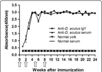
Following primary immunization, the hens produced comparable antibody response that was detected in serum by day 8. Although the pre-booster response was low in the serum, after the first booster there was a sharp increase in antibody titer both in serum and egg yolk. Three weeks after primary immunization, transfer-ence of antibodies specific to venom from serum to egg yolk was observed. At the 4th week, the antibody re-sponse both in serum and yolk reached the highest level and maintained thereafter at least for 20 weeks (Fig. 1).
Extraction, purification and biochemical identification of IgY
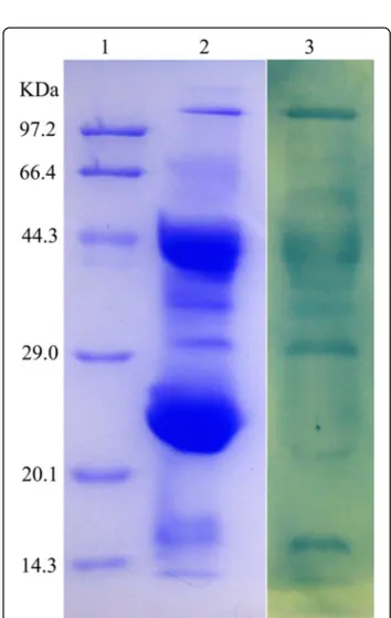
The partially purified antibody preparation (crude extract) was obtained from the egg yolk by the water-soluble extraction method; then, it was fractioned by affinity chromatography into two peaks with strong IgY activity. They exhibited only one band (about 182 kDa) on SDS-PAGE under non-reducing condi-tions. However, under reducing conditions, they pre-sented two bands, a heavy chain of IgY of about 66 kDa and a light chain of IgY of about 25 kDa (Fig. 2). Western blot showed that the purified sample could be recognized by HRP-rabbit anti-chicken IgY (Fig. 2). The average recovery of venom-specific IgY from 131 eggs was about 18.47% (Table 1). The venom-specific activities of IgY on different fractions–
including water-soluble fraction (WSF), salting-out fraction (SOF) and thiophilic-chromatography fraction (TCF)–were compared by ELISA. The results showed that the titer of TCF is twofold higher than that of SOF, and 16 times higher when compared with to
WSF. Obviously, the three purification steps used re-sulted in the enrichment of venom-specific IgY.
Immunological identification of IgY’s crude extract
Thirty micrograms ofD. acutusvenom was added to the central well, and the serial dilutions of the crude extract were added to the peripheral wells, respectively. The white precipitation lines occurred between the central well and each peripheral well indicated the immuno-logical activities of IgY (Fig. 3). The titer of the crude ex-tract by immunodiffusion was about 1/8. About 1200μg
of crude extract was added to the central well and the five different snake venoms (stored in our laboratory) were placed in the peripheral wells. The results indicated that the venoms of three species (T. albolabris, D. acu-tus and D. saxatilis in China) of Viperdae could react with the anti-D acutus IgY, but the venoms of two spe-cies (B. multicinctus and N. atra in China) of Elapidae could not (Fig. 3).
Evaluation of purified IgY titer
According to the results of ELISA, the titer of specific anti-D. acutusvenom IgY (0.3 mg/mL), which had been concentrated and desalted by ultrafiltration, was 1:40000 (Fig. 4).
Antigen recognition repertoire of IgY
Western blot analysis was carried out by using anti-D. acutusvenom IgY as the first antibody and using HRP-rabbit anti-chicken IgY as the second antibody. The results obtained demonstrated that not all protein components of D. acutus venom were recognized by IgY (Fig. 5).
Fig. 1Primary and secondary antibody response in serum and egg yolk of hens immunized withD. acutusvenom. The activity against
D. acutusvenom of serum and WSF was assessed by ELISA. The
white arrowsindicate immunization
Fig 2IgY samples from egg yolk analyzed by 12% SDS-PAGE and identified by Western blot. Lane 1 and lane 2: 10μg of IgY after
affinity chromatography (reducing and non-reducing conditions, respectively). Lane 3: 15μg of IgY’s crude extract. Lane 4: molecular
Neutralization studies of anti-D. acutusvenom IgY
Anti-D. acutus venom IgY that was in incubation with the venom prior to injection was capable of neutralizing the toxic components of venom. There was an increase in the survival rate of mice with increase in antivenom IgY. The proportion of 40 mg of IgY/kg of mouse body weight could produce 100% protection against a 3 LD50
dose (3 × 2.93 mg/kg) of venom and the value of ED50
was 14.14 mg IgY/kg. There were no survivals in the control group.
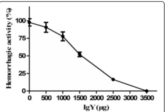
Concerning hemorrhagic activity, about 10 μg of D.
acutus venom produced a hemorrhagic spot of 10 mm diameter (MHD). It was estimated that 350 μg of IgY
was able to completely neutralize the hemorrhage in-duced by the challenge doses of venom (3 MHD), whereas 150μg of IgY was able to neutralize about 50%
of the hemorrhagic effects. Control mice injected with physiological saline or IgY showed no hemorrhage, re-spectively (Fig. 6).
Regarding the edema-forming activity, mice immu-nized withD. acutusvenom showed increase in footpad weight. About 1.3μg ofD. acutusvenom induced edema
formation within 3 h, which is considered as MED value. When anti-D. acutusvenom IgY was incubated with the challenge doses of venom (3 × MED) for 30 min prior to injection, IgY antibodies were capable of inhibiting edema-forming activity induced by venom in a
dose-Table 1Recovery ratio of proteins and titer of IgY fractions
Fractions Titer of IgY by
ELISA (× 104) Yolk proteins in131 eggs (g) Recovery ratioof proteins (%)
WSF 3.3 31.83 100
SOF 25.7 12.59 39.56
TCF 51.3 5.88 18.47
WSFWater-soluble fraction, SOF Salting-out fraction,TCF
Thiophilic-chromatography fraction
Fig 3Immunological assessment of anti-D. acutusvenom IgY against venom from five species of snakes found in China by immunodiffusion assay.aIn the central well (0):D. acutusvenom (30μg); in the peripheral wells: (1) anti-D. acutusIgY crude extract
(1200μg); (2–5) the serial dilutions (1:2, 1:4, 1:8, and 1:16, respectively)
of the crude extract; (6) physiological saline.bandcIn the central well (0) anti-D. acutusIgY crude extract (1200μg) whereas the the
peripheral wells contained 30μg of the following: (1)T. albolabris
venom; (2)D. acutusvenom; (3)D. saxatilisEmelianov venom; (4)B. multicinctusvenom; (5)N. atravenom; and (6) physiological saline
Fig 4Titer profile of IgY raised againstD. acutusvenom by ELISA. When the value of P/N is bigger than 2.1, the sample was positive (n= 3)
Fig 5Antigen recognition repertoire of anti-D. acutusvenom IgY by Western blot. Lane 1: Molecular weight marker on SDS-PAGE. Lane 2:D. acutusvenom (50μg) on SDS-PAGE. Lane 3: the
protein components ofD. acutus venom(50μg) on Western blot
dependent manner (Fig. 7), and ED50 of IgY was about
124.68μg.
In myotoxic activity, mice immunized with D. acutus venom showed increase in plasma CK activity. About 9.5 μg of D. acutus venom induced myotoxic activity,
which is considered a MMD value. When anti-D. acutus venom IgY was incubated with the challenge doses of venom (3 × MMD) for 30 min prior to injection, IgY
antibodies were capable of inhibiting CK activity induced by venom in a dose-dependent manner (Fig. 8), and ED50of IgY was about 766.43μg.
Discussion
The venom ofD. acutus, a snake endemic to China, pos-sess proteins and peptides whose activity is mainly hemotoxic [28]. At the bite site, swelling, bruising, blis-tering and necrosis usually develop within a few minutes or hours and spread rapidly, sometimes the whole limb is affected [6]. The persistent bleeding from the fang marks and other previous partially healed wounds indi-cates coagulopathy, which is caused by the abundant metalloproteinases and serine proteases of D. acutus venom [5, 6]. Its toxicity and pathophysiologic effects can be completely reversed by specific antivenom, which is conventionally produced from blood of large animals (horses or sheep) immunized with snake venoms [8, 10]. As an alternative to the conventional antivenom with various side effects and the disadvantages of its prepar-ation and production, antivenom IgY antibodies from egg yolk of chicken have considerable advantages [8, 16, 29]. Previous studies have shown that chicken immu-nized with snake venom produces IgY that may neutralize the toxic and lethal effects of venom and may serve to treat domestic animals affected by snakebites [16, 21, 30]. The current study for the first time de-scribed the creation of anti-D. acutus venom IgY anti-bodies in chicken egg yolk and assessed their efficacy in neutralizing the lethal effect and other activities of D. acutusvenom.
The antibody production started in serum eight days after the first injection of D. acutus venom and in egg
Fig 6Neutralization of hemorrhagic effects ofD. acutusvenom by IgY (mean ± SD,n= 4). Mixtures containing a constant amount of venom and various dilutions of IgY were incubated at 37 °C for 30 min. Afterwards, aliquots of the mixtures (10μL containing 3
MHD of venom) were injected subcutaneously into the dorsal skin of mice, whereas control group received the same amount of venom with normal yolk. Hemorrhage was expressed as a percentage, taking as 100% the diameter of the lesions induced by inoculating 10 MHD of venom alone
Fig 7Neutralization of edema-forming activity ofD. acutusvenom by IgY (mean ± SD,n= 4). Mixtures containing a constant amount of venom and various dilutions of IgY were incubated at 37 °C for 30 min. Afterwards, aliquots of the mixtures (20μL containing three
MED of venom) were injected into the right footpad of mice, whereas the left foot pad received 20μL of physiological saline.
The weight of both feet was estimated 3 h after injection with an electronic scale and edema was expressed as the percentage increment in weight of the right footpad when compared to the left one
Fig 8Neutralization of myotoxic activity ofD. acutusvenom by IgY (Mean ± SD,n= 4). Mixtures containing a constant amount of venom and various dilutions of IgY were incubated at 37 °C for 30 min. Then, aliquots of the mixtures (50μL containing 3 MMD of
venom) were injected into the right gastrocnemius muscle of mice, whereas control group received 50μL of physiological saline. CK
yolk at the 15th day, increasing progressively along the immunization procedure, and attaining a plateau after the second booster, which was maintained thereafter (Fig. 1). This antibody response induced by D. acutus venom is for the first time obtained and is in good ac-cordance with the antibody response of other snake venoms reported [21, 29, 30].
As shown by Duan et al. [21], three steps were chosen to extract and purify the specific IgY antibodies from egg yolk. The average recovery of venom-specific IgY from 131 eggs was about 18.47% and 5.88 g of pure IgY was obtained (Table 1). The anti-D. acutus IgY antibodies generated were pure and specific to HRP-rabbit anti-chicken IgY, which was revealed by SDS-PAGE and Western blot analysis (Fig. 2). It was further confirmed by Western blot analysis that the anti-D. acutusIgY anti-bodies were able to recognize and bind to most protein components of D. acutus venom (Fig. 5). This suggests that the obtained IgY is a specific polyclonal antibody againstD. acutusvenom.
The immunodiffusion assay indicated the anti-D. acu-tus IgY not only reacted against D. acutus venom, but also againstT. albolabrisand D. saxatilisvenoms. How-ever, it did not recognize B. multicinctus or N. atra venoms (Fig. 4). The results indicated that venoms ofD. acutus, T. albolabrisandD. saxatilis of the same Viperi-dae family possibly share some common antigen epi-topes. Conversely, they did not share any antigen epitopes with venoms of B. multicinctus or N. atra of the Elapidae family. These preclinical observations will provide some reference for clinicians to use antivenom or antibodies to treat bites caused by snakes from of the same family [5, 6].
According to the proteomic analysis, there were about 128 kinds of proteins and peptides identified in the venom of D. acutus [31]. The Western blot analysis showed that anti-D. acutusvenom IgY antibodies mainly recognized a molecular weight range of the protein com-ponents in the venom (including > 97.2 kDa, 66.4-29 kDa, 18–14.3 kDa), but did identify not other protein components (such as 20.1-29 kDa) (Fig. 6). Generally, protein or polypeptide components in such a mixture as crude venoms differ considerably in their abilities to elicit antibody response in immunized animals [16].
Our results indicated that protein or polypeptide com-ponents with molecular weight between 20.1 and 29 kDa in D. acutus venom possibly lacked some immunogen-icity for chickens. According to their biochemical characterization, the identified proteins in D. acutus venom were divided into three groups: serine proteases; P-I class snake venom metalloproteinases (SVMPs); and other proteins [32–34]. It was reported that the serine proteases and metalloproteinases in D. acutus venom, those whose molecular weight was distributed in the
range of > 97.2 kDa, 66.4-29 kDa and 18–14.3 kDa, con-tributed to the major immunogenicity ofD. acutusvenom and underpinned the hemorrhagic, edema-forming and myotoxic acitivities [31]. Our observations confirmed that the polyvalent IgY antibodies raised against D. acutus venom are effective in the neutralization of the most im-portant toxic effects including lethal, hemorrhagic, edema-forming and myotoxic acitivities of D. acutus venom (Fig. 6, 7 and 8). In addition, the neutralization showed a characteristic dose-dependent relationship.
Conclusion
In summary, IgY antibodies againstD. acutusvenom with high purity and titer were for the first time raised success-fully in egg yolk by immunizing hens with snake venom. They were effective in neutralizing lethal, hemorrhagic, edema-forming and myotoxic acitivities of D. acutus venom. IgY could be an effective source to develop alter-native treatment for snakebite victims in the future, either humans or other animals. However, further studies are re-quired for testing the safety and efficacy of IgY.
Abbreviations
CK:Creatine kinase; FCA: Freund’s complete adjuvant; FIA: Freund’s incomplete adjuvant; MED: Minimum edema-forming dose; MHD: Minimum hemorrhagic dose; MMD: Minimum myotoxic dose; OD: Optical density; PVDF: Polyvinylidene difluoride; SDS-PAGE: Sulfate-polyacrylamide gel electrophoresis; SOF: Salting-out fraction; SVMP: Snake venom
metalloproteinase; TCF: Thiophilic-chromatography fraction; TMB: 31,31,51,51 -tetramethylbenzidine; WSF: Water-soluble fraction.
Acknowledgments
The authors would like to thank the Natural Science Foundation of Chongqing (cstc2014yykfA0221, cstc2015shmszx1225), Chongqing Forestry Department (Yulin keyan 2015–5) for supporting the present study.
Funding
This work was supported by Natural Science Foundation of Chongqing (cstc2014yykfA0221, cstc2015shmszx1225), Chongqing Forestry Department (Yulin keyan 2015–5).
Authors’contributions
J-HL, Q-YH and W-WW equally and mainly contributied to this study, X-DY provided the idea, gave supervision for experimental designs and finalized the manuscript, other authors helped in all the experiments during the study period. All authors read and approved the final manuscript.
Competing interests
The authors declare that they have no competing interests.
Consent for publication
Not applicable.
Ethics approval and consent to participate
All experimental procedures involving animals were carried out in accordance with the Chinese Animal Welfare Act. The present study was approved by Chongqing Municipal Public Health Bureau (protocol n. cstc2015).
Publisher’s Note
Author details
1Animal Toxin Group, Chongqing Key Laboratory of Animal Biology, Chongqing Engineering Research Center of Bioactive Substances, Engineering Research Center of Active Substances and Biotechnology, Ministry of Education, College of Life Science, Chongqing 401331, China. 2Library, Chongqing Normal University, Chongqing 401331, China.
Received: 7 November 2016 Accepted: 28 March 2017
References
1. Kasturiratne A, Wickremasinghe AR, de Silva N, Gunawardena NK, Pathmeswaran A, Premaratna R, et al. The global burden of snakebite: a literature analysis and modelling based on regional estimates of envenoming and deaths. PLoS Med. 2008;5(11):1591–604.
2. Chippaux JP. Snake-bites: appraisal of the global situation. Bull World Health Organ. 1998;76(5):515–24.
3. Habib AG. Public health aspects of snakebite care in West Africa: perspectives from Nigeria. J Venom Anim Toxins incl Trop Dis. 2013;19(1):27. doi:10.1186/1678-9199-19-27.
4. Theakston RD, Warrell DA, Griffiths E. Report of a WHO workshop on the standardization and control of antivenoms. Toxicon. 2003;41(5):541–57. 5. Zhao EM. Snakes of China. Hefei: Anhui Science and Technology Publishing
House; 2006. p. 1–369.
6. Qin GP. China poisonous snake research. Nanning: Guangxi Science and Technology Press; 1998.
7. Bochner R. Paths to the discovery of antivenom serotherapy in France. J Venom Anim Toxins incl Trop Dis. 2016;22:20. doi:10.1186/s40409-016-0074-7. 8. Meenatchisundaram S, Parameswari G, Michael A, Ramalingam S. Studies on
pharmacological effects of Russell’s viper and Saw-scaled viper venom and its neutralization by chicken egg yolk antibodies. Int Immunopharmacol. 2008;8(8):1067–73.
9. Polson A, von Wechmar MB, Fazakerley G. Antibodies to proteins from yolk of immunized hens. Immunol Commun. 1980;9(5):495–514.
10. Carroll SB, Thalley BS, Theakston RD, Laing G. Comparison of the purity and efficacy of affinity purified avian antivenoms with commercial equine crotalid antivenoms. Toxicon. 1992;30(9):1017–25.
11. Karlson-Stiber C, Persson H. Antivenom treatment inVipera berus
envenoming–report of 30 cases. J Intern Med. 1994;235(1):57–61. 12. Ferreira Júnior A, Santiago FM, Silva MV, Ferreira FB, Macêdo Júnior AG,
Mota CM, et al. Production, Characterization and Applications for
Toxoplasma gondii-Specific Polyclonal Chicken Egg Yolk Immunoglobulins. PLoS One. 2012;7(7):e40391. doi:10.1371/journal.pone.0040391.
13. Cova L. DNA-designed avian IgY antibodies: novel tools for research, diagnostics and therapy. J Clin Virol. 2005;34(Suppl):70–4.
14. Carlander D, Kollberg H, Wejåker PE, Larsson A. Peroral immunotheraphy with yolk antibodies for the prevention and treatment of enteric infections. Immunol Res. 2000;21(1):1–6.
15. Aguilar I, Sanchez EE, Giron ME, Estrella A, Guerrero B, Rodriguez-Acosta FA. Coral snake antivenom produced in chickens (Gallus domesticus). Rev Inst Med Trop Sao Paulo. 2014;56(1):61–6.
16. Almeida CM, Kanashiro MM, Rangel Filho FB, Mata MF, Kipnis TL, da Silva WD. Development of snake antivenom antibodies in chickens and their purification from yolk. Vet Rec. 1998;143(21):579–84.
17. Schade R, Calzado EG, Sarmiento R, Chacana PA, Porankiewicz-Asplund J, Terzolo HR. Chicken egg yolk antibodies (IgY-technology): a review of progress in production and use in research and human and veterinary medicine. Altern Lab Anim. 2005;33(2):129–54.
18. Karlsson M, Kollberg H, Larsson A. Chicken IgY: utilizing the evolutionary advantage. Worlds Poult Sci J. 2004;60(3):341–8.
19. Dias da Silva W, Tambourgi DV. IgY: a promising antibody for use in immunodiagnostic and in immunotherapy. Vet Immunol Immunopathol. 2010;135(3–4):173–80.
20. Zhang WW. The use of gene-specific IgY antibodies for drug target discovery. Drug Discov Today. 2003;8(8):364–71.
21. Duan HL, He QY, Zhou B, Wang WW, Li B, Zhang YZ, et al. Anti-Trimeresurus albolabrisvenom IgY antibodies: preparation, purification and neutralization efficacy. J Venom Anim Toxins incl Trop Dis. 2016;22(1):23. doi:10.1186/ s40409-016-0078-3.
22. Lowry OH, Rosebrough NJ, Farr AL, Randall RJ. Protein measurement with the Folin phenol reagent. J Biol Chem. 1951;193(1):265–75.
23. Laemmli UK. Cleavage of structural proteins during the assembly of the head of bacteriophage T4. Nature. 1970;227(5259):680–5.
24. Towbin H, Staehelin T, Gordon J. Electrophoretic transfer of proteins from polyacrylamide gels to nitrocellulose sheets: procedure and some applications. Proc Natl Acad Sci U S A. 1979;76(9):4350–4.
25. Ouchterlony O. Antigen-antibody reactions in gels. Acta Pathol Microbiol Scand. 1949;26(4):507–15.
26. W.H.O. Progress in the Characterization of Venoms and Standardization of Antivenoms; Offset Publication, Ed. Geneva: World Health Organization; 1981;58:1-44.
27. Gutiérrez JM, Gené JA, Rojas G, Cerdas L. Neutralization of proteolytic and hemorrhagic activities of Costa Rican snake venoms by a polyvalent antivenom. Toxicon. 1985;23(6):887–93.
28. Van Dong L, le Quyen K, Eng KH, Gopalakrishnakone P. Immunogenicity of venoms from four common snakes in the South of Vietnam and development of ELISA kit for venom detection. J Immunol Methods. 2003;282(1–2):13–31. 29. Rojas E, Quesada L, Arce V, Lomonte B, Rojas G, Gutiérrez JM. Neutralization
of four PeruvianBothropssp. snake venoms by polyvalent antivenoms produced in Perú and Costa Rica: preclinical assessment. Acta Trop. 2005; 93(1):85–95.
30. Paul K, Manjula J, Deepa EP, Selvanayagam ZE, Ganesh KA, Subba Rao PV. Anti-Echis carinatusvenom antibodies from chicken egg yolk: isolation, purification and neutralization efficacy. Toxicon. 2007;50(7):893–900. 31. Chunhong H, Xiaodong H, Yi K, Wutong W. Coupled chromatography for
assay of the venom proteome of the snakeAgkistrodon acutus: an effective strategy for discovery of active components. Chromatographia. 2009;69(9–10): 879–86.
32. Zhu Z, Gong P, Teng M, Niu L. Purification, N-terminal sequencing, partial characterization, crystallization and preliminary crystallographic analysis of two glycosylated serine proteinases fromAgkistrodon acutusvenom. Acta Crystallogr D Biol. 2003;59(Pt 3):547–50.
33. Wang YM, Wang SR, Tsai IH. Serine protease isoforms ofDeinagkistrodon acutusvenom: cloning, sequencing and phylogenetic analysis. Biochem J. 2001;354(Pt 1):161–8.
34. Lou Z, Hou J, Liang X, Chen J, Qiu P, Liu Y, et al. Crystal structure of a non-hemorrhagic fibrin (ogen) olytic metalloproteinase complexed with a novel natural tri-peptide inhibitor from venom ofAgkistrodon acutus. J Struct Biol. 2005;152(3):195–203.
• We accept pre-submission inquiries
• Our selector tool helps you to find the most relevant journal • We provide round the clock customer support
• Convenient online submission • Thorough peer review
• Inclusion in PubMed and all major indexing services • Maximum visibility for your research
Submit your manuscript at www.biomedcentral.com/submit