Corresponding author: Dra. Fabiana da Rocha Oliveira.
e-mail: [email protected]
Received 25 October 2016
Accepted 11 April 2017
Biological and molecular properties of yellow venom of the
Amazonian coral snake Micrurus surinamensis
Fabiana da Rocha Oliveira
[1], Maria das Dores Nogueira Noronha
[2]and Jorge Luis Lopez Lozano
[2][1]. Laboratório de Ecologia e Biotecnologia de Microrganismos da Amazônia, Instituto Nacional de Pesquisas da Amazônia, Manaus, AM, Brasil.
[2]. Centro de Oidismo da Amazônia, Fundação de Medicina Tropical Doutor Heitor Vieira Dourado, Manaus, AM, Brasil.
Abstract
Introduction: The coral snake Micrurus surinamensis, which is widely distributed throughout Amazonia, has a neurotoxic venom. It is important to characterize the biological and molecular properties of this venom in order to develop effective antitoxins. Methods: Toxins from the venom of M. surinamensis were analyzed by two-dimensional polyacrylamide gel electrophoresis and their neurotoxic effects in vivo were evaluated. Results and Conclusions: Most proteins in the venom had masses < 14kDa, low
phospholipase A2 activity, and no proteolytic activity. The toxins inhibited the coagulation cascade. The venom had neurotoxic effects in mice, with a median lethal dose upon intravenous administration of 700 µg/kg. Immunogenic studies revealed abundant cross-reactivity of antielapidic serum with 14kDa toxins and limited cross-reactivity with toxins < 10kDa. These results indicate
that antielapidic serum against M. surinamensis venom has weak potency (0.35mg/ml) in mice.
Keywords: Micrurus surinamensis. Neurotoxins. Phospholipase A2. Biological activities.
INTRODUCTION
Approximately 800 species of reptiles have been identiied
throughout the Brazilian territory; more than 50% of these
are snakes including coral snakes, which are members of
the Elapidae family1 comprising approximately 40 genera2. Only the genera Micruroides Schmidt, 1928 (North America); Leptomicrurus Schmidt, 1937 (South America); and Micrurus
Wagler, 1824 are found in the Americas3,4 in fossorial or aquatic
habitats. Coral snakes are small animals, ranging from 20cm to just over 1m in length, and have ixed venom inoculators of the proteroglyphous type. In general, they do not attack; poisoning by these snakes only occurs when they are handled or trampled5.
Hence, envenomation is rare although the effects can range from
mild to severe, since the venom is highly neurotoxic; symptoms
include myasthenia (e.g., weakness and ptosis) that can evolve
to paralysis and respiratory failure6–8.
The venom components responsible for its toxicity include phospholipase (PL) A2 and neurotoxins. Both of these have low molecular masses (< 15kDa) and are neuro- and myotoxic9–11. PLA2 may also have coagulant/anticoagulant effects12–16.
Neurotoxins are categorized as β-neurotoxins (presynaptic),
α-neurotoxins (postsynaptic), cardiotoxins, and weak neurotoxins depending on their mode of action. β-neurotoxins block acetylcholine (ACh) release after neurotransmission and thereby prevent action potentials, whereas α-neurotoxins
compete with ACh at nicotinic cholinergic receptors of motor endplates. Both act at the neuromuscular junction where they
block nerve impulses and cause total paralysis of skeletal
muscle17–21. Cardiotoxins induce muscle cell depolarization and contraction and cell membrane disruption, damaging erythrocytes and epithelial cells22; weak neurotoxins have effects
similar to those of α-neurotoxins but are less toxic23.
Six species of coral snake have been identiied as medically
important in the State of Amazonas in Brazil: Micrurus averyi, Micrurus iliformes, Micrurus hemprichii, Micrurus
lemniscatus, Micrurus spixii, and Micrurus surinamensis. These
species are associated with low mortality rates in humans24,25.
Given the diversity and abundance of coral snakes across the
northern region of Brazil, biochemical studies of their venom have attracted wide interest.
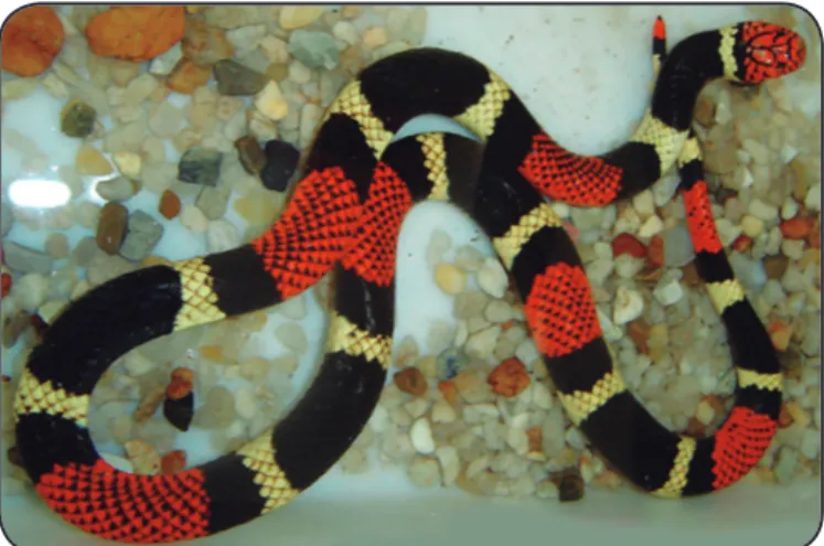
Micrurus surinamensis has red cephalic scales with black
borders and a red/black/yellow ring pattern, with the black rings arranged in perfect triads (Figure 1). Micrurus surinamensis
lives in swampy areas (igapós) and streams in the primary forest area and feeds primarily on ish, but has a broad geographic distribution that includes southeastern Venezuela; Guianas; and
the Amazonian regions of Colombia, Ecuador, Peru, Bolivia,
FIGURE 1 - Micrurus surinamensis (Cuvier, 1817). Photo: Prof. Paulo F. Bürnheim, 1998.
of other Micrurus species in terms of enzymatic composition28,29,
as evidenced from the protein expression proi le. In the present
study, we investigated the molecular composition and biological and neurotoxic activities of the venom of M. surinamensis, as well as the capacity of antielapidic serum to neutralize the neurotoxicity and lethal effects of this venom.
METHODS
Animals and venom
Mice (BALB/cJ) weighing 18-22g were provided by
Professor Paulo Friederich Bührnheim of the Experimental
Animal Lab of Molecular Toxicology Laboratory Snakebite Center, Tropical Medicine Doctor Heitor Vieira Dourado Foundation, Amazonas, Brazil. The animals were maintained in
plastic cages under a controlled temperature ranging from 22°C
to 25°C with free access to water and food. The experiments
were carried out according to local guidelines for the care and use of laboratory animals.
We extracted venom from snakes by pressing the head against a small glass vial covered with a Parai lm membrane,
with microcapillary tubes under the teeth. Lyophilized crude yellow venom from M. surinamensis (pooled from adult animals)
provided by the Molecular Toxicology Laboratory of Venoms bank and developed by the Snakebite Center was stored −20°C.
Tris-tricine polyacrylamide gel electrophoresis
Tris-tricine polyacrylamide gel electrophoresis (PAGE)
was carried out according to a previously described method30.
The stacking gel consisted of 1.1ml glycerol; 3.3ml gel buffer composed of 3M Tris-HCl and 0.3% sodium dodecyl sulfate (SDS) (pH 8.45); 3.3ml of 46.5% acrylamide/3% bisacrylamide solution; 2.3ml ultrapure water; 0.7% ammonium persulfate [(APS); 100mg/ml]; and 0.7% N,N,N',N'-tetramethylethylenediamine (TEMED) in a i nal volume of 10ml. The separating gel consisted of 2.5ml gel buffer, 1.5ml 48% acrylamide/1.5% bisacrylamide solution, 3.5ml ultrapure water, 0.7% APS, and 0.7% TEMED in a i nal volume of 7.5ml.
Samples were dissolved in 0.05M Tris-HCl sample buffer (pH 6.8), and a protein load of 20µg was separated by PAGE. Samples were reduced by applying dithiothreitol at a i nal
concentration of 0.1M. Bothrops atrox venom toxin was used
as molecular mass standards (50, 23, and 14kDa). Gels were
stained with Coomassie Blue R-250 to reveal protein bands.
Two-dimensional (2D) gel electrophoresis
Nonlinearly immobilized pH gradient (IPG) Immobiline DryStrips (24cm in length with a pH gradient of 3.0-10.0 and 13cm in length with a pH gradient of 4.0-7.0 or 7.0-11.0) (GE Healthcare, Little Chalfont, UK) were hydrated for 12h with 500µg
of M. surinamensis venom in sample buffer. Isoelectric focusing
was carried out using an IPGphor III system (GE Healthcare). The
IPG strips were separated according to mass by PAGE.
Discontinuous gels were obtained for the 24-cm strips
with a pH gradient of 3.0-10.0. The 4% stacking gel and 15% separating gel were prepared from a 30% acrylamide solution
and 0.8% N, N methylene-bisacrylamide dissolved in ultrapure
water. The separating gel was prepared using 1.5M Tris-HCl buffer (pH 8.8) containing 0.1% SDS to solubilize the samples and 0.7% APS and 0.7% TEMED to polymerize the gel. The stacking gel was prepared after polymerization using 0.5M Tris-HCl buffer (pH 6.8), 0.1% SDS, 0.7% APS, and 0.7% TEMED. Gradient gels (5%-20% polyacrylamide) were prepared using 13-cm IPG strips with pH gradients of 4.0-7.0 and 7.0-11.0. The resolving gel was prepared using acrylamide stock solution and 3.0 M Tris-HCl buffer (pH 8.8), to which 400µl of sucrose (1% by volume) were added. The spacer gel consisted of acrylamide stock solution and 3.0M Tris-HCl buffer (pH 8.8). The stacking gel was prepared from acrylamide stock solution and 0.5M Tris-HCl buffer (pH 6.8). B. atrox venom
toxins (10µg) were used as molecular mass markers (50, 23, and 14kDa). All gels were scanned and analyzed using the Image Master 2D Platinum 6.0 system (GE Healthcare) according to
the manufacturer’s instructions.
Intracranial injection of mice
The neurotoxic effects of M. surinamensis venom on the
mammalian central nervous system (CNS) were evaluated. Four groups of three mice were intracranial (i.c.) injected with 0.2, 1, 2, or 4µg venom diluted in physiological saline (0.15M NaCl). Samples were preincubated in a water bath at 37°C for 10 min
prior to injection. Physiological saline was used as a control.
A 1-ml insulin syringe equipped with a 0.3-mm needle (Ultra-Fine II; BD Biosciences, Franklin Lakes, NJ, USA) was used for injection. The needle size was modii ed to a length of 3mm
to accommodate the depth of insertion into the rat cranium.
The animals were immobilized with their heads in a horizontal position, and the venom in a i nal volume of 20µl was injected
into the dorsal region of the frontal lobe at the longitudinal
i ssure. After inoculation, the animals were observed for 48h
and neurological symptoms and mortality were documented.
Calculation of 50% lethal dose
Eight groups of four mice each were intravenously
the Probit analysis method31 based on the number of mice that died within 48h at each dose of venom.
Venom neutralization by antielapidic serum
The potency of the antielapidic serum was predetermined by calculating the 50% effective dose, deined as the dose at which
50% of the lethal effect exerted by a particular concentration of venom was neutralized with 1ml of the serum. Different dilutions of antielapidic serum produced at the Butantan Institute were mixed with 5× LD50 venom according to the recommendations
of the World Health Organization32. Speciically, 1:1, 1:2, and
1:3 venom:serum dilutions were prepared and incubated for 30 min at 37°C in a water bath; the mixture (inal volume of 200µl) was i.v. injected into four groups of three mice each.
M. surinamensis venom was administered at 5× LD50 as a
control. The animals were observed for 48 h, and the number of animals that died was noted. The potency of venom
neutralization by the serum was determined by Probit analysis31.
Evaluation of deibrinogenating and
hemorrhagic activity
The deibrinogenating activity of M. surinamensis venom was evaluated according to a previously described method33, with
some modiications. Three groups of four mice were i.v. injected with 10, 15, or 20µg of venom diluted in saline solution (0.15M NaCl) for a inal volume of 200µl per sample. The animals were
anesthetized with ether and exsanguinated via cardiac puncture
1h after injection. Blood samples (1ml) in hemolysis tubes were
allowed to stand at 26 °C for evaluation of blood coagulability. One animal per group was selected for assessment of hemorrhagic
activity; the animals were anesthetized and sacriiced 2h after
venom injection, and their thoracic cavities were opened to determine if bleeding was present.
Evaluation of PLA2 activity
PLA2 activity was detected by 1% agarose gel electrophoresis
using 3% egg yolk phosphatidylcholine in 0.04 PBS buffer (pH 8.1) as a substrate. The agarose solution and egg yolk were
homogenized, and the mixture was applied to a plastic plate and
allowed to gel at room temperature. Eight wells (4mm in diameter)
were created in the gel to form four columns. Column 1 served as a control for PLA2 activity and contained B. atrox venom (10µg
venom/20µl physiological saline solution); and columns 2-4
contained M. surinamensis venom at concentrations of 10, 20, and
40µg/20µl physiological saline, respectively. The samples were incubated at 37°C in a humid chamber for 24h. After incubation, the area forming a clear halo — relecting PLA2 activity — was
measured in millimeters using digital calipers.
Immunoneutralization of PLA2 activity
Inhibition of PLA2 activity was determined on plates
containing 1% agarose gel using 3% egg yolk phosphatidylcholine in 0.04M PBS buffer (pH 8.1) as a substrate. Different amounts
mixtures were incubated in a water bath at 37°C for 30 min. Venom from M. surinamensis (20µg/µl) and B. atrox (10µg/
µl) without antivenom served as controls. A 20-μl volume of the solution (venom + serum) was applied to each well of the gel, and the plates were incubated for 24h at 37°C in a humid
chamber. Gel areas were analyzed for the presence or absence
of PLA2 activity. Speciically, the diameters of the halos (mm) were measured with a digital caliper. The inhibitory activity of the serum was quantiied as the percent difference in PLA2
activity in the experimental group relative to the control group
according to the following formula: % inhibition = (100 − PLA2 related halo diameter) × 100/(control PLA2 activity-related halo diameter).
Evaluation of proteolytic activity by zymography
Proteolytic activity was assessed by zymography using a previously described method34. A 15% SDS-PAGE gel was
prepared, and 1% (w/v) casein, ibrinogen, and gelatin substrates
were separately added to each gel before polymerization. After
electrophoresis, gels were washed with Triton X-100 for 1h
at room temperature followed by ultrapure water for several
minutes, then placed in glycine buffer (pH 8.3) (gels with casein or ibrinogen) or collagenase buffer (pH 7.5) (gels with gelatin) in an oven at 37°C for 24h. B. atrox venom (30µg) was used as a
control for proteolytic activity and as a molecular mass marker.
In vitro evaluation of anticoagulant activity
(recalciication time)
The anticoagulant activity of the venom was evaluated as human plasma recalciication time35. Blood mixed with 3.8% sodium citrate at a 1:9 ratio was centrifuged at 1610 g for 15 min, and the plasma was separated and refrigerated at 4°C.
A 200-μl volume of human plasma was added to hemolysis tubes and maintained in a water bath at 37°C. Samples with 20, 40, or
80µg of M. surinamensis venom diluted in physiological saline
were prepared and 20μl were separately transferred to the human
plasma-containing tubes. At the predetermined concentration, each mixture was homogenized and then combined with 20µl
of 0.4M calcium chloride (CaCl2). Triplicate samples were
evaluated by observing the recalciication (clot formation) start time. The control consisted of plasma alone combined with
20µl of 0.4M CaCl2.
Western blot analysis of competitive interaction
Western blotting was performed as previously described36,
with some modiications. Samples were subjected to 5%-20%
SDS-PAGE and the proteins were transferred to a nitrocellulose membrane in transfer buffer with a constant current of 260mA
and 60V. The membrane was stained with Ponceau S to verify the eficiency of the transfer and then cut into strips that were
washed thoroughly with distilled water to remove excess dye
20 (TBS-T) for 2h. The strips were washed three times for 5 min each with TBS-T solution. To quantify the number of
antibodies capable of binding to venom toxins, samples were
pre-incubated in a water bath at 37°C for 30 min in mixtures
containing 5, 20, or 80µg of M. surinamensis venom with 4µl
of antielapidic serum in 5ml TBS. Each preparation was applied
to a nitrocellulose membrane strip and incubated for 1.5h. As
a control, a strip treated with antielapidic serum only (diluted 1:1,000 in TBS) was incubated for 1.5h at room temperature. Nitrocellulose membranes were washed three times with TBS-T
for 5 min each and then incubated for 1.5h at room temperature with the immunoenzyme conjugate [peroxidase-labeled
anti-horse immunoglobulin G (IgG)] diluted 1:2,000 in TBS. After washing three times with TBS-T and twice with TBS for 5 min
each, protein bands were detected by adding the peroxidase
substrate (1.5mg of 4-α-chloro-1-naphthol in 24µl of H2O2,
0.5ml methanol, and 17.5ml TBS). Antigen-antibody binding
ability was determined by competition assays.
RESULTS
Molecular proi les of Micrurus surinamensis
venom proteins
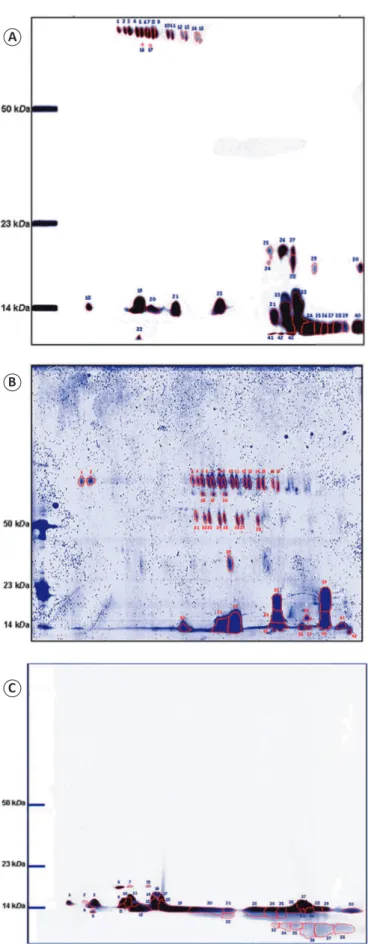
Two-dimensional gel electrophoresis of venom samples in gels with a pH range of 3-10 revealed 43 spots, including 22
and 21 in the acidic and basic regions, respectively. A total of
26 spots had a mass below 23kDa, and 17 had approximately the same mass above 50kDa but had different pH ranges (Figure 2A). Gels with a pH range of 4-7 had 42 spots, most of
which were in the pH range of 5-6. Fourteen spots had a mass below 23kDa, and 28 had a mass above 50kDa; the majority of spots in this acidic pH range had similarly high masses (Figure 2B). Spots detected in gels with a pH range of 7-11 had
low masses below 23kDa (Figure 2C).
Biological activities of toxins in
Micrurus surinamensis venom
Micrurus surinamensis venom at doses greater than 20µg was highly toxic, causing immediate muscle and respiratory paralysis
and death in mice within a few minutes of administration. The
following symptoms were observed following i.c. injection of 0.2, 1, 2, and 4µg of venom: total muscle paralysis and
difficulty breathing (< 1 min); leaping, increased energy,
hypersensitivity to touch and sound, compulsive scratching
(30 min); and convulsions followed by death at the higher doses.
Symptoms caused by i.c. injection of 20µl of physiological
saline solution (as a control for activity) were wheezing and
apathy that ceased within 10 min of injection without leading
to death. The following symptoms were observed following
i.v. injection of 1, 10, 15, or 20µg of M. surinamensis venom:
difi culty breathing; muscle hypotonia; unilateral and bilateral ptosis (30 min); compulsive scratching (legs, genitalia, tail, head, nose, and eyes); muscle stretching in the hind legs (1:30h);
and death due to respiratory arrest at the higher doses.
Probit analysis revealed that the LD50 of i.v. administered
venom was 14µg (16.8-11.3) per 20g body weight (700μg/kg). The neutralizing capacity of the antielapidic serum was very
FIGURE 2 - Electrophoretic proi le of Micrurus surinamensis venom. (A): 12.5% SDS-PAGE with an IPG strip in the pI range of 3.0-10.0. (B): 5%-20% SDS-PAGE with an IPG strip in the pI range of 4-7. (C): 5%-20% SDS-PAGE with an IPG strip in the pI range of 7-11. Spots were analyzed using the ImageMaster 2D Platinum 6.0 system. Molecular mass markers were Bothrops atrox toxins. Gels were stained with Coomassie Blue R-250. SDS-PAGE: sodium dodecyl sulfate polyacrylamide gel electrophoresis; IPG: immobilized pH gradient; pI: isoelectric point.
A
B
low, with a potency of 0.3 (0.5-0.02)mg/ml (venom/serum). In animals i.v. injected with 7.5, 10, or 13µg of venom there
was no hemorrhage in the thoracic cavity, nasal cavity, or genitalia. Blood collected from these animals coagulated within
a normal time interval for mouse blood (< 60s). However, in vitro experiments revealed a delay in the coagulation time of venom-treated plasma as compared to that of control plasma.
Speciically, human plasma clotted after 10 and 30 min in the presence of the lowest (20µg) and highest (80µg) doses
of venom, respectively. Animals that received 15 or 20µg of venom were autopsied immediately after death and exhibited no hemorrhaging in the thoracic cavity or in other visible areas
of the body, such as the external genitalia and nose (Table 1). Evaluation of serum-mediated neutralization of Micrurus surinamensis venom enzymatic activity
Micrurus surinamensis venom exhibited low PLA2 activity
compared to the control (10µg of B. atrox venom) at doses of
10, 20, and 40µg (Figure 3A). The antielapidic serum inhibited 100% of the PLA2 activity of M. surinamensis venom when
administered at a ratio between 1:1 and 1:0.05 (Figure 3B).
The antielapidic serum (maximum and minim doses of 20 and 1µl, respectively) also inhibited 100% of the PLA2 activity
of M. surinamensis venom. The venom had no caseinolytic,
ibrinolytic, or gelatinolytic protease activity according to the zymography results (data not shown).
Western blot analysis of competitive interaction
A western blotting analysis revealed that the antielapidic
serum had low potency (Figure 4). Antivenom (4µl) + venom
(5, 20, and 80µg) mixtures preincubated at 37°C for 30 min showed several low-intensity bands at 7-20kDa and especially around 14kDa, although these exhibited variable signal intensity. The proteins were detected after just 1h of reaction with peroxidase-speciic substrates.
DISCUSSION
The venom of snakes in the genus Micrurus contains a complex mixture of proteins, approximately 90% of which are
neurotoxins with low molecular masses of around 7-84kDa (< 12kDa, 43%; 14-32kDa, 47%; and > 50kDa, 10%)37-40. The
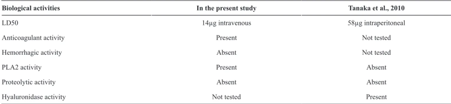
LD50 14µg intravenous 58µg intraperitoneal
Anticoagulant activity Present Not tested
Hemorrhagic activity Absent Not tested
PLA2 activity Present Absent
Proteolytic activity Absent Absent
Hyaluronidase activity Not tested Present
LD50: 50% lethal dose; PLA2: phospholipase A2 activity.
A
B
FIGURE 3 - (A): PLA2 activity of Micrurus surinamensis surinamensis
venom proteins separated in agarose gels. Duplicate: 10, 20, and 40µg/20µl venom. Control: 10µg of Bothrops atrox venom. (B): Neutralization of the PLA2 activity of Micrurus surinamensis venom with antielapidic serum at different dilutions (venom/serum) prepared in duplicate. Controls: 10µg
Bothrops atrox atrox venom and 20µg Micrurus surinamensis venom alone (duplicates). PLA2: phospholipase A2 activity.
venom of M. surinamensis differs from that of other Micrurus
species owing to its abundance of peptides between 6-7kDa, which was conirmed in the present work.
using this approach identiied approximately 30 proteins within the pH range 3-10, with the most intense spots observed in the range of 11-25kDa and only one spot in the range of 30-150kDa
that showed high similarity to l-aspartate oxidase41. In the
present study, we identiied 43 spots within the pH range of 3-10. All of the spots in the basic pH range had a low mass, whereas more than 60% of the spots in the acidic pH range had a high mass. A total of 17 spots had similar masses above 50kDa and an acidic pH, suggesting that they were isoenzymes. In some species, isoenzymes with basic pH and masses of 8-13kDa
have been isolated that exhibit various biological activities
(e.g., cardiotoxic, neurotoxic, PLA2, and hemorrhage-inducing)
and, in some cases, lethality in association with hemoptysis and hemoglobinuria42-44.
Despite their similar proiles, each type of snake venom has
peculiarities in terms of protein composition and abundance45,46.
Additionally, differences in snake venom proteomes within the
same species inhabiting different areas are not uncommon47. Proteomic variations in the venom of Micrurus species may be associated with differences in their geographic location and/or habitat as well as seasonal variations, dietary factors, age, sexual dimorphism, and even evolutionary history.
In this study, toxins from M. surinamensis did not cause bleeding and produced symptoms typical of venom from this genus, including uni- and/or bilateral ptosis, muscular hypotonia, spasms, respiratory failure, and exophthalmos. M. surinamensis
venom exhibited high neurotoxicity, causing death via respiratory
paralysis within minutes of injection. Toxins present in Micrurus
venoms generally do not cause localized or systemic bleeding and induce mild myonecrosis48,11, while M. surinamensis venom
is not known to induce myotoxicity, hemorrhage, or edema49. At high concentrations, M. altirostris venom does not cause bleeding, dermonecrosis, or coagulant activity37; venoms of
M. averyi and M. fulvius cause hemorrhaging, renal damage,
FIGURE 4 - Proile of Micrurus surinamensis venom proteins in a 20µg sample. (1) Sample subjected to 5%-20% gradient SDS-PAGE under
non-reducing conditions. (2) Immune proiles of polyclonal antibodies
against antielapidic serum. Control and antivenom (4µl) × venom (5, 20, or 80µg) were preincubated at 37°C for 30 min. Molecular mass markers of
Bothrops atrox venom toxins are shown to the left. Gels were stained with Coomassie Blue R-250. SDS-PAGE: sodium dodecyl sulfate polyacrylamide gel electrophoresis; IPG: immobilized pH gradient.
and severe inlammation49,50. In some cases, PLA2 is responsible for inducing severe bleeding and myoglobinuria51, but owing to the lethality of the neurotoxins present in the venom, these symptoms are often not manifested.
In general, snake toxins are associated with less bleeding and more neurotoxic effects; however, the venoms of snakes in the Viperidae and Crotalidae families cause serious
bodily injury due to bleeding within vital organs and blood circulation impairment caused by clot formation, edema, and
necrosis. These symptoms are mainly attributed to serine and
metalloproteases responsible for proteolytic degradation in mammalian blood plasma and tissues, which may function as blood coagulation activators or inhibitors15,52-54.
The venoms of some species of the genera Naja, Bungarus, and Micrurus lack proteolytic activity against gelatin, casein,
and ibrinogen55,56; this was also true of the venom of the species
examined in the present study. However, high concentrations of
M. surinamensis venom caused an increase in human plasma coagulation time, suggesting that it has anticoagulant activity.
Proteases are known to inhibit platelet aggregation57 and PLA2
inactivates blood clotting factors to block the coagulation
cascade58. Thus, PLA2 concentration may also be related to anticoagulant activity44,59.
Venom toxins of most coral snakes exhibit PLA2 activity
along with neurotoxic and myotoxic effects60. Both α- and β-type neurotoxins are responsible for the lethality of the venom in mice and are 10 times more potent when directly injected into
the CNS. Thus, although these neurotoxins have no effects in the peripheral nervous system (PNS), even small amounts
are highly toxic in the CNS61, causing damage in the cerebral cortex, rostrocaudal region of the brain, hemispheric white matter, corpus callosum, fornix, and hippocampus14. Similar
damage is caused by some three-inger neurotoxins that bind to rat hippocampus-speciic muscarinic acetylcholine and A-type γ-aminobutyric acid receptors following jaw paralysis and
intense seizures induced by intracerebroventricular injection of venom61,62.
Four fractions (7-22kDa) exhibiting neurotoxic PLA2
activity isolated from M. lemniscatus venom caused severe symptoms of envenomation in the mouse CNS63. Small
doses (1-2.1μg/μl) induced a variety of detectable symptoms,
including spasms, breathing difficulty, limbic seizures, cortical and hippocampal epileptic discharges, episodes of
convulsion, and ultimately death (in 80% of animals); high doses (4.5μg/μl) caused symptoms consistent with severe intoxication
such as reduced motor activity, hypersensitivity to touch and
sound, aggression, and death. Histological analysis revealed massive hippocampal neuronal loss. These results demonstrate
that M. surinamensis venom is highly neurotoxic to both the PNS and CNS. Clarifying the molecular mechanisms underlying these effects can lead to the development of new drugs for the treatment of neurological diseases based on neuropeptides
produced by these snakes64.
The species examined in the present work showed low
only weakly detected by western blotting. Our in vitro tests revealed the presence of antibodies in the antielapidic serum that recognized and neutralized proteins of approximately
14kDa in M. surinamensis venom, but not bands corresponding
to neurotoxins smaller than 10kDa. These results demonstrate that individual snakes of the same species have unique venom protein components and immunological proiles.
Our in vivo analysis showed that the antielapidic serum had
low eficacy in neutralizing M. surinamensis venom components
that exhibited high neurotoxicity by i.v. injection (14µg/20g venom/mouse), indicating that high doses of serum are required
for complete neutralization66,67. The low recognition of these
neurotoxins is a signiicant limitation given their abundance
and toxicity68.
The immune proile of M. surinamensis was distinct from that of other species. In particular, the toxic components of its venom showed limited reactivity with the antielapidic serum65,69 produced in Brazil by hyperimmunizing horses with Micrurus
corallinus and Micrurus frontalis venoms obtained from the Butantan Institute. Despite their small size and corresponding low venom inoculation capacity, high doses of serum are
recommended for envenomations involving coral snakes7. The
intra-species variability of coral snake venom raises concerns
regarding the effectiveness of sera developed using venoms
produced by a limited number of coral snake species70. A monoclonal antibody against M. nigrocintus nigrocintus reacted with M. surinamensis venom, indicating that the former belongs
to a group with a different antigenic proile than other Micrurus
species of medical importance in Brazil39. Geographic and dietary variations as well as phylogenetic factors and mutations
account for the diversity in snake venom composition. Although
diet is the main contributing factor71, other evolutionary forces driven by natural selection such as allelic mutations in an enzyme that leads to structural changes in the protein may be equally important72.
A recent study evaluated the immunogenic potential of venoms of different Micrurus species to ensure good cross-reactivity with antielapidic sera produced in experimental animals and to establish an antigenic mixture for generating
polyvalent antivenoms with higher eficacy than those used
to treat envenomation in humans69. The results were positive for most but not all of the species investigated; in fact, the
M. surinamensis venom was less eficiently recognized by the
antivenom, underscoring the dificulty of this task. Additional
basic and clinical studies are needed to generate a monovalent serum or venom that includes these species in the pool used to
produce commercial antivenom. Snake bites remain a serious
public health problem that have generally been neglected by the authorities and the general population73. Thus, there are many outstanding challenges and issues that must be addressed
to promote snake bite prevention and to develop effective
treatments for envenomation.
Conlict of interest
The authors declare that have no conlicts of interest.
Financial support
This study was supported by Amazonas State Research Foundation [Fundação de Amparo à Pesquisa do Estado do Amazonas (FAPEAM)], Financier of
Studies and Projects [Financiadora de Estudos e Projetos (FINEP)], National
Council for Scientiic and Technological Development [Conselho Nacional
de Desenvolvimento Cientíico e Tecnológico (CNPq)], and the Proteome Network of Amazon.
REFERENCES
1. Costa HC, Bérnils RS. Répteis brasileiros: lista de espécies 2015.
São Paulo: Sociedade Brasileira de Herpetologia; 2015; 4(3):75-93.
[Internet]. [updated 2015 August 10; cited 2017 March 24]. Available from: http://www.sbherpetologia.org.br.
2. Orr RT. Biologia dos Vertebrados. 5th ed. São Paulo: Roca; 1986. 508 p.
3. Roze JA. New world coral snakes (Elapidae): a taxonomic and biological summary. Mem Inst Butantan. 1982;46(1):305-38.
4. Hoge AR, Romano-Hoge SARWL. Sinopse das serpentes
peçonhentas do Brasil. Mem Inst Butantan. 1978/79;42/43: 373-496.
5. Santos MC, Martins M, Boechat AL, de Sá Neto RP, Oliveira ME.
Serpentes de Interesse Médico da Amazônia: Biologia, Venenos e Tratamento de Acidentes. Manaus: Universidade do Amazonas;
1995. 70p.
6. Bucaretchi F, Capitani EM, Vieira RJ, Rodrigues CK, Zannin M, Da
Silva Jr NJ, et al. Coral snake bites (Micrurus spp.) in Brazil: a review
of literature reports. Clin Toxicol (Phila). 2016;54(3):222-346
7. Bucaretchi F, Hyslop S, Vieira RJ, Toledo AS, Madureira PR,
Capitani EM. Bites coral snakes (Micrurus spp.) in Campinas, state
of São Paulo, southeastern Brazil. Rev Inst Med Trop Sao Paulo. 2006;48(3):141-5.
8. Ministério da Saúde. Manual de diagnóstico e tratamento de
acidentes ofídicos. Brasília: Fundação Nacional de Saúde; 2001.
120 p.
9. Urdaneta AH, Bolaños F, Gutierrez JM. Feeding behavior and
venom toxicity of coral snake Micrurus nigrocinctus (Serpentes:
Elapidae) on its natural prey in captivity. Comp Biochem Physiol C Toxicol Pharmacol. 2004;138(4):485-92.
10. Seraim FG, Reali M, Cruz-Holing MA, Fontana MD. Action of Micrurus dumerilii carinicauda coral snake venom on the
mammalian neuromuscular junction. Toxicon. 2002;40(2):167-74.
11. Gutiérrez JM, Chaves F, Rojas E, Bolaños R. Efectos locales inducidos por el veneno de la serpiente coral Micrurus nigrocinctus
em ratón blanco. Toxicon. 1980;18(5-6):633-9.
12. Arni RK, Ward RJ. Phospholipase A2–a structural review. Toxicon.
1996;34(8):827-41.
13. Carredano E, Westerlund B, Persson B, Saarinen M, Ramaswamy
S, Eaker D, et al. The three-dimensional structures of two toxins from snake venom throw light on the anticoagulant and neurotoxic
14. Clapp LE, Klette KL, Decoster MA, Bernton E, Petras JM, Dave
JR, et al. Phospholipase A2-induced neurotoxicity in vitro and
in vivo in rats. Brain Res. 1995;693(1-2):101-11.
15. Kini RM. Structure-function relationships and mechanism of anticoagulant phospholipase A2 enzymes from snake venoms.
Toxicon. 2005;45(8):1147-61.
16. Wickramaratna JC, Fry BG, Aguilar MI, Kini RM, Hodgson WC. Isolation and pharmacological characterization of a phospholipase A2 myotoxin from the venom of the Irian Jayan death adder
(Acanthophis rugosus). Br J Pharmacol. 2003;138(2):333-42.
17. Cruz-Höling MA, Rodrigues-Simioni L, Vital-Brazil O.
Ultrastructure changes in neuromuscular junctions of mouse diaphragm caused by the venom of the coral snake Micrurus
corallinus. Mem Inst Butantan. 1983/84;47/48:95-105.
18. Huang LF, Zheng JB, Xu Y, Song HT, Yu CX. A snake venom phospholipase A2 with high afinity for muscarinic acetylcholine
receptors acts on guinea pig ileum. Toxicon. 2008;51(6):1008-16.
19. Jolkkonen M, Giersbergen PLMV, Hellman U, Wernstedt C, Oras
A, Satyapan N, et al. Muscarinic toxins from the black mamba Dendroaspis polylepis. Eur J Biochem. 1995;234(2):579-85.
20. Karlsson E, Jolkkonen M, Mulugeta E, Onali P, Adem A. Snake toxins with high selectivity for subtypes of muscarinic acetylcholine
receptors. Biochimie. 2000;82(9-10):793-806.
21. Vital-Brazil O. Coral snake venoms: mode of action and
pathophysiology of experimental envenomation. Rev Inst Med Trop Sao Paulo. 1987;29(3):119-26.
22. Kumar TKS, Pandian SK, Srisailam S, Yu C. Structure and function
of snake venom cardiotoxins. J Toxicol Toxin Rev. 1998;17(2):183-211. 23. Nirthanan S, Gopalakrishnakone P, Gwee MCE, Khoo HE, Kini
RM. Non-conventional toxins from elapid venoms. Toxicon. 2003;41(4):397-407.
24. Noronha MDN, Souza ARB, Bührnheim PF. Estudo epidemiológico
dos acidentes ofídicos atendidos na FMT/IMT-AM, de janeiro de 1995 a outubro de 1999. Rev Soc Bras Med Trop. 2000;33(Suppl. I):162-3.
25. Buhrnheim PF, Lima HCL, Oliveira MEES. Ocorrência de serpentes
peçonhentas na Amazônia e acidentes ofídicos no Amazonas. Rev Soc Bras Med Trop. 1988;21(Suppl. I):119.
26. Cunha OR, Nascimento FP. Ofídios da Amazônia X: as cobras da
região leste do Pará. Publicações Avulsas. Museu Paraense Emílio Goeldi. 1978;31:1-218. Disponível em:
http://repositorio.museu-goeldi.br/handle/mgoeldi/904.
27. Morais DH, Ávila RW, Kawashita-Ribeiro RA, Carvalho MA. Squamata, Elapidae, Micrurus surinamensis (Cuvier, 1817): new
records and distribution map in the state of Mato Grosso, Brazil,
with notes on diet and activity period. Check List. 2011;7(3):350-1.
28. Da Silva Jr NJ, Aird SD. Prey speciicity, comparative lethality and
compositional differences of coral snake venoms. Comp Biochem Physiol C Toxicol Pharmacol. 2001;128(3):425-56.
29. Aird SD, Da Silva Jr NJ. Comparative enzymatic composition of
Brazilian coral snake (Micrurus) venoms. Comp Biochem Physiol
B. 1991;99(2):287-94.
30. Schägger H, Von Jagow G. Tricine-sodium dodecyl
sulfate-polyacrylamide gel electrophoresis for the separation of proteins in
the range from 1 to 100 kDa. Anal Biochem. 1987;166(2):368-79. 31. Finney DJ. Probit analysis. 3rd ed. New York: Cambridge University
Press; 1971. 333p.
32. World Health Organization. Progress in the characterization of venoms and standardization of antivenoms. WHO Offset Publ. 1981;(58):1-44. PMID: 7245916.
33. Theakston RDG, Reid HA. Development of simple standart assay procedures for the characterization of snake venoms. Bull World Health Organ. 1983;61(6):949-56.
34. Heussen C, Dowdle EB. Eletrophoretic analisys of plasminogen
activators in poliacrilamide gels containg sodium dodecyl sulfate
and copolymerized substrates. Anal Biochem. 1980;102(1):196-202. 35. Stocker KF, Meier J. Thrombin-like snake venom enzymes.
In: Pirkle H, Markland FS, editors. Hemostasis and animal venoms. New York: Marcel Dekker; 1988. p. 67-84.
36. Towbin H, Staehelin T, Gordon J. Electrophoretic transfer of proteins
from polyacrylamide gels to nitrocellulose sheets: procedure and
some applications. Proc Natl Acad Sci USA. 1979;76(9):4350-4. 37. Moraes FV, Sousa-E-Silva MCC, Barbaro KC, Leitão MA,
Furtado MFD. Biological and immunochemical characterization of Micrurus altirostris venom and serum neutralization of its toxic
activities. Toxicon. 2003;41(1):71-9.
38. Alape-Girón A, Gustafsson B, Lomonte B, Thelestam M, Gutiérrez JM. Immunochemical characterization of Micrurus nigrocinctus nigrocinctus venom with monoclonal and polyclonal antibodies.
Toxicon. 1994;32(6):695-712.
39. Alape-Girón A, Lomonte B, Gustafsson B, Da Silva Jr NJ, Thelestam
M. Electrophoretic and immunochemical studies of Micrurus snake
venoms. Toxicon. 1994;32(6):713-23.
40. Da Silva Jr NJ, Grifin PR, Aird SD. Comparative chromatography
of Brazilian coral snake (Micrurus) venoms. Comp Biochem
Physiol.1991;100B(1):117-26.
41. Olamendi-Portugal T, Batista CVF, Restano-Cassulini R, Pando V,
Villa-Hernandez O, Vargas AZM, et al. Proteomic analysis of the venom from the ish eating coral snake Micrurus surinamensis: novel
toxins, their function and phylogeny. Proteomics. 2008;8(9):1919-32.
42. Vergara I, Pedraza-Escalona M, Paniagua D, Restano-Cassulini
R, Zamudio F, Batista CVF, et al. Eastern coral snake Micrurus
fulvius venom toxicity in mice is mainly determined by neurotoxic phospholipases A2. J Proteomics. 2014;105:295-306.
43. Chang LS, Huang HB, Lin SR. The multiplicity of cardiotoxins from
Naja naja atra (Taiwan Cobra) venom. Toxicon. 2000;38(8):1065-76.
44. Francis BR, Da Silva Jr NJ, Seebart C, Silva LLC, Schimidt JJ, Kaiser II.
Toxins isolated from the venom of the Brazilian coral snake (Micrurus
frontalis frontalis) include hemorrhagic type phospholipases A2 and
postsynaptic neurotoxins. Toxicon. 1997;35(8):1193-203.
45. Takasaki C, Suzuki J, Tamiya N. Puriication and properties of several phospholipases A2 from the venom of Australian king brown
snake (Pseudechis australis). Toxicon. 1990;28(3):319-27.
46. Kulkeaw K, Chaicumpa W, Sakolvaree Y, Tongtawe P, Tapchaisri P. Proteome and immunome of the venom of the thai cobra, Naja kaouthia. Toxicon. 2007;49(7):1026-41.
47. Tan NH, Ponnudurai G. The biological properties of venoms of
some American coral snakes (genus Micrurus). Comp Biochem
Physiol. 1992;101B (3):471-4.
48. Remuzgo C, Alvarez MP, Rodriguez E, Lazo F, Yarleque A.
Micrurus spixii (peruvian coral sanke) venom - preliminary
biochemical and enzymatic characterization. J Venom Anim Toxins Incl Trop Dis. 2002;8(1):1-6.
49. Barros ACS, Fernandes DP, Ferreira LCL, Santos MC. Local effects
induced by venoms from ive species of genus Micrurus sp. (coral
snakes). Toxicon. 1994;32(4):445-52.
50. Gutiérrez JM, Lomonte B, Portilla E, Cerdas L, Rojas E. Local
from elapid snake venoms. Toxicon. 2007;49(8):1200-7.
53. Kini RM, Evans HJ. Inhibition of platelet aggregation by a ibrinogenase from Naja nigricollis venom is independent of
ibrinogen degradation. Biochim Biophys Acta. 1991;1095(2):117-21.
54. Teixeira CFP, Fernandes CM, Zuliani JP, Zamuner SF. Inlammatory
effects of snake venom metalloproteinases. Mem Inst Oswaldo Cruz. 2005;100(1):181-4.
55. Zhang Y, Xiong YL, Bom C. An activator of blood coagulation
factor X from the venom of Bungarus fasciatus. Toxicon. 1995;33(10):1277-88.
56. Tambourgi DV, dos Santos MC, Furtado MFD, de Freitas MCW, da
Silva WD, Kipnis TL. Pro-inlammatory activities in elapid snake venoms. Br J Pharmacol. 1994;112(3):723-7.
57. Kerns RT, Kini RM, Stefansson S, Evans HJ. Targeting of venom phospholipases: the strongly anticoagulant phospholipase A2 from
Naja nigricollis venom binds to coagulation factor Xa to inhibit the prothrombinase complex. Arch Biochem Biophys. 1999;369(1):107-13.
58. Cecchini AL, Marcussi S, Silveira LB, Borja-Oliveira CR, Rodrigues-Simioni L, Amara S, et al. Biological and enzymatic activities of Micrurus sp. (coral) snake venoms. Comp Biochem
Physiol A Mol Integr Physiol. 2005;140(1):125-34.
59. Terra ALC, Moreira-Dill LS, Simões-Silva R, Monteiro JRN, Cavalcante WLG, Gallacci M, et al. Biological characterization of the Amazon coral Micrurus spixii snake venom: isolation of a new
neurotoxic phospholipase A2. Toxicon. 2015;103:1-11.
60. Rosso JP, Vargas-Rosso O, Gutiérrez JM, Rochat H, Bougis PE.
Characterization of α-neurotoxin and phospholipase A2 activities from Micrurus venoms: determination of the amino acid sequence
and receptor-binding ability of the major α-neurotoxin from Micrurus nigrocinctus nigrocinctus. Eur J Biochem. 1996;238(1):231-9.
61. Silva DC, Medeiros WAA, Batista IFC, Pimenta DC, Lebrun I, Abdalla FMF, et al. Characterization of a new muscarinic toxin
from the venom of the Brazilian coral snake Micrurus lemniscatus in rat hippocampus. Life Sci. 2011;89(25-26):931-8.
62. Rosso JP, Schwarz JR, Diaz-Bustamante M, Céard B, Gutiérrez JM,
Kneussel M, et al. MmTX1 and MmTX2 from coral snake venom
lemniscatus coral snake venom: bahavioral, electroencephalographic and neuropathological aspects. Brain Res Bull. 2008;75(5):629-39.
64. Goswami PK, Samant M, Srivastava RS. Snake venom,
anti-snake venom & potential of anti-snake venom. Int J Pharm Pharm Sci. 2014;6(5):4-7.
65. Tanaka GD, Furtado MFD, Portaro FCV, Sant’Anna OA, Tambourgi
DV. Diversity of Micrurus snake species related to their venom
toxic effects and the prospective of antivenom neutralization. PLoS
Negl Trop Dis. 2010;4(3):e622.
66. Fry BG, Wickramaratna JC, Jones A, Alewood PF, Hodgson WC. Species and regional variations in the effectiveness of antivenom against the in vitro neurotoxicity of Death adder (Acanthophis)
venoms. Toxicol Appl Pharmacol. 2001;175(2):140-8.
67. Silva ARBP, Yamagushi IK, Morais JF, Higashi HG, Raw I, Ho
PL, et al. Cross reactivity of different speciic Micrurus antivenom
sera with homologous and heterologous snake venoms. Toxicon. 2001;39(7):949-53.
68. Ciscotto PHC, Rates B, Silva DAF, Richardson M, Silva LP,
Andrade H, et al. Venomic analysis and evaluation of antivenom
cross-reactivity of South American Micrurus species. J Proteomics.
2011;74(9):1810-25.
69. Tanaka GD, Sant’Anna OA, Marcelino JR, Lustoza da Luz
AC, Teixeira da Rocha MM, Tambourgi DV. Micrurus snake
species: venom immunogenicity, antiserum cross-reactivity and
neutralization potential. Toxicon. 2016;117:59-68.
70. Carvalho AV, David CF, Pessoa AM, da Silva Jr NJ. Um estudo do rendimento do veneno de cobras corais brasileiras e seu uso na
avaliação do soro antielapídico. Sci Med. 2014;24(2):142-9.
71. Daltry JC, Wüster W, Thorpe RS. Diet and snake venom evolution.
Nature. 1996;379(6565):537-40.
72. Sasa M. Diet and snake venom evolution: can local selection alone
explain intraspeciic venom variation. Toxicon. 1999;37(2):249-52. 73. Gutiérrez JM. Current challenges for confronting the public health