Vol.57, n.4: pp. 523-531, July-August 2014 http://dx.doi.org/10.1590/S1516-89132014005000020
ISSN 1516-8913 Printed in Brazil
BRAZILIAN ARCHIVES OF BIOLOGY AND TECHNOLOGY
A N I N T E R N A T I O N A L J O U R N A L
Production and Characterization of IgY against Canine IgG:
Prospect of a New Tool for the Immunodiagnostic of Canine
Diseases
Fernanda Nunes Santos
1*, Beatriz Coutinho Brum
1, Paula Borba Cruz
2, Claudia Moraes
Molinaro
3, Valmir Laurentino Silva
1and Sérgio Augusto de Miranda Chaves
41Departamento de Ciências Biológicas; Escola Nacional de Saúde Pública Sérgio Arouca; Fiocruz - Rio de Janeiro
- RJ - Brasil. 2Instituto Oswaldo Cruz; Fiocruz; Rio de Janeiro - RJ - Brasil. 3Laboratório de Tecnologia
Diagnóstica-Bio Manguinhos; Fiocruz; Rio de Janeiro - RJ - Brasil. 4Departamento de Endemias Samuel Pessoa;
Escola Nacional de Saúde Pública Sérgio Arouca; Fiocruz; Rio de Janeiro - RJ - Brasil
ABSTRACT
This study describes the production of a new avian polyclonal antibody (IgY) against canine IgG, as another tool for the immunodiagnostic using IgY technology. The immunization protocol caused neither deaths nor pathologies, and no decline in egg laying capacity was detected. The total concentration of isolated IgY was constant, without significant difference (p> 0.05), with average of 97.55 mg of IgY/yolk. The IgY revealed a strong sensitivity and specificity in recognition against canine IgG by ELISA. After the immunization, there was a significant increase in the production of IgY specific from the first to the second month (p <0.05), reaching a stable peak without decrease in the production by the end of the analysis period (p> 0.05). The IgY demonstrated a suitable specificity in Western blot against the purified and serum canine IgG, not enabling recognition of canine IgM or IgG of other animal species. The specific IgY in the egg yolks of immunized hens proved to be a molecule with an appropriate purity and desired specificity against the immunizing antigen. Moreover, its constant production in large quantities during the four months analyzed indicated that IgY antibody production technology could be considered as an excellent alternative to the standard methods.
Key words: IgY polyclonal antibodies, chicken antibodies, hen immunization, canine IgG
*Author for correspondence: [email protected]
INTRODUCTION
Antibody production against the canine
Imunoglobulin G (IgG) is very important for the elaboration of diagnostic kits, which are intended to detect the serological markers of infection. Polyclonal antibodies from the mammals are extensively used for this purpose, involving the ethical issues due to invasive collection methods and euthanasia, as well as operational issues such as costly maintenance of the animal antibody donors and laborious purification methods usually
resulting in low production yield (Leenaars et al. 1999). The antibody production should be performed in a manner imposing the least possible stress in the animals involved, combined with the maximum yield for long periods with a simple, efficient and economically viable purification process (Witkowski et al. 2009).
mammals, birds transmit immunity to their offspring transferring immunoglobulins from the serum to the egg yolk (Kowalczyk et al. 1985). The most abundant immunoglobulin in chicken serum is IgY, which is transferred to and accumulated in great amounts in the egg yolk (Rose et al. 1974).
Immunoglobulin Y (IgY) is an excellent
immunological reagent, presenting some
beneficial features when compared to the mammalian IgG. Due to the phylogenetic distance between the birds and mammals, chickens are able to produce specific antibodies against highly conserved mammalian antigens, unlike rabbits, which are the typical source of antibody production. IgY binds neither to Fc receptors of mammals nor to the mammalian rheumatoid factor, as well as not activating the complement factor, features that reduce interpretation errors, or false positive results (Schade et al. 2000). The economic and operational advantages are related to their production, which is continuous and in large quantities throughout the laying period (Schade et al. 2000), about two years (Pauly et al. 2009). There are also ethical advantages, since the production does not require invasive collection methods, or euthanasia (Schade et al. 2000). IgY can be applied in the same way as the mammalian IgG in immunodiagnostic tests (Schade et al. 2000). There are many studies describing IgY as an immunological reagent with appropriate sensitivity and specificity, revealing a performance equal, or superior to the mammalian immunoglobulins (Tini et al. 2002; Chalghoumi et al. 2009; Dias da Silva and Tambourgi 2010). Chicken antibodies can also be produced against many types of antigens and utilized in different methods and for different purposes (Narat 2003). IgY was produced against immunoglobulins from different animal species, displaying excellent performance when compared with the mammal
immunoglobulin (Bizanov et al. 2004;
Kritratanasak et al. 2004; Nikbakht et al. 2009). It has also been successfully adopted in passive immunization of the dogs against parvovirus (Nguyen et al. 2006). Griot-Wenk et al. (1998), using recombinant proteins derived from the heavy chain of canine IgE, produced IgY capable of effectively recognizing canine IgE. In view of all these advantages and the absence of scientific reports regarding IgY against canine IgG on the immunodiagnosis of canine diseases, deeper studies related to the production and performance
against the immunizing antigen are necessary and justified. This study describes IgY production against the canine IgG as a tool for the
immunodiagnosis with IgY technology,
emphasizing the evaluation of the immunization procedure, isolation, purification, characterization and the assessment of the specificity against the immunizing antigen and cross reaction presence.
METODOLOGY
Experimental design
Two Isa-Brown hens, 20-week-old, weighing 1.8Kg were used and maintained individually
receiving the ration and drinkable water ad
libitum. The experimental protocol was reviewed
and approved by the Ethics Committee on the Use of Animals -CEUA-Fiocruz. License Number: LW-19/10.
Immunization schedule
For the immunization, 200 µg of ultrapurified
canine IgG (Dog gamma-globulin ultrapurified Rockland-Inc) emulsified in 0.1mg/mL of Freund´s complete adjuvant was used. Two more inoculations in the same conditions were made using Freund´s incomplete adjuvant. The three inoculations were administered in the intervals of
one week, intramuscular in the pectoral
musculature with a final volume of 0.5 mL, distributed in various points. After an interval of 20 days from the last inoculation, the eggs were collected daily during four months and stored at 4-8°C. At the inoculation sites, the immunization adverse effects were observed by the palpation, tissue damage, or edema presence.
IgY Isolation
280 nm, according to Lambert-Beer law using an extinction coefficient of 1.33 (Leslie and Clem 1969).
IgY Purification by affinity chromatography
After the isolation, IgY was submitted to a purification process using thiofilic adsorption in Hitrap IgY purification column (GE, Healthcare). The sample and the column were submitted to a preparation procedure in accordance with the manufacturer instructions. A total of 100 mg of IgY was applied. A binding solution (20 mM NaH2PO4 and 0.5M K2SO4, pH 7.5) was used for the removal of non-bound material. Purified IgY was obtained using an elution solution (20 mM NaH2PO4, pH 7.5). To remove the non-bound
material a cleaning solution (20 mM NaH2PO4 ,
30% isopropanol, pH 7.5) was used. The fractions containing IgY were separated through higher readings at 280 nm.
IgY characterization by SDS-PAGE
After the isolation and purification procedures, IgY was characterized by the SDS PAGE (Laemmli 1970). The IgY was diluted at 1:4 and examined in 10% SDS-PAGE under reduction conditions. The electrophoretic run was performed at 200 V for 80 min. For visualization, the Comassie Blue staining solution (0.4%) was used.
Verification of IgY specificity by ELISA
ELISA plates (Maxi Sorb, Nunc) were covered with different concentrations of canine IgG (3.9-0.007 µg/mL) in a carbonate bicarbonate buffer (20% 0.06 M Na2CO3 and 80% 0.06 M NaHCO3, pH 9.6), and incubated at 4-8°C for 16 h. After two washings (PBS 0.05% Tween-20), different concentrations of purified IgY (56.4-0.3 µg/mL) were added in dilution solution (PBS 0.05% Tween-20 with 1% casein), followed by incubation at 37°C for 1h. After three washings, anti-IgY conjugated to peroxidase developed in the rabbits (Promega Corporation, USA) in dilution solution 1:1000 was added, followed by incubation at 37°C for 1h. Development was carried out using, the solution (0.83 M Na2HPO4, 0.33 M citric acid, pH 4.9-5.2, 0.05 M Ortho phenylene diamine, 30% H2O2). After 30 minutes, the reaction was stopped with 1M H2SO4 (Voller et al. 1976). For the analysis, a reference filter of 490nm and contrast of 630 nm were used. Aiming to analyze the production of isolated specific IgY anti-canine IgG, Elisa was used in the same
conditions with fixed concentrations of 0.5 µg IgG/mL and 0.8 µg IgY/mL with samples of all IgY isolated from eggs collected. The graphical representation was performed using average absorbance during the four months after immunization.
Verification of IgY specificity by Western blot
IgG of dog, cat, guinea pig, rabbit, goat, sheep, horse (Gamma-globulin ultrapurified, Rockland-Inc), IgM dog (whole molecule, Rockland-Inc)
and L. (L.) chagasi infected dog serum were
separated by the SDS-PAGE. Electro-transference was carried out at 100 V for 120 min (Towbin et al. 1979). Membranes were incubated in a blocking solution (PBS 0.05% Tween-20 and casein 5%) for 16 h, followed by a five minutes washing three times with a washing solution (PBS 0.05% Tween-20). Membranes were incubated with IgY purified in the dilution of 1:250 at 37ºC for 2 h, followed by a new washing and incubation with secondary antibody (rabbit IgG anti-chicken IgY - Sigma-Aldrich, USA) in the concentration of 1:5000 at 37ºC for 1 h. Afterwards, membrane was washed as described above and submitted to protein A peroxidase (Sigma-Aldrich, USA) incubation in concentration of 1:500 at 37ºC for 1h. For exposure, a developing solution (3.3 Diamine benzidine, 30% H2O2, 1% CoCl2 in PBS) was used until the bands appeared. The reaction was stopped with distilled water.
RESULTS AND DISCUSSION
During the immunization process, the hens remained healthy without any abnormality in the development. No deaths occurred and all the animals presented normal behavior of the species. At the inoculation sites, there was neither pain nor discomfort, based on the reaction of the animal to palpation, or edema, and no tissue damage was evident. The brief manipulation time (five minutes per hen) of the immunization process afforded low stress levels and calm behavior during the procedures.
granulomatous lesions in inoculation sites (Bollen and Hau 1999), only in the first inoculation, replaced by Freund´s incomplete adjuvant (FIA), which causes a less adverse effect in subsequent immunizations. The absence of adverse effects has been described in many studies with chickens as antibody donors, even with different types of immunizing antigens (Levesque et al. 2007; Witkowski et al. 2009; de Paula et al. 2011). Contrastingly with the mammals, adverse effects and lesions are usually described in the immunization process (Leenaars et al. 1999). In this study, there was no decline in egg laying capacity after the immunization process, which remained stable during four months, with an average of one egg per day. This has been described for chickens with respect to antibody production, since the chicken laying capacity is usually slightly affected by the antigen injection (Schade et al. 2005; Witkowski et al. 2009; de Paula et al. 2011; Matheis and Schade 2011). However, some studies have reported a reduction, or even an interruption in egg laying capacity for the chickens injected with toxic antigens (Schniering et al. 1996; Schade et al. 2005; Levesque et al. 2007), an occurrence not evident in the present study. Stress acts directly, decreasing the egg laying capacity and therefore, reducing IgY production (Shade et al. 2000; Schade et al. 2005). In this study, the immunization procedures involved low stress levels and absence of anesthesia, or invasive methods for the sample collection, usually associated with blood collection when mammals are recruited as antibody donors (Leenaars et al. 1999). Thus, the IgY production from the immunized chicken egg yolk was not in the least affected (Schade et al. 2000).
The adjuvant use in the immunization process can directly decrease the egg laying capacity (Schade et al. 2005). Usually, the most incriminated is the FCA (Bollen and Hau 1999). This was not observed in this study with FCA in association with FIA. In order to avoid the reduced egg laying capacity due to the immunization process, de Paula et al. (2011) immunized the chickens before the start of the laying period, the first inoculation being administered on the fourteenth day of life and the last, thirty days prior to the start of laying, and no decrease in laying capacity, or side effects due to the immunization process were detected (de Paula et al. 2011). In the present study, however, the first immunization was at twenty weeks of
age, near the start of the laying period (approximately twenty-two weeks of age) and the remaining two weeks during the laying period, with laying capacity decrease neither during nor after the immunization process. The results demonstrated that the combination of the immunizing antigen to FCA and FIA generated neither laying decrease nor adverse effects, which indicate that this combination could be a good option for chicken immunization affording polyclonal antibody production.
In the samples extracted, the IgY yield showed concentrations from 14.28 to 65.48 mg/mL, with 39.02 mg/mL as an average. The volume of total IgY isolated from each egg was 2.5 mL, with an average of 97.55 mg of IgY/yolk. Considering the total concentration of specific IgY produced per inoculated antigen was up to 10% (Mine and Kovacs-Nolan 2002; Pauly et al. 2009), the maximum concentration of IgY producing an average of 9.75 mg specific IgY would be obtained from one egg yolk. Analyzing the concentration of isolated IgY of each egg yolk collected after the complete immunization, there was a constant profile during all the months, with no significant difference (Kruskal-wallis, H =
2.28, p> 0.05) and without oscillation during the
period.
The successful production of polyclonal
chickens are more resistant than the mammals (Schade et al. 2000), and the utilization of FIA as a replacement is effective (Chalghoumi et al. 2009). The FCA in the first immunization in combination with FIA in subsequent inoculations is preferable to prevent the adverse effects and yet induce high levels of IgY (Kapoor et al. 2000; Li et al. 2006; Chalghoumi et al. 2009).
Even though not all yolk contained IgY could be isolated by polyethylene glycol (PEG) (Stalberg and Larsson 2001), the results displayed a good level of IgY production, similar to the levels described with PEG precipitation in other studies (Pauly et al. 2009; de Paula et al. 2011). However, more efficient methods of purification have been reported (Akita and Nakai 1993; Stalberg and Larsson 2001; Bizanov et al. 2004), and the effectiveness of PEG precipitation is debatable. Different recovery rates ranging from 15 to 150 mg of IgY/egg have been established (Bizanov and Vyhniauskis 2000; Stalberg and Larsson 2001; Kitaguchi et al. 2008), which coincide with the IgY rates recovered in the present study with this method (97.55 mg of IgY/egg). Regarding IgG extracted from the rabbit blood, a maximum volume of 40 mL could be collected at four week
intervals, involving the risk of death,
corresponding to 20 mL of serum with approximately 12 mg/mL of IgG (Matheis and Schade 2011). According to this information, in a four month period using a rabbit, 960 mg of IgG in 160 mL of blood would be produced, while in this study, during this same period, one single chicken produced approximately two times that amount in IgY without invasive methods of collecting samples, or euthanasia. Even though more efficient methods have been described for IgY isolation (Akita and Nakai 1993; Stalberg and Larsson 2001; Bizanov et al. 2004), the precipitation method with PEG was chosen for isolating IgY with a high purity, low cost and ease of sample processing.
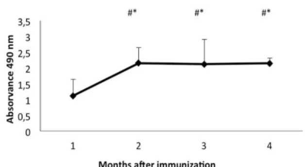
The production of isolated specific IgY after the immunization is shown in Figure 1, demonstrating a high level of immunoglobulin production twenty days after the complete immunization. There was a significant increase of IgY production from the first month after the immunization in relation to all the months analyzed (Kruskal-wallis test,
H=17.86; p<0.05), with a production peak in the
second month, stable until the end of the egg collection. There was no significant difference in IgY production during the last three months of egg
collection, indicating that the antibody level remained constant, without presenting a drop in production during this entire period
(Kruskal-wallis test, H= 17.86; p>0.05).
* Different from month 1, p< 0.05. # Without difference among months, p> 0.05.
Figure 1 - Production of isolated IgY specific anti-
canine IgG, by ELISA, after complete immunization. The bars represent standard deviation.
The kinetics of chicken antibody production usually demonstrates a transitory increase titer after the first immunization, and in subsequent immunizations, there may be an initial increase with approximately 10 days, generating a plateau for another ten days and a decline thereafter (Schade et al. 2005). Different results were demonstrated in this study, since 20 days after the last immunization, there were high titers of specific IgY with a significant increase for the second month after the immunization
(Kruskal-wallis test, H=17.86; p<0.05), after which there
was a peak of production remaining stable for three months after the immunization
(Kruskal-Wallis test, H = 17.86, p> 0.05). This confirmed
against pig IgG with FCA in the first inoculation
and FIA in the two subsequent ones,
demonstrating an earlier peak production, than in this work, in the first month after the complete immunization process. However, there was also a decline in IgY production two months after the last pig IgG inoculation, in contrast to the present study (Bizanov and Jonauskiené 2003).
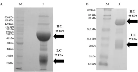
The SDS-PAGE of IgY in reduction conditions is depicted in Figure 2A, line 1 indicating isolated IgY by the PEG. Various bands with different molecular weights could be seen, varying from 220-25 kDa. The heavy chain of IgY weighed 68
kDa and the light chain 27 kDa, the visualized accessory protein bands represented the impurities that were not fully eliminated in the isolation process, justifying a purification process by thiofilic adsorption. The SDS-PAGE of the purified IgY is shown in Figure 2B, line 1 displaying purified IgY with a similar previous profile, the IgY presenting a heavy chain with 68 kDa and the light chain with 27 kDa. The reduction of accessory bands could be visualized. However, the bands between 56.2 and 35.8 kDa were not eliminated even after the purification process.
Figure 2 - Analysis by SDS-PAGE in reduced conditions of IgY, after isolation and purification
process. A: (HC)- Heavy chain; (LC)- Light chain. Lines: (M)- Molecular weight (BenchMark Protein ladder -Invitrogen); (1)–IgY isolated by PEG. B: (HC)- Heavy chain; (LC)- Light chain. Lines: (M)- Molecular weight (Prestained SDS-PAGE Standards, Broad Range (Bio-Rad Laboratories,Inc); (1)- Purified IgY by thiofilic adsorption.
Despite the consensus dispute about the molecular weight of IgY by most authors, the IgY was within agreement for the standard structure described (Warr et al. 1995; Schade and Hlinak 1996). There was a good result in the isolation process utilizing the PEG precipitation; however, with the presence
of accessory proteins in small amounts
representing the impurities not removed in this procedure. Because of this, a further purification process was justified for their removal, which directly interfered in the IgY recognition of the antigen for which it was produced, besides directly interfering in the process of conjugation with different types of enzymes and fluorochromes (Schade et al. 2000). The purification procedure resulted in a pure antibody, which could be
verified removing most of said accessory protein bands without changing the electrophoretic profile after isolation. The presence of bands between 56.2 and 35.8 kDa, not removed after the purification procedure, has been described in other studies (Pauly et al. 2011; Matheis and Schade 2011), which probably corresponded to the C-terminal fragment of the vitellogenin II precursor (Klimentzou et al. 2006). These fragments encumbered IgY recognition neither of the immunizing antigen adopted in this study nor any of the previously described (Matheis and Schade 2011; Pauly et al. 2011).
revealing an excellent specificity of produced IgY against the immunizing antigen. These results showed the excellent recognition between IgY and IgG with the proclivity of linkage between both the immunoglobulins, even in the dilutions with lower concentrations of IgG and IgY, respectively. The production of IgY against the mouse IgG with FCA and FIA presented stable titers up to 1: 250 000 (Kritratanasak et al. 2004). Pauly et al. (2009) demonstrated in ELISA stable titers of up to 1000 000 IgY with FCA in the first and FIA in
subsequent immunizations. Tu et al. (2006) with
FCA in the first and FIA in subsequent immunizations produced an IgY with stable titers
of 1.68 x 10⁸, after the first injection until the
sixteenth week. The developments of IgY titers in the present study were derived from only three immunizations, unlike the results described above with higher amounts of immunizing doses. Tu et al. (2006) used seven inoculations (one per week for seven weeks) and Pauly et al. (2009) used thirteen inoculations with an interval of four to
eight weeks between them. However,
Kritratanasak et al. (2004) applied three inoculations at intervals of two weeks each, resulting in superior IgY titers, compared to the present study (Kritratanasak et al. 2004).
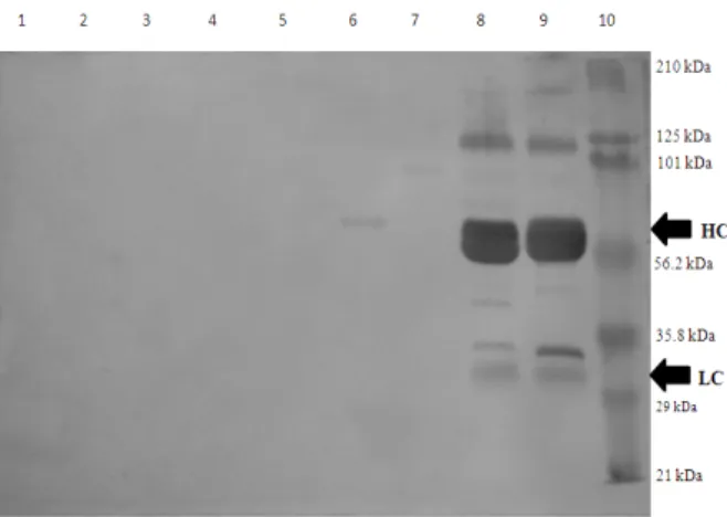
In western blot (Fig. 3), line 9 demonstrated that the purified IgY was capable of specifically recognizing the purified canine IgG. The bands of 210 kDa to 29 kDa were evident, and both the heavy chain as well as the light chain were detected. Similarly, this antibody also recognized
IgG of serum from a Leishmania (Leishmania)
chagasi infected dog (line 8). The purified IgY did
not recognize IgG of other animal species such as cat IgG (line 6), guinea pig IgG (line 5), rabbit IgG (line 4), goat IgG (line 3), sheep IgG (line 2) or horse IgG (line 1). Furthermore, no recognition was detected for dog IgM (line 7).
In the western blot, the purified IgY was able to recognize effectively and specifically both the specific IgG canine used as immunizing antigen, as present in the serum of infected dog, due to IgY ability to recognize the epitopes more effectively when mammalian proteins were used with the antigens (Svendsen et al. 1996). There was no binding of IgY to other animal species
immunoglobulins, a characteristic usually
described in the mammal antibodies, which have cross reactivity with different immunoglobulins species (Dias da Silva and Tambourgi 2010).
Figure 3 - Specificity analysis by western-Blot of
purified IgY. (HC)–Heavy chain; (LC)– Light chain. Lines: (1) Horse IgG ;(2)– Seep IgG;. (3)– Goat IgG; (4)–Rabbit; (5)–Guinea pig IgG; (6)– Cat IgG; (7)– Dog IgM; (8)– L. (L.) chagasi infected dog serum; (9)-Dog IgG; (10)- Molecular weight (Prestained SDS-PAGE Standards, Broad Range (Bio-Rad Laboratories).
Similar results were found in this study, in which the IgY was able to recognize the immunizing antigen specificity, were common in the literature (Gassmann et al. 1990; Tini et al. 2002; de Paula et al. 2011). However, different results were described by Nikbakht et al. (2009) when they produced an IgY against the camel IgG. They obtained strong western-blot recognition against the heavy and light chain of camel serum IgG. However, the IgY produced was capable of also recognizing the IgG heavy chain of bovine, horse and sheep serum, indicating that this IgY was produced with the different epitopes of the IgG of these other species in western blot analysis (Nikbakht et al. 2009). The results in this work showed the excellent potential of produced IgY as an immunological reagent, which could be used as a capture antibody, or conjugate in the kits for the immunological diagnosis of different canine diseases.
CONCLUSIONS
collection methods, able to recognize both serum IgG from the infected dog and purified IgG used with antigen with effectiveness without cross-reactivity with other species immunoglobulins and isotypes. Hence, it presented itself as an excellent tool for detecting the specific antibodies, which might be adopted as efficient immunological reagent for canine diagnosis of different diseases.
ACKNOWLEDGMENT
This study was financed by the National Counsel for Scientific and Technological Development
(CNPq, Edict MCT/CNPq
15/2007-Universal/Edital MCT/CNPq, process Number: 476052/2007-6. Edict MCT-
CNPq/MS-SCTIE-DECIT–Nº25/2006, process Number:
410571/2006-7, and the Foundation of Research Support of the State of Rio de Janeiro (Fundação de Amparo a Pesquisa do Estado do Rio de Janeiro) with Doctor Scholarship, process number: E-26/100.242/2010). The English was reviewed and revised by Mitchell Raymond Lishon, native of Chicago, Illinois, USA-UCLA, 1969.
REFERENCES
Akita EM, Nakai S. Comparison of four purification methods for the production of immunoglobulins from egg laid by hens immunized with an enterotoxigenic
E. coli strain. J Immunol Methods. 1993; 160:
207-214.
Bizanov G, Jonauskiené I. Production and purification of IgY from egg yolk after immunization of hens with pig IgG. Bull Vet Inst Pulawy. 2003; 47: 403-410. Bizanov G, Jonauskiené I, Hau J. A novel method,
based on lithium sulfate precipitation for purification of chicken egg yolk immunoglobulin Y, applied to immunospecific antibodies against Sendai virus. J
Lab Anim Sci. 2004; 31 (3): 121-130.
Bizanov G, Vyshniauskis G. A comparison of three methods for extracting IgY from the egg yolk of hens immunized with Sendai virus. Vet Res Commun.
2000; 24 (2): 103-113.
Bollen LS, Hau J. Freund's complete adjuvant has a negative impact on egg laying frequency in immunised chickens. In vivo.1999; 13 (1):107-108. Chalghoumi R, Beckers Y, Portetelle D, Théwis A. Hen
egg yolk antibodies (IgY), production and use for passive immunization against bacterial enteric infections in chicken: a review. Biotechnol Agron Soc
Environ. 2009; 13 (2): 295-308.
de Paula VS, da Silva AS, de Vasconcelos GA, Iff ET, Silva ME, Kappel LA, et al. Applied biotechnology for production of immunoglobulin Y specific to hepatitis A virus. J Virol Methods. 2011; 171 (1): 102-106.
Dias da Silva W, Tambourgi DV. IgY: a promising antibody for use in immunodiagnostic and in immunotherapy. Vet Immunol Immunopathol. 2010;
135 (3-4): 173-180.
Gassmann M, Thömmes P, Weiser T, Hübscher U. Efficient production of chicken egg yolk antibodies against a conserved mammalian protein. FASEB J.
1990; 4 (8): 2528-2532.
Griot-Wenk ME, Marti E, Racine B, Crameri R, Zurbriggen A, Weck A.L, et al. Characterization of two dog IgE-specific antibodies elicited by different recombinant fragments of the epsilon chain in hens.
Vet Immunol Immunopathol. 1998; 64: 15-32.
Hau J, Hendriksen CF. Refinement of polyclonal antibody production by combining oral immunization of chickens with harvest of antibodies from the egg yolk. ILAR J. 2005; 46 (3):294-299.
Kapoor P, Compton MM, Howarth B. Immunization of chickens with quail and turkey perivitelline membrane proteins: production of antibodies and their effects on fertility. Poult Sci. 2000; 79 (2): 245-256.
Kitaguchi K, Osada K, Horio F, Murai A. Exclusion of polymeric immunoglobulins and selective immunoglobulin Y transport that recognizes its Fc region in avian ovarian follicles. Vet Immunol
Immunopathol. 2008; 121 (3-4): 290-299.
Klimentzou P, Paravatou-Petsotas M, Zikos C, Beck A, Skopeliti M, Czarnecki J, et al. Development and immunochemical evaluation of antibodies Y for the poorly immunogenic polypeptide prothymosin alpha.
Peptides. 2006; 27 (1):183-193.
Kowalczyk K, Daiss J, Helpern I, Routh TE. Quantitation of maternal-fetal IgG transport in chicken. Immunology. 1985; 19: 157-167.
Kritratanasak S, Chiampanichayakul S, Kasinrerk W. Production of IgY anti-mouse IgG antibodies from chicken eggs. Asian Pac J Allergy Immunol. 2004; 22 (1): 61-68.
Laemmli UK. Cleavage of structural proteins during assembly of the head bacteriophage T4. Nature.
1970; 227: 680-685.
Leenaars MPPA, Hendriksen CFM, Leeuw WA, Carat F, Delahaut P, Fischer R, et al. The Production of Polyclonal Antibodies in Laboratory Animals. ATLA.
1999; 27: 79-102.
Levesque S, Martinez G, Fairbrother JMPS. Improvement of adjuvant systems to obtain a cost-effective production of high levels of specific IgY.
Poult Sci. 2007; 86 (4): 630-635.
Li XL, Shuai JB, Fang WH. Protection of Carassius
auratus Gibelio against infection by Aeromonas
hydrophila using specific immunoglobulins from hen
egg yolk. J Zhejiang Univ Sci B. 2006; 7 (11): 922-928.
Matheis W, Schade R. Development of an IgY-based rocket-immunoelectrophoresis for identity monitoring of Pertussis vaccines. J Immunol Methods.2011; 369 (1-2): 125-132.
Mine Y, Kovacs-Nolan J. Chicken egg yolk antibodies as therapeutics in enteric infectious disease: a review.
J Med Food. 2002; 5 (3): 159-169.
Narat M. Production of Antibodies in Chickens. Food
Technol Biotechnol. 2003; 41 (3): 259–267.
Nguyen SV, Umeda K, Yokoyama H, Tohya Y, Kodama Y. Passive protection of dogs against clinical disease due to Canine parvovirus-2 by specific antibody from chicken egg yolk. Can J Vet Res.
2006; 70 (1): 62-64.
Nikbakht GHB, Tabatabaei S, Khormali M, Ashrafi I. Characterization of IgY antibodies, developed in hens, directed against camel immunoglobulins. Int J
Vet Res. 2009; 3: 37-41.
Pauly D, Chacana PA, Calzado EG, Brembs B, Schade R. IgY technology: extraction of chicken antibodies from egg yolk by polyethyleneglycol (PEG) precipitation. J Vis Exp [Internet]. 2011[cited 2012 Dez. 10]; 1 (51): 3084. Avaliable from: http://www.jove.com/video/3084/igy-technology-
extraction-chicken-antibodies-from-egg-yolk?ID=3084.
Pauly D, Dorner M, Zhang X, Hlinak A, Dorner B, Schade R. Monitoring of laying capacity, immunoglobulin Y concentration, and antibody titer development in chickens immunized with ricin and botulinum toxins over a two-year period. Poult Sci.
2009;88 (2): 281-290.
Polson A, Coetzer T, Krugar Maltzahn von E, Merwe van der KJ. Improvements in the isolation of IgY from the yolk of eggs laid by immunized hens.
Immunol Invest. 1985; 14: 323-327.
Polson A, Von Wechmar MB, Van Regenmortel MH. Isolation of viral IgY antibodies from yolks of immunized hens. Immunol Commun. 1980; 9: 475-493.
Rose ME, Orlans E, Bujuttress N. Immunoglobulin classes on the hen’s egg: their segregation in yolk and white. Eur J Immunol. 1974; 4: 521-523.
Schade R, Behn I, Erhard M, Hlinak A, Staak C. Chicken egg yolk antibodies, production and application: IgY-Technology. Germany: Springer; 2000.
Schade R, Calzado EG, Sarmiento R, Chacana PA, Porankiewicz-Asplund J, Terzolo HR. Chicken egg yolk antibodies (IgY-technology): A review of progress in production and use in research and in human and veterinary medicine. ATLA. 2005; 33 (2):129-154.
Schade R, Hlinak A. Egg yolk antibodies, state of the art and future prospects. ALTEX. 1996; 13 (5): 5-9. Schniering A, Schade R, Hiepe T. Development of an
IgY-based assay for the detection of ascaris-suum antigens. ALTEX. 1996; 13 (5): 62-65.
Silva VL. Imunobiologia aviária e aplicação da imunoglobulina Y (IgY) na soroepidemiologia das leishmanioses caninas [PhD Thesis]. Seropédica, Rio de Janeiro: Rural Federal University of Rio de Janeiro; 1999.
Stalberg J, Larsson A. Extraction of IgY from egg yolk using a novel aqueous two-phase system and comparison with other extraction methods. Ups J
Med Sci. 2001; 106 (2): 99-110.
Svendsen L, Crowley A, Stodulski G, Hau J. Antibody production in rabbits and chickens immunized with human IgG. A comparison of titre and avidity development in rabbit serum, chicken serum and egg yolk using three different adjuvants. J Immunol
Methods. 1996; 191 (2): 113-120.
Tini M, Jewell UR, Camenisch G, Chilov D, Gassmann M. Generation and application of chicken egg-yolk antibodies. Comp Biochem Physiol A Mol Integr
Physiol. 2002;131 (3): 569-574.
Towbin H, Staehelin T, Gordon J. Electrophoresis transfer of proteins from polyacrylamide gels to nitrocellulose sheets: procedure and some applications. Proc Natl Acad Sci USA. 1979; 76: 4350-4354.
Tu YY, Ma CY, Ho SB, Chen CC, Chang HM. Affinity Measurement of Lactoferrin (LF)-Anti-LF Immunoglobulin in Yolk (IgY) Complexes by Competitive Indirect Enzyme-Linked Immunosorbent Assay (CI-ELISA). J Food Drug Anal. 2006; 14 (4): 379-384.
Voller A, Bidwell DE, Bartlett ANN. Enzime immunoassays indiagnostic medicine. Theory and practice. Bull World Health Organ. 1976; 53: 55-65. Warr W, Magor KE, Higgins DA. IgY: clues to the
origins of modern antibodies. Immunology Today.
1995; 16 (8): 392-398.
Witkowski PT, Bourquain DR, Hohn O, Schade R, Nitsche A. Gene gun-supported DNA immunisation of chicken for straightforward production of poxvirus-specific IgY antibodies. J Immunol
Methods. 2009; 341 (1-2): 146-153.