Rev Bras Med Esporte _ Vol. 13, Nº 1 – Jan/Fev, 2007
47e
1. Departamento de Biomecânica, Medicina e Reabilitação do AparelhoLocomotor, Faculdade de Medicina de Ribeirão Preto, USP, Ribeirão Preto, SP.
2. Interunidades Bioengenharia, Escola de Engenharia de São Carlos, Fa-culdade de Medicina de Ribeirão Preto, Instituto de Química de São Carlos, USP, Ribeirão Preto, SP.
Received in 14/9/05. Final version received in 27/6/06. Approved in 2/8/06. Correspondence to: João Paulo Chieregato Matheus, Rua Afonso Rato, 852, Bairro Mercês – 38060-040 – Uberaba, MG. Tels.: (34) 3312-7318 / (34) 9195-1011. E-mail: [email protected]
Effects of neuromuscular electric stimulation during
immobilization in the mechanical properties
of the skeletal muscle
João Paulo Chieregato Matheus1, Liana Barbaresco Gomide2, Juliana Goulart Prata de Oliveira2,
José Batista Volpon1 and Antônio Carlos Shimano1
O
RIGINALA
RTICLEKeywords: Immobilization. Muscular atrophy. Traction resistance. Mechanical
prop-erties. Electric stimulation therapy. ENGLISH VERSION
ABSTRACT
The neuromuscular electric stimulation (NMES) is an important tool used in sports medicine to accelerate the recovery process. The objective of this study was to analyze the effects of NMES during immobilization of the gastrocnemius muscle, in lengthened (LP) and shortened positions (SP). Sixty young female Wistar rats were distributed into six groups and followed for 7 days: control (C); electric stimulation (ES); immobilized in shortening (ISP); im-mobilized in lengthening (ILP); imim-mobilized in shortening and tric stimulation (ISP + ES) and immobilized in lengthening and elec-tric stimulation (ILP + ES). For the immobilization, a tubular mesh and cotton rolls together with the plaster were wrapped around the rat’s right posterior paw. NMES in a frequency of 50 Hz was used 10 minutes a day, totaling 20 contractions in each session. After 7 days the animals were sacrificed and their gastrocnemius muscles of the right paw were submitted to a mechanical test of traction in a universal test machine (EMIC®). From the load versus
elongation curves the following mechanical properties were ob-tained: elongation in the yield limit (EPL), load in the yield limit (LPL) and stiffness. The SP and LP immobilizations promoted sig-nificant reductions (p < 0.05) in the EPL and LPL properties, being more remarkable in the ISP group. When the NMES was used, there was significant increase (p < 0.05) of these properties only in the ISP group. As for stiffness, significant reduction was observed (p < 0.05) only of the C group for the ISP group. When the NMES was used, the stiffness of the ILP + EE group was significantly (p < 0.05) higher and closer to the C group than of the ISP + EE group. We conclude that in this experimental model the immobili-zation of the muscles in the lengthened position delayed the atro-phy process and the electric stimulation during the immobilization contributed to the maintenance of the mechanical properties dur-ing the immobilization period, mainly for the ILP + ES group.
INTRODUCTION
In modern sports medicine, there is a tendency to early immobi-lize an injured segment with the aim to fast rehab it, although strict immobilization has already been used as treatment for several in-juries, either musculo-skeletal or ligamentar. However, it is possi-ble to observe muscular atrophy even in the first seven days in which the individuals remain immobilized(1-4).
The intensity in which atrophy occurs is directly influenced by the position in which the limb is immobilized(5). Immobilization of a
muscle in lengthening (LP) delays the atrophy process by disuse; however, the antagonist muscle, immobilized in shortening (SP), suffers more rapidly the deleterious effects of immobilization(6).
The time in which the individuals remain immobilized as well as the prevention of the negative effects derived from the immobili-zation, especially in the sports scenario, has been a crucial factor for the recovery of athletes. With the fast improvement of rehabil-itation programs, resources which minimize the deleterious effects secondary to surgeries as well as immobilization have been searched. One of the procedures is the use of the neuromuscular electrical stimulation (NMES) which next to kinesiotheraphy has been one of the resources most widely used in muscular strength-ening, as well as in the prevention of atrophy before, during and after the injury episodes.
Since the Montreal Olympic Games (1976), when Russian phy-sician Yakov Kots obtained strength gains of 30 to 40% in elite athletes, several other studies have suggested NMES, especially during rehabilitation processes, as in the post-surgery period of knee surgeries(7). The utilization of NMES in a trial to prevent the
deleterious effects of immobilization processes and surgical pro-cedures, have great applicability, since it reduces the rehabilitation time as well as promotes the return of the individuals to their usual activities in a shorter time.
Considering the lack of research concerning the application of the electric stimulation as prevention of muscular atrophy, the aim of the present study was to analyze the effects of the neuromus-cular electric stimulation (NMES) during the immobilization of the gastrocnemius muscle of rats, in lengthening (LP) and shortening positions (SP).
METHODS
Animals
Sixty Wistar, Rattus norvegicus albinus female rats were used for the development of this study. The animals from the Central bio cemetery of the City Hall of the Ribeirão Preto University of São Paulo Campus (USP) were kept during the experimental pro-tocols in the bio cemetery of the Bioengineering Laboratory of the Medicine School of Ribeirão Preto – USP. The rats were kept in collective cages with three animals per cage, at controlled room temperature of 25oC, photo period of 12 h clear/12 h dark,
48e
Rev Bras Med Esporte _ Vol. 13, Nº 1 – Jan/Fev, 2007Experimental groups
The animals were randomly distributed in the following experi-mental groups:
Group (1) – Control (C): composed of 10 animals kept in stan-dard cages for seven days.
Group (2) – Electro stimulated (ES): composed of 10 animals which had the right gastrocnemius muscle submitted to electric stimulation for seven days.
Group (3) – Immobilized-SP (ISP): composed of 10 animals which had the right posterior limb (pelvis, hip and knee) immobilized in extension and the ankle in plantar flexion, which kept the gastroc-nemius muscle in shortening for seven days.
Group (4) – Immobilized-LP (ILP): composed of 10 animals which had the right posterior limb (pelvis, hip and knee) immobilized in extension and the ankle in dorsal flexion, which kept the gastroc-nemius muscle in elongation position during seven days.
Group (5) – Immobilized-SP/Electro stimulated (ISP + ES): com-posed of 10 animals immobilized according to model used in group 3. However, they were submitted to the electric stimulation proto-col during the seven days in which they were kept immobilized.
Group (6) – Immobilized-SP/Electro stimulated (ILP + ES): com-posed of 10 animals immobilized according to model used in group 4. However, they were submitted to the electric stimulation proto-col during the seven days in which they were kept immobilized.
Immobilization technique
Prior to the casting apparel manufacturing, the animals were anesthetized with association of ketamine hydrochloride (80 mg/ Kg) and xylazine hydrochloride (15 mg/Kg), in the dose of 0,6 ml of the solution for each 100 grams of body weight.
The right posterior limb, from the hip to the ankle, was initially wrapped in a tubular mesh together with cotton plasters in order to avoid ischemia in the limb and prevent the formation of pres-sure ulcers. For the immobilization, fast-drying plaster cast with approximately three centimeters wide was used.
Prior to the complete cast drying of groups 5 and 6, an opening of approximately one centimeter of diameter was made on the region of the motor point of the gastrocnemius.
The immobilization model used in this study was based on the one proposed by Booth and Kelso(8), with adaptation for one limb
only.
Electric stimulation
An equipment of electric stimulation by BIOSET®, model
Phys-iotonus Four, with generator units of low frequency, double phased (depolarized) and pulsing of short duration, applied under controlled frequency was used.
Stimulation electrodes
In order to stimulate the gastrocnemius muscle of groups 2, 5 and 6 the manufacturing of two electrodes was necessary. A dis-perse electrode with 6 cm2 area which connects to the lumbar
region and another one active, pen-shaped with 0,5 cm diameter which connects on the motor point of the right gastrocnemius muscle through the opening manufactured on the cast.
Stimulation protocol
The muscles were submitted to electric stimulation with 50 Hz frequency, with 8-second contraction and 22-second rest. There were 20 muscular contractions electrically-induced in each daily session of 10 minutes during seven consecutive days.
In order to allow better contact between the skin and the elec-trodes, protect the animal from a possible burn, as well as facili-tate the electric current conduction, a gel coating was applied be-tween the electrodes and the contact regions.
In groups 5 and 6, the electric stimulation was performed after two hours of the cast apparels manufacturing.
Preparation of the gastrocnemius muscle
After the experimental protocols ending, the animals were sub-mitted to euthanasia by intraperitoneal administration of excessive dose of sodium thiopental, in order to have their gastrocnemius dissected and submitted to the traction mechanical essay.
The gastrocnemius was removed from the right posterior limb of each animal by skin and some other soft parts removal, followed by the disarticulation of the ankle and hip. Extra caution was kept in order to keep the muscle integrity, preserving its origin in the distal third of the femur and insertion on the calcaneus. The bone origin and insertion were kept to facilitate the fixation of the piece to the essay machine.
After dissection, the pieces were immersed in a Ringer lactate solution during 30 minutes in room temperature until the essays performance.
Traction mechanical essay
The universal essay machine (EMIC®brand name, model
DL10000) of the Bioengineering Laboratory of the Medicine School of Ribeirão Preto – USP, equipped with load cell of 50 kgf was used for the traction essay of the gastrocnemius muscle.
The machine used has interface direct to a microcomputer with Tesc® software capable to generate a load versus lengthening graph
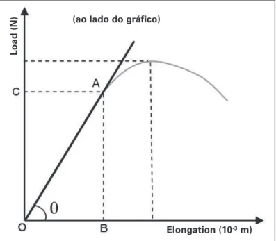
for each essay (figure 1).
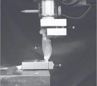
Two accessories were manufactured for the fixation of the piece to be tested; being one for the fixation of the femur and the other for fixation of the calcaneus, keeping the knee and the ankle in a 90o angle (figure 2). At the time of the essay, the muscle was
con-nected to the machine, and according methodology established by the laboratory, a pre-load of 200 g was given during an accommo-dation time of 30 seconds, with the aim to promote accommoda-tion to the system.
After the pre-load, the essay continued for an average of 8 min-utes with pre-established velocity for a 10 mm/minute essay. The load applied was registered by the software in regular lengthening intervals until the rupture moment of the muscle.
From the load versus lengthening graphs of each essay, the fol-lowing mechanical properties were obtained and analyzed:
– elongation in the yield period (EPL): it is the lengthening value of the initial point (O) until the representation point of the maximal elastic lengthening (OB in figure 1). It is represented in meters (x10-3 m).
Figure 1 – Standard graph of load versus lengthening
(ao lado do gráfico)
Load (N)
Rev Bras Med Esporte _ Vol. 13, Nº 1 – Jan/Fev, 2007
49e
– load in the yield period (LPL): it is the maximal load value reg-istered in the elastic phase (OC in figure 1). It is represented in Newtons (N).
– stiffness: it corresponds to the angle tangent (θ). It is
repre-sented in Newtons/meter (N/m).
Statistical analysis
The statistical analysis was performed through the BioEstat®
program v. 2.0. The Kolmogorov Smirnov normality test was per-formed. For simultaneous analysis of the groups the ANOVA test was used and for comparison between groups the Turkey-Kramer test was used, both with pre-established significance levels of 5%.
RESULTS
Sixty muscles were essayed, being the values expressed in means and standard deviations for each of the properties of the six analyzed groups.
Elongation in the yield period (EPL)
The means and standard deviations concerning the EPL of the six groups are presented in figure 3.
Significant difference (p < 0.05) of the Control group was ob-served for the Immobilized groups, both SP and LP. In the compar-ison between the Immobilized SP and Immobilized SP + ES groups significant difference (p < 0.05) was also observed, which did not occur in the comparison between the Immobilized LP and Immobi-lized LP + ES groups (p > 0.05).
Significant difference (p < 0.05) was observed between the Im-mobilized SP + ES and ImIm-mobilized LP + ES groups. Conversely, between the Immobilized SP + ES and the Control group this dif-ference was not observed (p > 0.05).
Load in the yield period (LPL)
The means and standard deviations concerning the LPL of the six groups are presented in figure 4.
Figure 2 – Setting for the performance of the traction essay on the essay universal machine. Muscle (A), accessories for muscle fixation (B).
Figure 3 – Mechanical property of elongation in the yield period of the Control; Electro Stimulated (ES); Immobilized SP; Immobilized LP; Immo-bilized SP + Electro Stimulated and ImmoImmo-bilized LP + Electro Stimulated groups.
Figure 4 – Mechanical property of the load in the yield period of the Con-trol; Electro Stimulated (ES); Immobilized SP; Immobilized LP; Immobilized SP+ Electro Stimulated and Immobilized LP + Electro Stimulated groups.
Significant difference (p < 0.05) of the Control and ES groups for the Immobilized groups, both SP and LP, was observed. In the com-parison between the Immobilized SP and Immobilized SP + ES groups significant difference (p < 0.05) was also observed, which did not occur in the comparison between the Immobilized LP and Immobilized LP + ES groups (p > 0.05).
It was not observed significant difference between the Immobi-lized SP + Electro Stimulated and ImmobiImmobi-lized LP + Electro Stimu-lated groups (p > 0.05).
Stiffness (S)
The means and standard deviations concerning stiffness of the six groups are presented in figure 5.
Figure 5 – Mechanical property of stiffness of the Control; Electro Stimu-lated (ES); Immobilized SP; Immobilized LP; Immobilized SP + Electro Stim-ulated and Immobilized LP + Electro StimStim-ulated.
EPL (x10
-3 m)
Control ES ISP ILP ISP + ES ILP + ES
Control ES ISP ILP ISP + ES ILP + ES
Stiffness (x10
3 N/m)
Control ES ISP ILP ISP + ES ILP + ES
50e
Rev Bras Med Esporte _ Vol. 13, Nº 1 – Jan/Fev, 2007Significant difference (p < 0.05) of Control group for the ES and Immobilized SP groups (p < 0.05) was observed. It was not possi-ble to observe significant difference (p > 0.05) between the Con-trol and Immobilized LP groups; whereas between the Immobi-lized SP + Electro Stimulated and ImmobiImmobi-lized LP + Electro Stimulated groups significant difference was observed (p < 0.05).
DISCUSSION
The findings of this study demonstrated that the casting immo-bilization of the gastrocnemius muscle for a period of seven days promoted significant biomechanical reductions, and that the posi-tion in which it was kept immobilized directly acted in the intensity of these alterations. In this study, as well as in the one by Järvinen et al.(5), the gastrocnemius muscles immobilized in shortening (SP)
had greater decrease of the properties than the ones immobilized in lengthening (LP). Tabary et al.(9), on the other hand, did no find
significant differences between the soleus muscles immobilized in SP and LP in their study.
According to Williams and Goldspink(10), the muscles
immobi-lized in LP are constantly submitted to tension, which stimulates the increase in the number of sarcomeres in series and reduces the intensity in which the atrophy occurs. As seen in our study, the immobilization in LP caused milder muscular alterations than in SP. Conversely, one may identify that the contracted muscle (SP) presents two basic characteristics: decrease of the sarcomeres in series and increase in the density of the conjunctive tissue. Thus, it is possible to understand that the shortened immobilized mus-cles have lower elasticity(11); what is observed in our study.
Anoth-er factor which may have contributed to the reduction of the prop-erties of the immobilized muscles was the decrease in diameter of the muscular fibers, already described by Appell(12).
Some authors affirm that it is possible to prevent the accumula-tion of conjunctive tissue(13) as well as atrophy occurrence(14) through
muscular contraction induced by electric stimulation.
Concerning the properties of EPL and LPL, the muscles of the Immobilized SP + ES group better responded to electric stimula-tion than the ones of the Immobilized LP + ES group. We believe that the occurrence of greater loss of properties in the Immobi-lized SP group than in the ImmobiImmobi-lized LP had contributed to it.
In the mechanical property of stiffness, lower mean was found in the Immobilized LP + ES group than in the Immobilized SP + ES group. According to Järvinen et al.(5), muscles stiffness is an
im-portant property to be studied, since their reduction indicates that the muscle is lengthening more in the presence of lower load, which makes it more susceptible to injury. Thus, it seems suitable to sup-pose that muscles immobilized in lengthening and electro stimu-lated are in better conditions, after the cast removal, than those immobilized in shortening and electro stimulated.
Some limitations as well as observations in the present study need to be mentioned. The most remarkable difficulty found in the pilot test was concerning the animals’ immobilization and their time in a cast. The method of wrapping using a tubular mesh, initially adopted, did not keep the animal immobilized for longer than two days. After some changes in the manufacturing methodology, an immobilization model similar to the one described by Booth and Kelso(8) was adopted. Such model is widely accepted by the
scien-tific community; however, instead of the two limbs, only the right posterior limb was kept in a cast. Moreover, the lack of current scientific literature concerning the mechanical properties of the
skeletal muscle and the use of electro stimulation as a tool in the rehabilitation processes makes the discussion of results difficult.
Several animals have been applied in experimental studies. In this one, we chose rats, since according to some authors(5,8), they
present musculo-skeletal structure similar to humans. Nonethe-less, the results obtained in these studies cannot be totally trans-ferred to humans; they should instead, guide further research for future practical application.
Therefore, the results of the present study suggest that immo-bilization of muscles in lengthening delays the process of atrophy and that electric stimulation performed during immobilization, con-tributes to prevention of the mechanical properties during the immobilization period.
ACKNOWLEDGMENTS
To CAPES (Coordenação de Aperfeiçoamento de Pessoal de Nível Su-perior – Coordination of Personal Improvement of Higher Level) for the financial support and concession of Master’s degree scholarships.
To BIOSET®Company – Industry of Electronics Technology Ltda. – which
on the person of Mr. Júlio César Bucalon, lent the electric stimulation ma-chine.
All the authors declared there is not any potential conflict of inter-ests regarding this article.
REFERENCES
1. Wills CA. Effects of immobilization of human skeletal muscle. Orthopaedic Re-view. 1982;11:57-64.
2. Appell HJ. Morphology of immobilized skeletal muscle and the effects of a pre and postimmobilization training program. Int J Sports Med. 1986;7(1):6-12. 3. Booth FW. Physiologic and biochemical effects of immobilization on muscle.
Clin Orthop Relat Res. 1987;(219):15-20.
4. Mercier J. Muscle plasticity and metabolism: effects of exercise and chronic diseases. Mol Aspects Med. 1999;20:319-73.
5. Järvinen MJ, Einola SA, Virtanen EO. Effect of the position of immobilization upon the tensile properties of the rat gastrocnemius muscle. Arch Phys Med Rehabil. 1992;73(3):253-7.
6. Herbert RD, Balnave RJ. The effect of position of immobilization on resting length, resting stiffness, and weight of the soleus muscle of the rabbit. J Orthop Res. 1993;11:358-66.
7. Stevens JE, Mizner RL, Snyder-Mackler L. Neuromuscular electrical stimulation for quadriceps muscle strengthening after bilateral total knee arthroplasty: a case series. J Orthop Sports Phys Ther. 2004;34(1):21-9.
8. Booth FW, Kelso JR. Production of rat muscle atrophy by cast fixation. J Appl Physiol. 1973;34(3):404-6.
9. Tabary JC, Tabary C, Tardieu C, Tardieu G, Goldspink G. Physiological and struc-tural changes in the cat’s soleus muscle due to immobilization at different lengths by plaster casts. J Physiol. 1972;224(1):231-44.
10. Williams PE, Goldspink G. Connective tissue changes in immobilized muscle. J Anat. 1984;138(2):343-50.
11. Salvini TF. Plasticidade e adaptação postural dos músculos esqueléticos. In: Mar-ques AP, editor. Cadeias musculares: um programa para ensinar avaliação fisio-terapêutica global. São Paulo: Manole, 2000;3-14.
12. Appell HJ. Muscular atrophy following immobilization. A review. Sports Med. 1990;10(1):42-58.
13. Williams PE, Catanese T, Lucey EG, Goldspink G. The importance of stretch and contractile activity in the prevention of connective tissue accumulation in mus-cle. J Anat. 1988;158:109-14.