Hypernatremic hemorrhagic encephalopathy: case
report and literature review
Encefalopatia hemorrágica hipernatrêmica: relato de caso e
revisão da literatura
CASE REPORT
A Caucasian 58 years-old male patient with previous high blood pressure and undeined psychiatric disorder was admitted to the Hospital Universitário Professor Edgard Santos of the Universidade Federal da Bahia (HUPES) on February 5, 2010, referred from another service. Twenty days before the admission he was found with reduced sensorial level, oliguria, piuria and increased blood urea nitrogen and creati-nine (BUN 180 mg/dL, Cr 5.0 mg/dL). Due to lack of both urinary and sensorial improvement in the previous service, was referred to a higher complexity hospital.
His medical history included systemic high blood pressure and a psychiatric disorder poorly characterized by his family, involving something similar to a bipo-lar disorder since 14 years-old. Were reported regubipo-lar use of Haloperidol, Chlor-promazine and Fenergan, prescribed by his treatment doctor. Family members reported that three days before being found with impaired consciousness level, he gone out of home during an agitation crisis.
By admission the patient had precarious general status, was eupneic, had good peripheral perfusion, was discolored +/IV, was restricted to bed and had no signs of trauma. His heart rate was 96 bpm, respiratory rate 24 ipm, and blood pressure 110/70 mmHg. he cardiovascular, respiratory and abdominal examinations evi-denced no changes. he neurological examination showed Glasgow 10 (4-1-5), with spontaneous eyes opening, aphasia, and eyes movement when commanded. Tetraparetic, with no signs of hyperrelexia. Isocoric and light relexive pupils. Other cranial nerves and sensitivity tests were not conducted, as the patient’s consciousness level was impaired.
he initial laboratory tests showed hemoglobin 9.5 g/dL, white blood cells count without deviations and platelet counts 210,000/mm3. Creatinine 5.1mg/ dL (reference range: 0.5-1.3 mg/dL), blood urea nitrogen 181 mg/dL (reference
Lucas Sampaio Mata1, Dimitri
Gusmão2, Antônio Raimundo Pinto
de Almeida3
1. Physician of the Internal Medicine Service of the Hospital Universitário Professor Edgard Santos of the
Universidade Federal da Bahia – UFBa – Salvador (BA), Brazil.
2. Physician of the Hospital Universitário Professor Edgard Santos – Universidade Federal da Bahia – UFBa – Salvador (BA), Brazil.
3. Professor for the Internal Medicine Department of the Faculdade de Medicina da Universidade Federal da Bahia – UFBa - Salvador (BA), Brazil.
ABSTRACT
Hypernatremia is a common elec-trolyte disorder in people with impaired thirst control mechanism or access to water, and may lead from minimal dis-orders until coma. Among the hyper-natremia morbidities, central nervous system hemorrhage is uncommon and poorly studied. We report a case
invol-ving a patient admitted to the intensive care unit with reduced consciousness level, hypernatremia and head comput-ed tomography scan showing bilateral parenchyma hemorrhage. A literature review of hypernatremia hemorrhagic encephalopathy was conducted.
Keywords: Hypernatremia; Cerebral hemorrhage; Brain diseases
his work was developed in the Internal Medicine Service of the Hospital Professor Edgard Santos of the Universidade Federal da Bahia – UFBa – Salvador (BA), Brazil.
Conlicts of interest: None.
Submitted on June 26, 2010. Accepted on September 8, 2010.
Author for correspondence: Dimitri Gusmão
Rua Eng. Celso Torres, 18 - Apt. 601 - Graça
Zip Code: 40150-280 - Salvador (BA), Brazil.
range: < 40 mg/dL). Ionogram revealed sodium 196 mEq/L (reference range: 135-145 mEq/L), potassium 3.3 mEq/L (reference range: 3.5-5.5 mEq/L) and blood gasometry with borderline PO2, and no metabolic changes.
he patient was transferred to the intensive care unit (ICU) on the 2nd hospitalization day, and put on mechanical ventilation and widened his antimicrobial covering, and then sent to a head computed tomography (CT) scan without administration of a contrast agent which evidenced bilateral cerebellum and external capsule hemorrhage (Figure 1).
Figure 1 – Head computed tomography scan without admi-nistration of a contrast agent evidencing bilateral cerebellum and external capsule hemorrhage and surrounding edema.
he patient progressed in the ICU with improved BUN and creatinine (Graph 1) in addition to increased urinary output following parenteral hydration with crys-talloids, with no renal replacement therapy need. After the volume correction, hypotonic solution therapy was started in order to correct the hypernatremia and com-plying with the daily maximal correction (Graph 1). During the hydroelectrolytic balance correction, and after sedation was withheld, the patient spontaneously opened his eyes and recovered the respiratory drive. Tracheostomized in February 24, was weaned from me-chanical ventilation and was discharged from the ICU on March 9, and was discharged from the hospital on April 14, alert and with no neurological deicits.
DISCUSSION
Plasma sodium concentration and serum osmolarity are controlled by the water homeostasis, mediated by thirst, vasopressin and the kidneys. Hypernatremia is a frequent hydroelectrolytic disorder, deined by a sodium plasma concentration above 145 mmol/L.(1) It may be caused either by water and hypotonic luids loss,
inap-Graph 1 - Sodium, creatinine, and blood urea nitrogen progression
Sodium progression
m
Eq
/L
ite
r
Sodium
Creatinine progression
m
g/
dL
Creatinine
BUN
Blood urea nitrogen (BUN) progression
m
g/
propriate water ingestion, or large hypertonic solutions administration. As it commonly involves more a deicit of water than properly sodium excess, hypernatremia is more frequent in groups with thirst mechanism impair-ment or restricted access to water. Among the high risk group should be emphasized the impaired mental status subjects, psychiatric patients, those under mechanical ventilation, elderly and children.
Being fundamentally extracellular, sodium contrib-utes for plasma hypertonism leading to water membrane migration. herefore, its plasma increase leads to hyper-tonic hyperosmoloarity causing cell dehydration due to water migration to the extracellular space.(1,2) his dis-order’s morbidities may range from minimal to very se-rious, including death. his severity is proportional to the disorder’s severity and how fast the sodium increases, however it is often diicult to identify the real hyperna-tremia contribution to the outcome, as other morbidities may be involved.
Hypernatremia and central nervous system hemor-rhage
he central nervous system (CNS) is frequently in-volved in hypernatremia cases. he clinical features de-scribed are several, and may include apathy, convulsion and even coma.
Hypernatremia as cause of acute neurological disor-der is both well-described, and frequent. However, hy-pernatremia as cause of CNS hemorrhage is not frequent in the literature. In 1919, Weed and McKibben demon-strated capillary retraction and congestion following hy-pertonic NaCl infusion in animals, with no hemorrhage evidence.(3) In 1979 Young reported a case of an hyper-natremia adolescent after inadvertent hypertonic solu-tion infusion and had seizures and coma. he head CT evidenced small hemorrhages, difused by the subcorti-cal area, and the necropsy conirmed these indings.(4) In 1997 Han reported on ultrasound and tomography indings in 2 children admitted with neurological signs and hypernatremia.(5) Both patients had similar imagery indings, showing cerebral parenchyma changes with multifocal hemorrhage and hemorrhagic infarction.
Luttrell, in 1958 described three cases in children with hypernatremia and changed consciousness level, ranging from lethargy to coma. he autopsy evidenced central nervous system hemorrhage as predominant pathologic change. Extensive subarachnoidal bleeding, as well as intradural, intracerebral and intraventricular were described in this study. Microscopic examination has shown severe vascular congestion in small and
capil-lary nervous system vessels, with brain tissue pethechiae. As reporting on children admitted with increased sodi-um and eventually already with consciousness changes, the Luttrell’s study did not allow stating that the sodium disorder preceded the hemorrhage, or even caused it.(2)
In 1959, the same author reported on an experimen-tal trial in cats, divided in three groups. On group I, so-dium hypertonic solution was injected intraperitoneally, on group II, hypertonic urea solution was injected in the peritoneum, and the group III was the used as control, with part of the group having luids restriction and the other part peritoneally injected with normal saline so-lution.3 It should be remarked that the animals infused hypertonic solutions had no hypertensive peaks during the trial. Marked drop in spinal pressure was identiied 60-80 minutes after the hypertonic injection for both intervention groups, and the pressure drop was shorter in the urea group. Cerebral tissue retraction and intra-cranial hemorrhage were consistently found in similar degrees in both groups, with no hemorrhagic signs in other organs. None of these changes was seen in the con-trol group animals. In six intervention groups animals (4 in the NaCl hypertonic solution and 2 in the urea group) their spinal pressure was maintained for 4 hours by means of cisternal solutions infusion. In this group, the arterial and venous pressure changes were similar to groups I and II without spinal pressure maintenance. he autopsy showed that none of these animals had evi-dence of intracranial hemorrhage.(2)
herefore, CNS hypernatremia determined hemor-rhage appears to involve several factors. Cell water out-low and sout-lowed sodium transportation between blood and cerebrospinal luid (CSF) would lead to reduced cerebral and CSF volumes. his results in a negative pressure around the brain tissues, leading to venous and capillary expansion and consequent vessels rup-tures.(3) hen, cerebral hemorrhage would result from cerebral retraction inside a rigid structure (the skull) which, associated to negative CSF pressure, would stress the vascular network, and produce hemorrhage from a congested, dilated and consequently fragile capillary network.(2,3) his rational corroborates the necropsy ind-ings where all organs but CNS were spared. he vascular factor as a causative of cerebral hemorrhage would be secondary, and consequent to the CSF pressure reduc-tion. It is admissible, however that vascular injuries in functionally relevant areas may have some primary role on the neurological damage caused by hypernatremia.(2)
hemor-rhage. In addition, by means of this trial it was possible to imply that CSF pressure reduction is fundamental for hemorrhage induction, as it was completely prevented by luid infusion keeping positive CSF pressure.
It should be noted that in this study by Luttrell, hy-pernatremia in the intervention group was exclusively induced by hypertonic solution administration (either sodium chloride or urea). herefore, studies showing that free water or hypotonic luids loss-induced hyper-natremia is a causative of CNS hemorrhage are miss-ing. Additionally, the study had an inappropriate size control group, too small versus the total study animals. However it should be highlighted the consistency of the hemorrhagic indings in the intervention group (48/53; 90.5%). In addition, it would have been interesting measurement of the plasma sodium before and after the intervention, to conirm its levels changes and even to show which sodium levels were associated to cerebral hemorrhage.
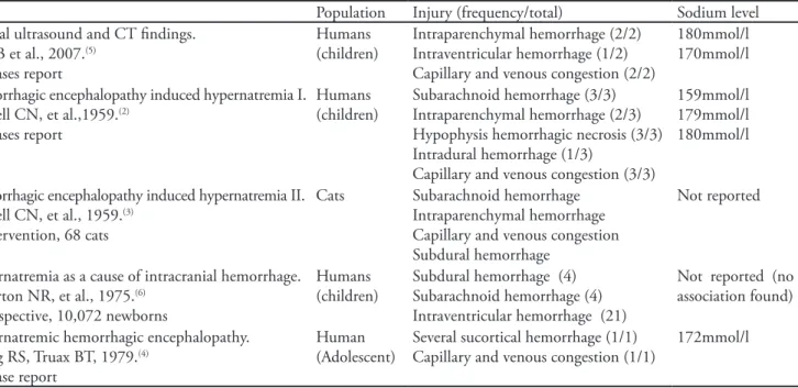
In the available studies, the sodium levels associated with intracranial hemorrhage are consistently above 160 mmol/L (Table 1). Most of the studies showing these values, were in children. As the children brains are softer and have higher water content, brain tissue retracts more than adult brains, justifying the higher incidence of hy-pernatremia induced hemorrhage in younger patients. It is therefore possible that in the adult, the sodium levels
associated with CNS hemorrhage are even higher than the commonly described for children.
Interestingly in none of the necropsy indings scriptions was pontine or extra-pontine myelinolisis de-scribed, likely because this change is exclusively associ-ated with excessive hyponatremia correction.
We could not ind any other hypernatremic hemor-rhagic encephalopathy reported in adults (published in English). herefore, this appears to be the irst report in an adult patient.
RESUMO
Hipernatremia é um distúrbio hidroeletrolítico frequente em pessoas nas quais o mecanismo da sede ou o acesso à água está comprometido podendo causar desde morbidades míni-mas até coma. Entre as morbidades causadas pela hipernatre-mia, a hemorragia do sistema nervoso central é infreqüente e pouco estudada. Relatamos um caso de paciente admitido na unidade de terapia intensiva com redução do nível de con-sciência, hipernatremia e tomograia computadorizada de crânio evidenciando hemorragia intraparenquimatosa bilat-eral. Foi realizada revisão de literatura de encefalopatia hemor-rágica hipernatrêmica.
Descritores: Hipernatremia; Hemorragia cerebral; Ence-falopatias
Table 1 – Central nervous system hemorrhage-associated hypernatremia – studies description
Study Population Injury (frequency/total) Sodium level
Cranial ultrasound and CT indings. Han B et al., 2007.(5)
2 cases report
Humans (children)
Intraparenchymal hemorrhage (2/2) Intraventricular hemorrhage (1/2) Capillary and venous congestion (2/2)
180mmol/l 170mmol/l
Hemorrhagic encephalopathy induced hypernatremia I. Luttrell CN, et al.,1959.(2)
3 cases report
Humans (children)
Subarachnoid hemorrhage (3/3) Intraparenchymal hemorrhage (2/3) Hypophysis hemorrhagic necrosis (3/3) Intradural hemorrhage (1/3)
Capillary and venous congestion (3/3)
159mmol/l 179mmol/l 180mmol/l
Hemorrhagic encephalopathy induced hypernatremia II. Luttrell CN, et al., 1959.(3)
Intervention, 68 cats
Cats Subarachnoid hemorrhage Intraparenchymal hemorrhage Capillary and venous congestion Subdural hemorrhage
Not reported
Hypernatremia as a cause of intracranial hemorrhage. Roberton NR, et al., 1975.(6)
Prospective, 10,072 newborns
Humans (children)
Subdural hemorrhage (4) Subarachnoid hemorrhage (4) Intraventricular hemorrhage (21)
Not reported (no association found)
Hypernatremic hemorrhagic encephalopathy. Young RS, Truax BT, 1979.(4)
1 case report
Human (Adolescent)
Several sucortical hemorrhage (1/1) Capillary and venous congestion (1/1)
REFERENCES
1. Adrogué HJ, Madias NE. Hypernatremia. N Engl J Med. 2000;342(20):1493-9.
2. Luttrell CN, Finberg L. Hemorrhagic encephalopathy induced by hypernatremia. I. Clinical, laboratory, and pathological observations. AMA Arch Neurol Psychiatry. 1959;81(4):424-32.
3. Luttrell CN, Finberg L, Drawdy LP. Hemorrhagic encephalopathy
induced by hypernatremia. II. Experimental observations on hyperosmolarity in cats. Arch Neurol. 1959;1:153-60.
4. Young RS, Truax BT. Hypernatremic hemorrhagic encephalopathy. Ann Neurol. 1979;5(6):588-91.
5. Han BK, Lee M, Yoon HK. Cranial ultrasound and CT indings in infants with hypernatremic dehydration. Pediatr Radiol. 1997;27(9):739-42.