16 artigo 556
ORIGINAL ARTICLE
1 – MSc in Medicine and Health Sciences, PUCRS, Porto Alegre, RS, Brazil.
2 – Biologist and Doctoral Student in Medicine and Health Sciences, PUCRS, Porto Alegre, RS, Brazil. 3 – Biologist and Master’s Student in Medicine and Health Sciences, PUCRS, Porto Alegre, RS, Brazil. 4 – PhD in Medicine and Health Sciences, PUCRS, Porto Alegre, RS, Brazil.
5 – Titular Professor of the Department of Surgery, School of Medicine, PUCRS, Porto Alegre, RS, Brazil. Work performed at the Pontificate Catholic University of Rio Grande do Sul (PUCRS), Porto Alegre, RS.
Correspondence: Av. Ipiranga 6690, conj. 216, Jardim Botânico, 90610-000 Porto Alegre, RS, Brazil. E-mail: [email protected] Work received for publication: July 7, 2011; accepted for publication: August 4, 2011.
VIABILITY OF VASCULARIzED BONE GRAFT FROM THE ILIAC CREST
USING THE ILIAC BRANCH OF THE ILIOLUMBAR
ARTERY: EXPERIMENTAL STUDY ON RATS
Fabian Maccarini Peruchi1, Alessandra Deise Sebben2, Martina Lichtenfels3, Marcos Ricardo de Oliveira Jaeger4, Jefferson Braga Silva5
AbSTRACT
Objective: Through an experimental model, our aim was to create inferences about the viability of vascularized bone grafts from the iliac crest in rats and investigate their histologi-cal features. Methods: Twenty-one rats were used, divided into two groups: the first consisted of animals that were subjec-ted to the technique of vascularized bone graft pedicled onto the iliac branch of the iliolumbar artery; the second (control group) underwent the same procedure as performed on the first group, with the addition of ligation of the vascular pedi-cle. The viability of the bone grafts was observed for three we-eks, by means of direct observation of the graft, histology and immunohistochemistry. Results: All the vascularized grafts evaluated in the first week showed viability according to direct
The authors declare that there was no conflict of interest in conducting this work
This article is available online in Portuguese and English at the websites: www.rbo.org.br and www.scielo.br/rbort
observation, histology and immunohistochemistry. However, in the second and third weeks, direct observation showed that 75% of the grafts were unviable, while histological analysis and immunohistochemistry showed that 50% were unviable. Conclusions: Some grafts that are designed to be vascularized became unviable and began to behave like non-vascularized grafts under direct observation and histology. Despite the possibility of failure, use of vascularized bone grafts should be encouraged, because descriptive histology shows greater cell density in the medullary bone portion, and osteocytes that function better regarding deposition of bone matrix, with preservation of the intraosseous vascular network.
Keywords - Bone Transplantation; Bone Matrix; Histology; Immunohistochemistry; Models, Animal
INTRODUCTION
Bone grafts continue to be used frequently for re-solving clinical situations that involve loss of bone substance. They are very widely applicable in recons-tructive surgery, especially within orthopedics, for re-pairing bone tissue. Such repairs might be consequent to trauma, pseudarthrosis, correction of deformities, tumor resection or stimulation of osteogenesis(1-4).
The viability of the bone cells transferred with the graft is one of the determining factors for the me-chanical properties of the graft, such as the resistan-ce to external forresistan-ces, porosity and bone density, and also physiological properties such as the capacity for
osteogenesis and osteoinduction. For this reason, fac-tors that contribute towards cell survival should be respected, in order to obtain superior-quality grafts, thereby leading to better prognoses in interventions that aim to reconstruct skeletal defects.
Figure 1 – Surgical access and location of the transition between the gluteal and abdominal musculatures, which were reference points for locating the pedicle.
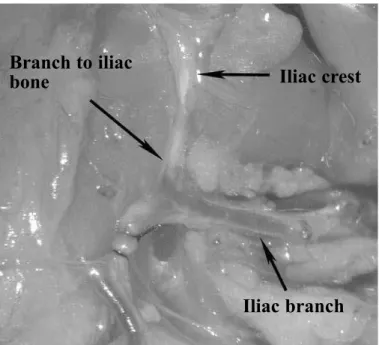
Figure 2 – Laparotomy with dissection of the iliac branch of the iliolumbar artery, towards its origin in the abdominal aorta.
Figure 3 – Ligation of the iliac branch distally to the emergence of the vessel to the iliac bone.
vascularized bone graft will be transformed into a non-vascularized graft.
Through an experimental model in rats, our aim here was to make inferences regarding the viabili-ty of a vascularized bone graft from the iliac crest, sustained by a vascular pedicle, and to investigate its histological characteristics.
METHODS
Animals
Twenty-one male Wistar rats (Rattus norvegi-cus) were used. They were from a research-dedi-cated stock maintained at the Medical Skills and Surgical Research Laboratory of PUCRS, under appropriate environmental conditions for the spe-cies, with controlled temperature, humidity, ven-tilation, light, noise, odor and social interaction. They were kept in individual cages, receiving water and food of quality and quantity that were appropriate for the species. The study protocol had previously been approved by the Ethics Committee for Animal Use of PUCRS.
The animals were divided into two comparison groups: Group 1: vascularized bone graft from the iliac crest (n = 12).
Group 2 (control): non-vascularized bone graft from the iliac crest (n = 9).
surgical procedure
Anesthesia was administered intramuscularly, con-sisting of a solution of 0.2 ml of chlorpromazine and 0.8 ml of ketamine, at a dose of 3 ml/kg. Maintenance doses of anesthetic were administered over the course of the surgical procedure, as necessary.
The surgical incision was longitudinal: parallel to and separated by 2 cm from the dorsal median line, starting 1 cm below the final costal arch and ending at the level of the hips (Figure 1).
In the subfascial layer, the iliac branch of the ilio-lumbar vein and artery were identified and carefully dissected in the proximal direction, as far as the point where they entered the abdominal cavity. To access the abdominal cavity, the insertion of the abdominal muscles was released at the iliac crest. The vascular pedicle was dissected as far as its origin in the aortic artery and vena cava inferior (Figure 2).
The branch to the iliac bone was then identified, and the artery was ligated distally to it (Figure 3).
Gluteal muscles
Abdominal muscles
Gluteal muscles
iliac crest
Paravertebral muscles
iliac branch
Abdominal cavity
Branch to iliac
bone iliac crest
377
The thoracolumbar fascia was incised at the lateral margin of the spine and the medial portion of the iliac bone was exposed. Parallel to the upper margin of the iliac bone and 1 cm from it, the gluteus maximus muscle was incised in the external face of the iliac and the iliac muscle was incised to expose the internal face of the iliac bone (Figure 4).
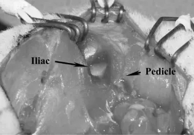
Bicortical osteotomy separated the upper third of the iliac bone from the lower two thirds, which were jointed to the sacrum and hips. The soft tissues that were inserted into the upper half were released, thus making it possible to produce a bone graft vascula-rized by the iliac branch of the iliolumbar vein and artery (Figure 5).
The bone graft was wrapped in a silicone lamina, thus creating a mechanical barrier that made
inva-sion difficult and diminished the influence of nei-ghboring tissues on bone graft viability, and making the graft dependent on its vascular pedicle for via-bility to be maintained.
Group 2 was used as a control group. In this, the same procedure as described for group was perfor-med, with the modification that the vascular pedicle was ligated and sectioned.
The graft, wrapped in the silicone lamina, was placed inside the abdominal cavity with the aim of minimizing the chances of extrusion to the ex-ternal environment.
Among the 21 animals, seven were sacrificed one week after the surgical procedure; seven, two weeks afterwards; and seven, three weeks afterwards. Among the seven animals sacrificed each week, four were in group 1 and the other three were in group 2.
Histology
The grafts were fixed in 10% buffered formalin for 24 hours, and then were washed with water and kept in water for 12 hours and then decalcified for 24 hours, using a solution of 22% formic acid and 10% sodium citrate. After this procedure, the speci-men was included in paraffin in accordance with the processing for conventional tissues, and histologi-cal sections of 3 µm were cut and then stained with hematoxylin-eosin (HE).
immunohistochemistry
The same specimens that had been fixed and de-calcified for histological analysis were used to pre-pare slides for immunohistochemical analysis. The anti-CD34 endothelial marker was used to assess the intraosseous vascularization.
STATISTICAL ANALYSIS
The variables were described in terms of absolute and relative frequencies.
To evaluate the concordance between the methods, the kappa concordance coefficient was applied. To estimate the magnitude of the kappa coefficient, the 95% confidence interval was calculated.
The significance level adopted was 5%, and the analyses were performed using the SPSS software (Statistical Package for the Social Sciences), version 13.0, and the PEPI software (Programs for Epidemio-logists), version 4.0.
Figure 4 – Vascularized bone graft from the iliac bone based on the iliac branch of the iliolumbar artery.
Figure 5 – Exposure of the external face of the iliac bone after incision and separation of the gluteal muscle.
OF THE ILIOLUMBAR ARTERY: EXPERIMENTAL STUDY ON RATS
Graft
Pedicle
iliac
RESULTS
direct observation
Differences in macroscopic characteristics between the vascularized and non-vascularized grafts were evident. In the vascularized grafts, a piece of bone of reddish tone was found, with abundant bone marrow that was bleeding. On manipulation, it presented plasticity similar to that of the bone encountered at the time when the initial surgical procedure had been performed, albeit with loss of resistance. The non-vascularized bone grafts presented a yellowish-white coloring, with sparse bone marrow, and they were less plastic and more friable, without any evidence of bleeding, and a dried-out appearance.
In all the rats whose procedure was aimed towards obtaining a non-vascularized graft, bone portions with the above-described characteristics were obser-ved via ectoscopy. However, among the rats whose procedure was aimed towards integrity of graft vas-cularization, all those sacrificed in the first week were characterized via ectoscopy as vascularized. Only one was classified as vascularized among those of the second and third weeks. Thus, vascularization was seen to be absent in 75% (three out of four) of the grafts that had initially been vascularized, in the second and third weeks.
Histology
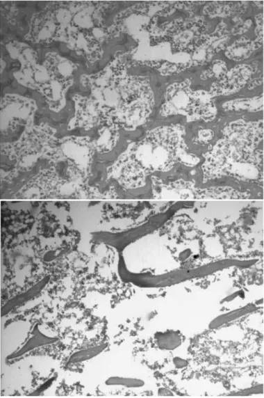
From observations on the slides stained with he-matoxylin-eosin (Figure 6), the bones could be clas-sified as vascularized or non-vascularized, according to the structures viewed. In the vascularized grafts, well-structured trabeculae were found in the bone marrow region, and large quantities of osteocytes with abundant deposition of osteoid matrix surroun-ding it, and with a high density of cellular elements in the bone marrow region. On the other hand, si-tuations in which a slide with smaller quantities of osteocytes were found, accompanied by smaller quantities of bone matrix deposited around them, and sparse bone marrow, were characterized as a non-vascularized bone pattern. All the slides from group 2 were classified as non-vascularized. The slides from the histological specimens from group 1 in the first week were classified as vascularized. However, only 50% (two out of four) of the slides obtained afterwards, in the second and third weeks, were classified as vascularized.
The slides stained with the immunohistochemical marker anti-CD 34 for endothelial tissue also showed differences in the histological descriptive analysis. In assessing the slides that were classified as presenting the vascularized bone pattern, a large quantity of structures marked with brown staining (staining due to DAB) were found, with the presence of a lumen containing erythrocytes, thus characterizing blood vessels. The slides with non-vascularized bone showed absence of the above-described structures. None of the slides from group 2 showed structures corresponding to blood vessels. Among the slides from vascularized grafts, all of those from the first week demonstrated signs of vascularization, while only 50% of the slides from the second and third weeks had blood vessels.
379
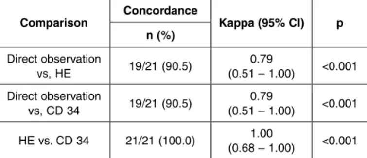
Correlation between direct observation and histology There was statistically significant concordance between all the methods (p < 0.001), as presented in Table 1. It was observed that the kappa coefficient presented good concordance between all the methods
(≥ 0.79), with complete concordance between the he -matoxylin-eosin and CD34 staining (k = 1.00). Only two of the 21 evaluations (9.5%) were discordant be-tween direct observation and histology using hema-toxylin-eosin and CD34 staining. In these two cases, direct observation demonstrated devascularization, while the other methods presented vascularization.
vascularized grafts in the cases with worse prognosis, these may also be used more frequently or even as the first choice in situations that are considered to be less severe.
One frequent question raised regarding vascula-rized bone grafts is in relation to the viability of the graft with the passage of time: would it be justifiable to choose a technique that prolongs the duration of the surgical procedure or requires referral to professio-nals with greater skills in performing this procedure? Grafts that initially were proven to be vascularized during the operation may cease to be so for a variety of reasons, for example: venous stasis, vascular mi-crotrauma, coagulation disorders, systemic abnorma-lities, external compression, edema and inflammatory or infectious processes.
In an experimental study on rats, Linsell et al(12)
reported that 88% of their vascularized grafts of the femur were viable after one week of follow-up, when they used arteriovenous microanastomosis.
Sempuku et al(13) detected bone neoformation
wi-thin two weeks, in a model for vascularized bone grafts in rat tails, such that in the group with non--vascularized grafts, complete necrosis was observed one week after the surgery. Bone neoformation sur-rounding the necrotic bone was detected five weeks after the surgery, thus suggesting the revascularization and osteogenesis were occurring through invasion by surrounding tissues.
Nasir et al(14) observed from histological analysis
on rats that, after one week, a bone graft from the iliac crest with vascularization from the lateral circumflex femoral artery was viable.
Ozkan et al(15) used an experimental model in rats,
for a vascularized bone graft based on the iliolumbar artery, and found that all the grafts examined were his-tologically viable. The experimental model of Ozkan et al(15), which was chosen for the present study, was
shown to be feasible and only presented small varia-tions in vessel path, vessel location and number of branches. This constant anatomy made construction of vascularized grafts reliable and simple. Because rats are small-sized animals and consequently, their vessels are small, use of a microsurgical microscope added security in dissecting and ligating the vessels. The main advantages of this model that they cited were the small functional loss for the animal wi-thout alterations in gait, the bone fragment of ample
Table 1 – Assessment of the concordance between direct observation, hematoxylin-eosin (HE) and CD34.
Comparison Concordance Kappa (95% CI) p n (%)
Direct observation
vs, HE 19/21 (90.5) (0.51 – 1.00)0.79 <0.001 Direct observation
vs, CD 34 19/21 (90.5) (0.51 – 1.00)0.79 <0.001 HE vs. CD 34 21/21 (100.0) (0.68 – 1.00)1.00 <0.001
DISCUSSION
Bone grafts are fundamental for reconstruction of skeletal defects. The success of the treatment may be influenced by the type of graft used, because of the differences in properties between vascularized and non-vascularized bone grafts.
The properties of vascularized bone grafts make it possible to achieve survival of a greater proportion of their cellular elements(5), earlier consolidation and
integration of the receptor bed(6,7), better maintenance
of bone mass(5), greater capacity for hypertrophy(5)
and higher resistance to infections in relation to non--vascularized bone grafts(8,9).
Physiologically, vascularized bone grafts present greater capacity for osteogenesis, osteoinduction and osteoconduction, compared with non-vascularized bone grafts, and it becomes clinically imperative to use them in certain situations in which the failure rate with conventional bone grafts would be far too high to justify using them, such as in cases of extensive bone defects, poorly vascularized or even devascu-larized receptor beds(10,11) and previous procedures
dimensions (1 x 1 cm) that was obtained, the possibi-lity of protecting the bone fragment in the abdominal cavity and the ease of execution.
In this experimental study, it was found during the operation that all the vascularized grafts presented active bleeding, with functional vascular pedicles. At the time of the surgical reintervention to collect ma-terial and analyze the results, the animals sacrificed in the first week had grafts that were considered to be vascularized according to both direct observation and histology. However, among the animals of the second week, only one was considered to be vascularized according to direct observation (25%) and, according to histology, two of them (50%). These results were repeated among the animals sacrificed in the third week. There was a correlation between the results from the ectoscopic and histological classifications.
CONCLUSION
The grafts that were initially vascularized became devascularized at a certain time and started to behave like non-vascularized grafts, under ectoscopic and histological analysis. Further studies would be needed in order to make inferences about the possible causes of the failures of vascularization.
Despite the possibility of failure, use of vascu-larized bone grafts should be encouraged, since the descriptive histological evaluation showed that these grafts had greater cell density in the bone marrow portion and osteocytes with greater func-tionality of bone matrix deposition, with a pre-served intraosseous vascular network. Thus, bone portions with inherent characteristics that promote greater chances of cure for patients could be used in reconstructive procedures.
REFERENCES
1. Yajima H, Tamai S, Mizumoto S, Inada Y. Vascularized fibular grafts in the treatment of osteomyelitis and infected nonunion. Clin Orthop Relat Res. 1993;(293):256-64.
2. Weiland AJ, Moore JR, Daniel RK. Vascularized bone autografts. Experience with 41 cases. Clin Orthop Relat Res. 1983;(174):87-95.
3. Wood MB, Cooney WP 3rd. Vascularized bone segment transfers for manage-ment of chronic osteomyelitis. Orthop Clin North Am. 1984 Jul;15(3):461-72.
4. Wood MB, Bishop AT. Massive bone defects of the upper limb: reconstruction by vascularized bone transfer. Hand Clin. 2007;23(1):49-56.
5. Tamai S. Experimental vascularized bone transplantations. Microsurgery. 1995;16(4):179-85.
6. Haw CS, O’Brien BM, Kurata T. The microsurgical revascularisation of resected segments of tibia in the dog. J Bone Joint Surg Br. 1978;60(2):266-9.
7. Weiland AJ, Phillips TW, Randolph MA. Bone grafts: a radiologic, histologic, and biomechanical model comparing autografts, allografts, and free vascularized bone grafts. Plast Reconstr Surg. 1984;74(3):368-79.
8. Cutting CB, McCarthy JG. Comparison of residual osseous mass between vascularized and nonvascularized onlay bone transfers. Plast Reconstr Surg. 1983;72(5):672-5.
9. Antonyshyn O, Colcleugh RG, Anderson C. Growth potential in onlay bone grafts: a comparison of vascularized and free calvarial bone and suture bone grafts. Plast Reconstr Surg. 1987;79(1):12-23.
10. Cooney WP, Linscheid RL, Dobyns JH, Wood MB. Scaphoid non-union: role of anterior interpositional bone grafts. J Hand Surg Am. 1988;13(5):635-50.
11. Green DP. The effect of avascular necrosis on Russe bone grafting for scaphoid nonunion. J Hand Surg Am. 1985;10(5):597-605.
12. Linsell M, Jablonski P, Howden B, Scott D, Marshall V. The thigh flap: an osteo-myocutaneous free-flap model in the rat. Plast Reconstr Surg. 1988;81(2):240-5.
13. Sempuku T, Tamai S, Mizumoto S, Yajima H. Vascularized tail bone grafts in rats. Plast Reconstr Surg. 1993;91(3):502-10.
14. Nasir S, Aydin A, Kayikçioğlu A, Sökmensüer C, Cobaner A. New experimental composite flap model in rats: gluteus maximus-tensor fascia lata osteomuscle flap. Microsurgery. 2003;23(6):582-8.