Non-alcoholic fatty liver disease (NAFLD) in different populations: A
clinical and epidemiological study – sample of São José do Rio Preto
GABRIEL COSTA DE ANDRADE¹*, LUCIANA HARUMI FUJISE², JAIME EUCLIDES DE SANTANA FILHO2, FABIANE OLIVEIRA3,
RITA DE CÁSSIA MARTINS ALVES DA SILVA4
1MD, Specialist degree in Ophthalmology from Associação Médica Brasileira (AMB) and Conselho Brasileiro de Oftalmologia (CBO), São Paulo, SP
2Gynecologist – Specialist degree in Gynecology and Obstetrics from AMB and Federação Brasileira das Associções de Ginecologia e Obstetrícia (Febrasgo), São Paulo, SP, Brazil 3Department of Nutrition – Faculdade de Medicina de São José do Rio Preto (Famerp), São José do Rio Preto, SP, Brazil
4PhD in Gastroenterology from Universidade Estadual de Campinas (Unicamp). PhD Professor, Faculdade de Medicina de São José do Rio Preto (Famerp), São José do Rio Preto, SP, Brazil
S
UMMARYStudy conducted at Faculdade de Medicina de São José do Rio Preto (Famerp), São José do Rio Preto, SP, Brazil
Article received: 8/31/2014
Accepted for publication: 11/3/2014
*Correspondence:
Address: Alameda Barros, 66 São Paulo, SP – Brazil Postal code: 01232-000 [email protected]
http://dx.doi.org/10.1590/1806-9282.62.03.218
Financial support: Research grant – National Council for Scientiic and Technological Development (CNPq)
Introduction: NAFLD is an heterogeneous condition that includes steatosis and non-alcoholic steatohepatitis (NASH), in the absence of signiicant alcohol con-sumption, reaching 30% of the population. The most common risk factors are: age, gender, ethnicity, diabetes mellitus (DM), obesity, predisposition, metabolic
syn-drome (MS), insulin resistance (IR), drugs, and polycystic ovary synsyn-drome. Objective: To describe the proile of patients with NAFLD seen at Hospital de Base of Rio Preto, in the state of São Paulo.
Method: Patients with NAFLD were assessed, with medical and epidemiologi-cal data collected after informed consent.
Results: Of the 62 patients evaluated, 76% were women, 73% Caucasians, and 71% were aged between 50 and 69 years and had no symptoms. Ultrasonogra-phy results showed steatosis in 84%. NASH was diagnosed in 61% of the sample. 21 patients underwent liver biopsy, of which 36% had cirrhosis, 1 had liver can-cer, and 1 pure steatosis (5% each). Risk factors were found in 70% of patients with metabolic syndrome, 87% with increased waist circumference, 63% with dyslipidemia, 61% (n=38) with high blood pressure (HBP), 28% with DM, 52% physically inactive, and 44% with insulin resistance (IR) (HOMA> 3.5). There was an association between IR and NASH (p=0.013), IR and obesity (p=0.027), IR and MS (p=0.006), and MS and steatosis on medical ultrasound (USG) (p=0.014). Conclusion: The most frequent risk factors were MS and its variables: increased waist circumference, dyslipidemia and HBP. This underscores the importance of metabolic control in NAFLD and conirms its role as the hepatic component of metabolic syndrome.
Keywords: fatty liver, diabetes mellitus, obesity.
INTRODUCTION
Non-alcoholic fatty liver disease (NAFLD) represents the spectrum of a heterogeneous condition that encompass-es steatosis and non-alcoholic steatohepatitis (NASH), in the absence of signiicant consumption of alcohol,27 which
may progress into cirrhosis. Histologically, fatty liver dis-ease is characterized predominantly by macrovesicular steatosis and NASH, and is recognized when, in associa-tion to the accumulaassocia-tion of fat, one or more of the fol-lowing aspects are found: lobular inlammation, hepato-cellular ballooning, Mallory’s hyaline bodies and zone 3 perisinusoidal ibrosis.8,32
Although NAFLD can remain stable and stationary for long periods of time, the condition can progress to advanced stages of cirrhosis and liver cancer.4,22,38,39,53,54
The predisposing factors to the progressive course of NAFLD remain unclear.
NAFLD prevalence is high, being reported in approx-imately 20 to 30% of the general population in studies based on imaging methods.6,56 For histological studies, in
patients.45 In patients with type 2 diabetes mellitus (DM2),
the frequency of fatty liver disease can reach 75%.17,29,48
Numerous individual characteristics or external con-ditions associated with NAFLD may play a role in the eti-ology, pathogenesis, natural history and progression of this disease, such as: age, gender, ethnicity,46,56diabetes mel-litus,2,15,38 obesity,2 family predisposition,50 metabolic
syn-drome26 and peripheral insulin resistance.29
Some drugs such as amiodarone, tamoxifen, diltiazem, cortisone and HAART have been associated with NAFLD, and the induction of NASH is associated with prolonged therapy (longer than 6 months) and drug accumula-tion.10,20,31,37,40,50-52 Procedures such as total parenteral
nu-trition, jejunoileal or gastric bypass have been linked to fat-ty liver disease. An association has been described between NAFLD and rare genetic disorders such as lipoatrophy, Mauriac syndrome,39 abetalipoproteinemia,36 Andersen
disease and Weber-Christian disease.55 Environmental
fac-tors such as various kinds of petrochemicals and sol-vents11-13,41 are related to the appearance of NAFLD. Data
from the literature, in the irst few studies and more, show-ing the variability of the risk factors for NAFLD accordshow-ing to gender, race and ethnicity,46,56 demonstrate the
multi-plicity of clinical, genetic and environmental factors asso-ciated with the heterogeneous presentation of NAFLD. There is still no consensus on the treatment of choice for this disease and there are several studies focused on the use of drugs for insulin resistance and antioxidants.24,34 In
this context, studies on clinical and epidemiological as-pects of the disease can add important information to the diagnostic and therapeutic management of these patients.
OBJECTIVE
To describe the epidemiological and clinical proile of pa-tients with NAFLD cared for at Hospital de Base in the city of São José do Rio Preto (SJRP).
To evaluate the risk factors, clinical and diagnostic aspects of NAFLD.
To analyze the histological aspects of NAFLD.
METHOD
Study design and patients selection
This clinical and epidemiological study was irst submit-ted to the approval of the local ethics committee. Adult patients with a deinitive diagnosis of NAFLD (as deined below) were evaluated. The medical records of these pa-tients with NAFLD, obtained during its investigation and routine follow-up during the period from June 2006 to April 2007, were reviewed with the objective of complet-ing a form created for collection of epidemiological,
clin-ical and laboratory data and results from imaging and histological examinations.
Diagnosis of NAFLD
NAFLD was deined in patients as steatosis proven by biopsy or imaging examination, such as medical ultra-sound (USG) and/or computed tomography (CT) and magnetic resonance imaging (MRI), associated with known risk factors, in the absence of alcohol intake great-er than 20 g pgreat-er day.
The diagnosis of probable NASH was deined as per-sistent and unexplained increase in transaminase accom-panied by steatosis on USG and/or CT and/or MRI ex-aminations of the upper abdomen, without any history of signiicant alcohol consumption (<20 g per day) and associated with one of the following risk factors: meta-bolic syndrome, overweight or obesity, diabetes mellitus,
pe-ripheral insulin resistance, drugs, environmental and oth-er factors demonstrably associated with NAFLD.
Deinitive diagnosis of NASH was based on the pres-ence of steatohepatitis on biopsy and in the abspres-ence of signiicant consumption of alcohol (<20 g per day).
Exclusion criteria
We excluded patients previously submitted to stomach reduction surgery such as jejunoileal or gastric bypass, as well as liver diseases, such as hepatitis B and C, Wilson’s disease, hemochromatosis and autoimmune hepatitis. Patients with incomplete or inconsistent mandatory in-formation were not included.
Study variables
Demographic and clinical data, imaging exams and lab-oratory tests of all individuals, carried out at the time of the NAFLD diagnosis, in the medical records were re-viewed for the purpose of completing the study protocol containing ields marked as either mandatory or option-al. Being overweight was deined as a BMI ≥ 25 kg/m2 and
obesity as a BMI ≥ 30 kg/m2. The diagnosis of diabetes mel-litus was based on the criteria of the American Diabetes
Association (2003). Metabolic Syndrome was deined based on the criteria of the ATP III (NCEP-ATP III, 2001).16
HBs-Ag and Anti-HCV. Complete serology for viral hep-atitis, HCV-RNA, autoantibodies to autoimmune hepa-titis, ceruloplasmin and HFE genetic testing for hemo-chromatosis were studied if clinically recommended.
Histological analysis
In patients undergoing liver biopsy, the corresponding slide was submitted to review by a single pathologist. For the diagnosis and grading of NASH we used the criteria created by Matteoni,32 while for ibrosis staging followed
the criteria of E. Brunt.8
Statistical analysis
Minitab software was used to evaluate the descriptive data, which was expressed as the mean and median with their variations and standard deviation, as appropriate.
Student’s t-test or Mann-Whitney U tests were ap-plied to compare continuous variables, and chi-squared test and Fisher’s exact test were used for categorical vari-ables. P value was considered as less than 0.05.
RESULTS
After signing the informed consent form, 98 adult pa-tients cared for at Hospital de Base, SJRP, were evaluat-ed. Only one patient refused to participate in the study. Thirty-six patients were excluded for the following rea-sons: 18 patients (50%) due to alcoholism > 20 g/day, 10 (28%) patients with hepatitis B, 4 (11%) patients with hep-atitis C, 1 (3%) with hemochromatosis and 8 (22%) due the lack of data in the medical records.
Characterization of the sample
Demographic, clinical and some of the laboratory data of the 62 patients with NAFLD are represented in Table 1. 47 of the 62 patients were women (76%). Regarding race, 45 patients (73%) were Caucasian, 15 (25%) Black and one (2%) Asian.
The main symptoms reported by the patients were: abdominal pain: 5 patients (8%); dyspepsia: 2 (3%); diar-rhea: 2 (3%); postprandial fullness: 2 (3%); increased ab-dominal volume: 2 (3%); jaundice: 1 (2%); borborygmus: 1 (2%); asthenia: 1 (2%); and bitterness in the mouth: 1 (2%). Forty-four patients (71%) referred to themselves as being asymptomatic.
There were changes to the physical examination in 28 patients (45%), with hepatomegaly evident in 21 pa-tients (34%), telangiectasia in 7 (11%), palmar erythema in 7 (11%), splenomegaly in 4 (6%), jaundice in 3 (5%), ede-ma of the lower limbs in 3 (5%) and ascites in 2 (3%).
AST and ALT values above the reference values were found in 70% and 34% of patients, respectively. Abnor-mal AST/ALT ratio was found in 47% of patients. 15 pa-tients (24%) did not have changes to ALT and AST.
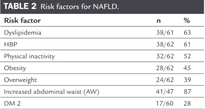
The risk factors found for NAFLD are listed in Ta-ble 2.
There were 24 (39%) overweight patients. 14 (23%) of 28 obese patients were classiied as grade I obesity, 10 (16%) as grade II, and 4 (6%) as grade III obesity.
The waist was measured in 47 of 62 patients and found compatible with metabolic syndrome in 41 cas-es (87%).
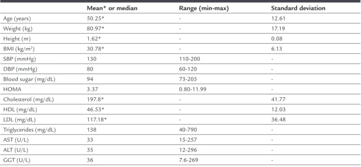
TABLE 1 Demographic, clinical and laboratory data of 62 patients with NAFLD.
Mean* or median Range (min-max) Standard deviation
Age (years) 50.25* - 12.61
Weight (kg) 80.97* - 17.19
Height (m) 1.62* - 0.08
BMI (kg/m2) 30.78* - 6.13
SBP (mmHg) 130 110-200
-DBP (mmHg) 80 60-120
-Blood sugar (mg/dL) 94 73-205
-HOMA 3.37 0.80-11.99
-Cholesterol (mg/dL) 197.8* - 41.77
HDL (mg/dL) 46.53* - 12.03
LDL (mg/dL) 117.18* - 36.48
Triglycerides (mg/dL) 138 40-790
-AST (U/L) 33 15-257
-ALT (U/L) 35 12-296
-GGT (U/L) 36 7.6-269
TABLE 2 Risk factors for NAFLD.
Risk factor n %
Dyslipidemia 38/61 63
HBP 38/62 61
Physical inactivity 32/62 52
Obesity 28/62 45
Overweight 24/62 39
Increased abdominal waist (AW) 41/47 87
DM 2 17/60 28
HBP: high blood pressure; DM 2: type 2 diabetes mellitus.
Abnormal high-density lipoprotein (HDL) cholesterol and triglycerides (TG) results were found in 52% and 44% of the 62 patients, respectively. We found changes to both HDL and TG values in 19 patients (31%). 13 patients (21%) showed changes to HDL alone, and 8 patients (13%) showed alterations to the TG values.
Evaluating the blood pressure (BP) measurement of 59 patients, 51% were classiied as having some degree of hypertension: 16 patients (27%) with mild hypertension (grade 1); 13 patients (22%) with moderate hypertension (grade 2); and 1 patient (2%) with severe hypertension (grade 3). Among the normotensive patients, 4 (7%) were classiied as having optimum BP, 16 (27%) as having nor-mal BP and 9 (15%) as borderline BP.
In the evaluation of physical activity, 30 patients claimed to be active, 3 of whom (10%) could not inform the frequency of exercises, 23 (77%) performed physical activity at least three days a week, 3 (10%) on 2 days of the week and 1 patient (3%) reported irregular exercise.
With regard to medication, a total of 94% of the patients reported use of at least one type of medication. The use of medication known to be associated with NAFLD was not-ed in 21% of patients and these drugs are listnot-ed in Table 3.
TABLE 3 Medications associated with NAFLD used by the 62 patients.
Medication n %
Estrogens 7 11.29
Tamoxifen 3 4.83
Acetylsalicylic acid (ASA) 2 3.22
Chloroquine 1 1.61
Imaging and biopsy methods
Evaluating the imaging methods used in the investiga-tion, medical ultrasound (USG) was conducted in 62 pa-tients, and the sole imaging method used for 57 patients (92%). USG associated with computed tomography (CT) was conducted in 2 patients (3%), nuclear magnetic
res-onance (NMR) imaging in 1 patient (2%) and in 2 patients (3%) it was associated with CT and MRI.
Steatosis was not evident on USG in 10 patients (16%). Steatosis was conirmed by biopsy in 9 of these patients (15%) and, in just one patient, there was advanced cryp-togenic cirrhosis subjected to liver transplantation, with diagnosis made after removal of the cirrhotic organ. Of 52 patients who had some degree of steatosis, 8 (13%) had grade 1 steatosis, 5 patients (8%) had grade 2 steatosis, 6 patients (10%) had grade 3, and 33 patients (53%) had ste-atosis that was not graded, with one of the patients pre-senting focal steatosis.
22 patients (35%) underwent liver biopsy, with ste-atosis described in all of them. Non-alcoholic steatohep-atitis (NASH) was found in 21 patients. Other indings in the biopsies were: hepatocellular ballooning in 16 pa-tients (72.72%), ibrosis in 16 papa-tients (72.72%), iron over-load in 13 patients (59.09%), presence of Mallory bodies in 11 patients (50%) and the presence of a tumor in one patient (4.54%).
Staging based on the degree of inlammation accord-ing to Matteoni32 was stage 1 in 1 patient (4.76%), 3 in 2
patients (9.53%) and 4 in 19 patients (90.47%).
Staging of the degree of ibrosis according to Brunt8
was: absent in 6 patients (27.27%), stage 1 in 4 patients (18.18%) 2 in 3 patients (13.63%) 3 in one patient (4.54%) and 4 in 8 patients (36.36%).
Other indings found in the biopsies by order of fre-quency were: cell retraction igures in 12 biopsies (55%), nuclear vacuolation in 8 (36%), acidophilus corpuscles in 6 (27%), standard biliary portal reaction in 4 (18%), duc-tal proliferation in 3 (14%), multifocal large cell hepato-cellular dysplasia in 2 (9%) and liver cell rosettes associ-ated with necrosis in 1 biopsy (5%).
Risk factors
Metabolic syndrome
Among the 36 patients in whom metabolic syndrome could be studied, the condition was found in 70%. In 26 (42%) of the 62 patients, evaluating metabolic syndrome was not possible due to incomplete data.
3 or more criteria indicating metabolic syndrome were found in 25 patients (70%), with 3 criteria found in 14 patients (39%), 4 criteria in 9 patients (25%) and 5 cri-teria in 2 patients (6%). Among the 11 patients (31%) that did not have 3 or more criteria, 5 patients (14%) had only 2 criteria, 5 patients (14%) had only 1 criterion, and 1 pa-tient (3%) had none.
which this information could be collected. The average was 106.11 cm (±10.48).
Regarding dyslipidemia, 26 (55%) of the 47 women studied and 6 (40%) of the 15 men studied were found to have abnormal HDL cholesterol values. With respect to the values for triglycerides, 27 (44%) of the 62 patients evaluated had abnormal values for the parameters of met-abolic syndrome.
With respect to mean arterial pressure (MAP) a val-ue compatible with metabolic syndrome was evident in 21 (36%) of the 59 patients, with the maximum values for systolic blood pressure (SBP) and diastolic blood pres-sure (DBP) at 200 and 120 mmHg, respectively, and min-imum values for SBP and DBP at 140 and 90 mmHg, and a median of 150 x 100 mmHg.
There was an association between metabolic syndrome and steatosis on USG (p=0.014). Metabolic syndrome was estimated in 69% of patients with steatosis on USG (95CI 54-81%) and there is evidence that the syndrome affects the majority of these patients (p=0.005).
Insulin resistance
Insulin resistance (IR) was found in 22 (44%) of the 50 pa-tients who had homeostatic model assessment (HOMA) index calculated. There was an association between IR and metabolic syndrome in 90% of patients with HOMA ≥ 3.5 (p=0.006). There was also evidence of an association be-tween IR and obesity (p=0.027) and bebe-tween IR and NASH (p=0.01). There was no evidence of an association between IR and waist circumference compared individually (p=0.23).
Presence of NASH
38 of the 62 patients studied (61%) were classiied as hav-ing NASH accordhav-ing to one of the criteria adopted. Of these 38 patients, 30/62 (48%) were classiied as having probable NASH and 21/62 (34%) as having deinitive NASH.
22 of the 30 patients with NASH had HOMA ≥ 3.5 (p=0.032). In these patients, abnormal ALT and AST val-ues were found in 1 (5%) and 4 (18%) of the patients, re-spectively. Both enzymes were found to be abnormal in 5 patients (23%).
DISCUSSION
NAFLD has gained epidemiological relevance in recent years, representing one of the leading causes of chronic liver disease in the 21st century, and may manifest itself in
different age ranges, ethnic groups and gender. The true prevalence of NAFLD is underestimated in the general population, because the disease is asymptomatic and the majority of patients start investigation due to incidental
indings on USG, increased liver enzymes or check-ups.1
Recent studies indicate prevalence at around 20 to 30% in the general population. In obese patients or with diabetes mellitus it can reach up to 50% of the population.33
Most of the available studies on NAFLD include pa-tients in hospital environments and, therefore, their char-acteristics may not exactly represent those expected in the general population. In Brazil, there are not many studies regarding the prevalence of NAFLD; however, among the 2,232 cases of NAFLD recorded by the Brazilian Society of Hepatology in 2004, 68% had NASH.13,14 This case series is
part of the second investigation conducted at several Bra-zilian centers, documenting this pathology in more detail. In the present study, there was a predominance of NAFLD in middle-aged women, as demonstrated in pre-vious studies.3,21 However, the actual participation of
gen-der as a determining factor in the development of NAFLD has not yet been established, given that some studies show a higher prevalence in men or an equal distribution be-tween the sexes.1,3,5,33
With regard to the ethnic groups studied, a higher prevalence of Caucasians (73%) was found, followed by Black patients (25%) and only one Asian patient (2%). Such indings contrast with previous studies that show a high-er prevalence in the Caucasian group, followed by His-panics and Asians.3,5 It is believed that the percentage of
black patients found is due to the high level of miscege-nation and dificulty in deining races in the group stud-ied and, possibly, throughout Brazil.
The vast majority of patients with NAFLD diagnosis is asymptomatic (71%), showing that the disease can prog-ress insidiously. Nonspeciic symptoms such as abdomi-nal pain, postprandial fullness and fatigue were found in 8, 3 and 2% of patients, respectively. In the physical ex-amination, 46% of patients had some kind of inding, with hepatomegaly being the abnormality found most frequently, as it was present in 21 patients (34%). Periph-eral signs found more often were: telangiectasia in 7 pa-tients (11%), palmar erythema in 7 (11%) and jaundice in 3 (5%) patients. Signs of liver decompensation, sugges-tive of more advanced degrees of disease, were also veri-ied with the following distribution: ascites in 2 patients (3%), splenomegaly in 4 patients (6%) and edema of the lower limbs in 3 patients (5%). Such indings are in agree-ment with previous studies, characterizing NAFLD as a disease that has few symptoms and with a nonspeciic clinical presentation.3,18,45
estrogens, tamoxifen, ASA and chloroquine. Despite these patients having risk factors for metabolic syndrome, we cannot rule out the possibility that the use of these drugs is correlated with the progression of NAFLD.
With respect to NASH, less than 2% of the causes are due to drug induction. The mechanisms of action can be divided into direct hepatoxicity and action on metabol-ic processes in the liver, such as mitochondrial ATP pro-duction and the metabolism of fatty acids.20,50,51
Tamoxifen, a drug used in patients with breast can-cer to inhibit estrogen receptors in this organ alone, was found in almost 5% of the patients. Studies show that tamoxifen raises the risk of developing NAFLD/NASH only in overweight and obese women with risk factors as-sociated with metabolic syndrome. Studies revealed that 43.2% of patients developed steatosis within 2 years of use and had their tests return to normal 1 year after the end of therapy.7,35
Knowledge of the group of medication used by these patients was also relevant, as it showed that 40% of the patients used at least one type of anti-hypertensive and 27% used at least one anti-diabetic drug, suggesting the presence of diseases directly associated with metabolic syndrome, and also related to NAFLD.3,56
Metabolic syndrome is represented by a set of risk factors, mainly related to a central deposition of fat and insulin resistance, which are found in almost 70% of pa-tients in the sample studied. According to a previous study, the presence of 3 or more criteria for metabolic syndrome raises the risk of developing severe ibrosis, chronic liver disease and cardiovascular disease by 3.5 times.19,25In this
study, three or more criteria for metabolic syndrome were found in 25 patients (70%).
The association between metabolic syndrome and steatosis on USG was found in 69% of the patients (p=0.014), corroborating the concept that NAFLD can be a hepatic component of metabolic syndrome.
Being overweight or obese was found in the vast ma-jority of the patients (84%). This inding is compatible among the patients with NAFLD and is consistent with the literature.9,30 Obesity, estimated using the BMI, was
found in 45% of patients studied. Previous studies have estimated NAFLD at 57.5% to 74% of the obese popula-tion, showing the importance of calculating the BMI and the intense relationship between obesity and NAFLD, as well as showing that there is a direct correlation between obesity and the severity of steatosis.44,45
Waist circumference relates to the amount of viscer-al adipose tissue and is predictive of comorbidities such as obesity, hypertension and diabetes mellitus, relating to
the pathogenesis of insulin resistance and glucose intol-erance. Abnormal WC values were found in 66% of the 62 patients, and were higher than those found in the litera-ture (42-47%).45 However, if considering the abnormal
WC values only in those cases where this datum could be measured, the percentage increases to 87%. This datum may be related to the fact that the measurements were carried out on the vast majority of the obese patients, em-phasizing the importance of anthropometric measure-ments, which is often neglected in the physical exam.
Dyslipidemia was found in 38 (62.30%) of the 61 pa-tients, and this prevalence is compatible with the results of other studies, which showed a frequency of 28 to 66%.9,30
The most common change found was low HDL values, which was found in 32 (52%) of the 62 patients. Hyper-triglyceridemia was found in 27 (44%) of the 61 patients from which this datum was collected. Such data differs from previous literature that indicates hypertriglyceride-mia as the main component of dyslipidehypertriglyceride-mia as risk
fac-tor for NAFLD.2,45 These indings can be explained by
change in lifestyle, with physical activity and proper diet, associated with effective drug therapy.
High blood pressure (HBP) was found in 38 of the 62 patients studied (61%), consistent with previous studies that describe this condition in most patients.5,9
In the literature, the frequency of insulin resistance was found in 47 to 98% of the patients, even in those with-out DM2.28,45 In the present study, insulin resistance based
on HOMA index was present in 90% of patients with met-abolic syndrome, considering HOMA ≥ 3.5. DM2 was found in 28% of the patients, and has been associated with NAFLD between 10 and 75% in previous studies.45
Elevated AST and ALT were found in 69% and 34% of the patients, respectively. Aminotransferase, when ab-normal, suggests the presence of inlammation and is in-dicated as a predictor of ibrosis in these patients, espe-cially when there is an AST/ALT ratio >1.34,43 However,
normal levels do not indicate the absence of inlamma-tion. AST/ALT ratio >1 was found in 47% of the 62 pa-tients. Among patients who underwent biopsy and had ibrosis, the ratio was abnormal in 44% of them.
In previous studies, NAFLD was diagnosed on USG in 20 to 40% of the patients.20 In this sample, steatosis
In the literature it is accepted that the mere presence of fat without inlammation in the liver may have a be-nign course, although the association of steatosis, inlam-mation, ballooning degeneration, Mallory bodies and i-brosis characterizes NASH, which can develop into cirrhosis and resulting complications, such as hepatocar-cinoma.34 The progression from steatosis to cirrhosis and
death due to its complications only occurs in less than 5% of cases; however, when steatohepatitis is present, that frequency of progression can reach up to 25%.32 Data about
the speed of the progression of this disease is scarce. 38 of the 62 patients studied (61%) were classiied as having NASH. Of these 38 patients, 30/62 (48%) were classiied as having probable NASH, that is, abnormal USG associated with increased transaminases and risk factors for metabolic syndrome, while 21/62 (34%) had deinitive NASH, that is, with a biopsy proving NASH.
The deinitive diagnosis of steatosis was described in the 22 biopsies performed, with ballooning degeneration in 73% and ibrosis also in 73% of the biopsies. Grade 4 ibrosis was found in 36% of the biopsies, demonstrating the presence of cirrhosis in most patients undergoing bi-opsy and the importance of carrying out a bibi-opsy in pa-tients being investigated for NAFLD, not only for diag-nosis, but also to assess the progression of the disease. In all patients that showed cirrhosis, we found clinical signs of chronic liver disease, and in patients with a severe lev-el of ibrosis, it is suggested that there is greater risk of progression to hepatocellular carcinoma.47,49
The indings of ballooning degeneration in 73% of patients and grade 4 ibrosis in 36% are the only data from biopsies in this study that were found in greater frequen-cy than described in the literature, which is around 33 and 6%, respectively. With respect to the presence of i-brosis of all grades, iron overload and hepatocellular car-cinoma were found at a frequency similar to previous studies. Lastly, the presence of Mallory bodies in 50% and grade 1 ibrosis in 18% represented values lower than those described previously, being found in the literature at around 80 and 27%.8,23,32
Other indings in the biopsy, such as standard bili-ary portal reaction and ductal proliferation found in 18 and 14% of the biopsies, respectively, suggest a progres-sive pattern of the disease.42
CONCLUSION
Considering the epidemic of metabolic syndrome in the modern world, the investigation of other components of this syndrome is necessary. Metabolic syndrome summa-rizes a large part of the systemic manifestations of
insu-lin resistance. In addition to the classic components of the syndrome, new components of great clinical relevance, such as NAFLD, have been demonstrated.
Current studies indicate a growing frequency of this disease in association with metabolic syndrome risk fac-tors, meaning that better diagnostic and prognostic in-vestigation of NAFLD is required. As there is an impor-tant association between NAFLD and metabolic syndrome, this emphasizes a need to control their component fac-tors and corroborates the idea that NAFLD may be a he-patic component of metabolic syndrome.
Screening for metabolic syndrome with laboratory and imaging exams should be supplemented with anthro-pometric measurements, which are often neglected in the physical exam.
As an insidious and progressive illness with nonspe-ciic symptoms, NAFLD can have a malignant course, pro-gressing to NASH, cirrhosis of the liver and hepatocellu-lar carcinoma. Thus, liver biopsy becomes an indispensable examination for evaluating the course of the disease.
We documented NAFLD in all of its clinical forms, predominantly steatohepatitis. The most frequent risk factors were metabolic syndrome and its variables: in-creased waist circumference, dyslipidemia and hyperten-sion. This underscores the importance of metabolic con-trol in NAFLD and confirms its role as the hepatic component of metabolic syndrome.
As an insidious and progressive illness with nonspe-ciic symptoms, NAFLD can have a malignant course, pro-gressing to NASH, cirrhosis of the liver and hepatocellu-lar carcinoma. Thus, liver biopsy becomes an indispensable examination for evaluating the course of the disease.
NASH was described in practically a third of the sam-ple, while hepatocellular carcinoma was described in about 5% of patients. The indings of grade 4 ibrosis in 36% of the sample, in addition to indings of NASH and hepa-tocellular carcinoma, are higher than the described in the literature. This may be explained by the fact that the sam-ple in the present study includes patients cared for at a reference center, many of whom are at an advanced stage of hepatic impairment.
RESUMO
Doença hepática gordurosa não alcoólica em diferentes populações: um estudo clínico e epidemiológico – Amos-tra de São José do Rio Preto
estea-tose e esteato-hepatite não alcoólica (NASH), na ausên-cia de consumo signiicante de álcool, podendo atingir 30% da população. Fatores de risco mais comuns são ida-de, gênero, etnia, diabetes mellitus (DM), obesidade,
predis-posição, síndrome matabólica (SM), resistência à insuli-na (RI), drogas e síndrome do ovário policístico. Objetivo: descrever o peril de portadores de DHGNA as-sistidos no Hospital de Base de São José do Rio Preto, SP. Método: foram avaliados pacientes com DHGNA e cole-tados dados clínico-epidemiológicos, após consentimen-to informado.
Resultados: dos 62 pacientes estudados, houve predo-mínio de mulheres (76%), caucasoides (73%), idade entre a quinta e sexta décadas e assintomáticos (71%). Exame de ultrassonograia (US) mostrou esteatose em 84%. NASH foi diagnosticada em 61% da casuística. Em 21 pacientes, biópsia hepática mostrou cirrose em 36% e câncer de fí-gado e esteatose pura em um paciente (5%) cada. Dos fa-tores de risco, 70% dos pacientes apresentavam SM; 87%, cintura abdominal alterada; 63%, dislipidemia; 61% (n=38), hipertensão arterial sistêmica (HAS); 28%, DM; 52%, se-dentarismo, e em 44% encontrou-se RI (HOMA>3,5). Hou-ve associação entre RI e NASH (p=0,013), RI e obesidade (p=0,027), RI e SM (p=0,006), SM e esteatose à US (p=0,014).
Conclusão: os fatores de risco mais frequentes foram SM e suas variáveis (cintura abdominal aumentada, dislipi-demia e HAS), o que ressalta a importância do controle metabólico na DHGNA e corrobora o seu papel como componente hepático da SM.
Palavras-chave: fígado gorduroso, diabetes mellitus,
obe-sidade.
REFERENCES
1. Adams LA, Lymp JF, Sauver JSP, Sanderson SO, Lindor KD, Feldstein A, et al. The natural history of nonalcoholic fatty liver disease: a population-based cohort study. Gastroenterology. 2005; 129(1):113-21.
2. Angulo P, Keach JC, Batts KP, Lindor KD. Independent predictors of liver ibrosis in patients with nonalcoholic steatohepatitis. Hepatology. 1999; 30(6):1356-62.
3. Angulo P. GI epidemiology: nonalcoholic fatty liver disease. Aliment Pharmacol Ther. 2007; 25(8):883-9.
4. Bacon BR, Farahvash MJ, Janney CG, Neuschwander-Tetri BA. Nonalcoholic steatohepatitis: an expanded clinical entity. Gastroenterology. 1994; 107(4):1103-9.
5. Bedogni G, Marchesini G, Tiribelli C, Bellentani S, Miglioli L, Masutti F. Prevalence of and risk factors for nonalcoholic fatty liver disease: the Dionysos nutrition and liver study. Hepatology. 2005; 42(1):44-52.
6. Browning JD, Szczepaniak LS, Dobbins R, Nuremberg P, Horton JD, Cohen JC, et al. Prevalence of hepatic steatosis in an urban population in the United States: impact of ethnicity. Hepatology. 2004; 40(6):1387-95.
7. Bruno S, Maisonneuve P, Castellana P, Rotmensz N, Rossi S, Maggioni M, et al. Incidence and risk factors for non-alcoholic steatohepatitis: prospective
study of 5408 women enrolled in Italian tamoxifen chemoprevention trial. BMJ. 2005; 330(7497):932.
8. Brunt EM, Janney CG, Di Bisceglie AM, Neusechwander-Tetri BA, Bacon BR. Nonalcoholic steatohepatitis: a proposal for grading and staging the histological lesions. Am J Gastroenterol. 1999; 94(9):2467-74.
9. Chalasani N. Fatty liver disease as a component of metabolic syndrome. Hepatology. 2006; 44(Suppl. 1):37A-187A.
10. Chitturi S, Farrell GC. Etiopathogenesis of nonalcoholic steatohepatitis. Semin Liver Dis. 2001; 21(1):27-41.
11. Cotrim HP, Andrade ZA, Paraná R, Portugal M, Lyra LG, Freitas LA. Nonalcoholic steatohepatitis: a toxic liver disease in industrial workers. Liver. 1999; 19(4):299-304.
12. Cotrim HP, Freitas LA, Freitas C, Braga L, Paraná R, Lyra L, Carvalho F. Nonalcoholic steatohepatitis in petrochemical workers: follow up of those removed from exposure area and those who remained. Hepatology. 2002; 36:983.
13. Cotrim HP, De Freitas LA, Freitas C, Braga L, Carvalho F, Paraná R, et al. Clinical and histopathological features of NASH in workers exposed to chemicals with or without associated metabolic conditions. Liver Int. 2004; 24(2):131-5.
14. Day CP. Natural history of NAFLD: remarkably benign in the absence of cirrhosis. Gastroenterology. 2005; 129(1):375-8.
15. Diehl AM, Goodman Z, Ishak KG. Alcoholic liver disease in non-alcoholics. A clinical and histological comparison with alcohol induced liver injury. Gastroenterology. 1988; 95(4):1056-62.
16. Executive Summary of the Third Report of the National Cholesterol Education Program (NCEP) Expert Panel on Detection, Evaluation, and Treatment of High Blood Cholesterol in Adults (Adult Treatment Panel III). JAMA 2001; 285(19):2486-7.
17. Falchuk KR, Fiske SC, Haggitt RC, Federman M, Trey C. Pericentral hepatic ibrosis and intracellular hyaline in diabetes mellitus. Gastroenterology. 1980; 78(3):535-41.
18. Farrel GC. Drugs and steatohepatitis. Semin Liver Dis. 2002; 22(2):185-94. 19. Gholam PM, Kotler DP, Flancbaum LJ. Liver pathology in morbidly obese patients undergoing Roux-en-Y gastric bypass surgery. Obes Surg. 2002; 12(1):49-51.
20. Grieco A, Forgione A, Miele L, Vero V, Greco AV, Gasbarrini A, et al. Fatty liver and drugs. Eur Rev Med Pharmacol Sci. 2005; 9(5):261-3.
21. Hashimoto E, Yatsuji S, Kaneda H, Yoshioka Y, Taniai M, Tokushige K, et al. The characteristics and natural history of Japanese patients with nonalcoholic fatty liver disease. Hepatol Res. 2005; 33(2):72-6.
22. Ong MD, Younossi ZM. Nonalcoholic fatty liver disease (NAFLD) – two decades later: are we smarter about its natural history? Am J Gastroenterol. 2003; 98(9):1915-7.
23. Kleiner DE, Brunt EE, Natta MV, Behling C, Contos MJ, Cummings OW, et al. Design and validation of a histological scoring system for nonalcoholic fatty liver disease. Hepatology. 2005; 41(6):1313-21.
24. Lima VMR. Esteatohepatite não-alcoólica (ENA) em camundongos obesos (ob/ob): avaliação do efeito citoprotetor do Yo Jyo Hen Shi Ko (YHK). São Paulo: Universidade de São Paulo-USP, 2007.
25. Lobo RA, Carmina E. The importance of diagnosing the polycystic ovary syndrome. Ann Intern Med. 2000; 132(12):989-93.
26. Lonardo A. Fatty liver and nonalcoholic steatohepatitis. Where do we stand and where are we going? Dig Dis. 1999; 17(2):80-9.
27. Ludwig J, Viaggiano TR, McGil DB, Oh BJ. Nonalcoholic steatohepatitis: Mayo Clinic experience with a hitherto unnamed disease. Mayo Clin Proc. 1980; 55(7):434-8.
28. Machado M, Pinto HC. Non-alcoholic fatty liver disease and insulin resistance. Eur J Gastroenterol Hepatol. 2005; 17(8):823-6.
29. Marchesini G, Brizi M, Moreselli-Labate AM, Bianchi G, Bugianesi E, McCullough AJ, et al. Association of nonalcoholic fatty liver disease with insulin resistance. Am J Med. 1999; 107(5):450-5.
30. Marchesini G, Brizi M, Bianchi G, Tomassetti S, Bugianesi E, Lenzi M, et al. Nonalcoholic fatty liver disease: a feature of metabolic syndrome. Diabetes. 2001; 50(8):1844-50.
31. Martinez E, Mocroft A, García-Viejo MA, Pérez-Cuevas JB, Blanco JL, Mallolas J, et al. Risk of lipodystrophy in HIV-1-infected patients treated with protease inhibitors: a prospective cohort study. Lancet. 2001; 357(9256):592-8. 32. Matteoni CA, Younossi ZM., Gramlich T, Boparai N, Liu YC, McCullough
33. Mattos AA. Nonalcoholic steatohepatitis. J Bras Gastroenterol. 2005; 5:160-5. 34. Miele L, Forgione A, Hernandez AP, Gabrieli ML, Vero V, Di Rocco P, Greco AV, et al. The natural history and risk factors for progression of non-alcoholic fatty liver disease and steatohepatitis. Eur Rev Med Pharmacol Sci. 2005; 9(5):273-7.
35. Nishino M, Hayakawa K, Nakamura Y, Morimoto T, Mukaihara S. Effects of tamoxifen on hepatic fat content and the development of hepatic steatosis in patients with breast cancer: high frequency of involvement and rapid reversal after completion of tamoxifen therapy. AJR Am J Roentgenol. 2003; 180(1):129-34.
36. Partin JS, Partin JC, Schubert WK, McAdams AJ. Liver ultrastructure in abetalipoproteinemia: evolution of micronodular cirrhosis. Gastroenterology. 1974; 67(1):107-18.
37. Poucell S, Ireton J, Valencia-Mayoral P , Downar E, Larrat L, Patterson J, et al. Amiodarone-associated phospholipidosis and ibrosis of the liver. Light, immunohistochemical and electron microscopic studies. Gastroenterology. 1984; 86(5 Pt 1):926-36.
38. Powell EE, Cooksley WG, Hanson R, Searle J, Halliday JW, Powell LW. The natural history of nonalcoholic steatohepatitis: a follow-up study of forty-two patients for up to 21 years. Hepatology. 1990; 11(1):74-80.
39. Powell EE, Searle J, Mortimer R. Steatohepatitis associated with limb lipodystrophy. Gastroenterology. 1989; 97(4):1022-4.
40. Pratt DS, Knox TA, Erban J. Tamoxifen-induced steatohepatitis. Ann Intern Med. 1995; 123(3):236.
41. Redlich CA, West AB, Fleming L, True LD, Cullen MR, Riely CA. Clinical and pathological characteristics of hepatotoxicity associated with occupational exposure to dimethylformamide. Gastroenterology. 1990; 99(3):748-57.
42. Richardson MM, Jonsson JR, Powell EE, Brunt EM, Neuschwander-Tetri BA, Bhathal PS, et al. Progressive ibrosis in nonalcoholic steatohepatitis: association with altered regeneration and a ductular reaction. Gastroenterology. 2007; 133(1):80-90.
43. Salgado Júnior W, Santos JS, Sankarankutty AK, Silva OC. Nonalcoholic fatty liver disease and obesity. Acta Cir Bras. 2006; 21(Suppl.1):72-8.
44. Santos RR, Cotrim HP. Relevância das medidas antropométricas na avaliação de pacientes com doença hepática gordurosa não alcoólica. Rev Bras Nutr Clin. 2006; 21(3):229-32.
45. Sanyal AJ; American Gastroenterological Association. AGA technical review on nonalcoholic fatty liver disease. Gastroenterology. 2002; 123(5):1705-25. 46. Schwimmer JB, McGreal N, Deutsch R, Finegold MJ, Lavine JE. Inluence of gender, race, and ethnicity on suspected fatty liver in obese adolescents. Pediatrics. 2005; 115(5):561-5.
47. Shimada M, Hashimoto E, Taniai M, Hasegawa K, Okuda H, Hayashi N, et al. Hepatocellular carcinoma in patients with non-alcoholic steatohepatitis. J Hepatol. 2002; 37(1):154-60.
48. Silverman JF, O Brien KF, Long S, Leggett N, Khazanie PG, Poris WJ, Norris HT, et al. Liver Pathology in morbidly obese patients with and without diabetes. Am J Gastroenterol. 1990; 85(10):1349-55.
49. Stravitz RT, Sanyal AJ. Drug-induced steatohepatitis. Clin Liver Dis. 2003; 7(2):435-51.
50. Struben VM, Hespenheide EE, Caldwell SH. Nonalcoholic steatohepatitis and cryptogenic cirrhosis within kindreds. Am J Med. 2000; 108(1):9-13. 51. Genuth S, Alberti KG, Bennett P, Buse J, Defronzo R, Kahn R, et al. Follow-up
report on the diagnosis of diabetes mellitus. Diabetes Care. 2003; 26(11):3160-7. 52. Van der Valk M, Bisschop PH, Romijn JA, Ackermans MT, Lange JM, Endert E, et al. Lipodystrophy in HIV-1 positive patients is associated with insulin resistance in multiple metabolic pathways. AIDS. 2001; 15(16):2093-100. 53. Wanless IR, Lentz JS. Fatty liver hepatitis (steatohepatitis) and obesity: an
autopsy study with analysis of risk factors. Hepatology. 1990; 12(5):1106-10. 54. Wanless IR, Shiota K.The pathogenesis of nonalcoholic steatohepatitis and other fatty liver diseases: a four-step model including the role of lipid release and hepatic venular obstruction in the progression to cirrhosis. Semin Liver Dis. 2004; 24(1):99-106.
55. Wasserman JM, Thung SN, Berman R, Bodenheimer HC Jr, Sigal SH. Hepatic Weber-Christian disease. Semin Liv Dis. 2001; 21(1):115-8.