ASSOCIATION BETWEEN MASTICATORY ACTIVITY AND
GROSS MOTOR FUNCTION, SPASTICITY AND TOPOGRAPHIC
CLASSIFICATION IN CEREBRAL PALSY
Associação da atividade mastigatória com a função motora ampla,
espasticidade e classiicação topográica na paralisia cerebral
Lilian Gerdi Kittel Ries (1), Kelly Cristine Schmidt (2), Marianne Briesemeister (3),
Camila Isabel Santos Schivinski (4)
(1) Department of Physical Therapy – UDESC, Florianópolis,
SC, Brazil;
(2) Department of Physical Therapy – UDESC, Florianópolis,
SC, Brazil;
(3) Department of Physical Therapy – UDESC, Florianópolis,
SC, Brazil;
(4) Department of Physical Therapy – UDESC, Florianópolis,
SC, Brazil;
Conlict of interest: non-existent
and biomechanical changes also contribute to disability in CP and their functional limitation3.
The most common type of cerebral palsy is the spastic form4. Spasticity is related to a
signiicant restriction in range of motion of the
affected muscles, an abnormal pattern in reciprocal inhibition between antagonist muscles and results in functional impairment5. Such disturbances of
movement, posture and tone can also affect oral-motor functions6-8. However, no studies were found
that evaluated how these disorders can inluence
these oral functions.
Although there are studies about the problems found in children with CP during mastication, still are sparse researches that aim to evaluate the activity and behavior of the muscles of the stomato-gnathic system in this pathology. This study may contribute to a better understanding of changes in the stomatognathic system, and thus aid the devel-opment of more appropriate intervention methods.
INTRODUCTION
Cerebral palsy (CP) or chronic non-progressive encephalopathy is a term that describes a group of non progressive disorders of movement and posture associated with an immature brain defect1. The CP
usually interferes in the functioning of the muscu-loskeletal system, including disorders of muscle tone, posture and voluntary movements2. Muscle
weakness, limited muscle synergisms, contractures
ABSTRACT
Purpose: to verify the existence of an association between the degree of spasticity, the level of motor function and wide topographical classiication of CP children with amplitude and time parameters of
electromyographic activation of temporalis (AT) and masseter (MA) muscles. Methods: muscle activity
during chewing task was evaluated in ifteen children with CP. The clinical instruments used were the Modiied Ashworth Scale for spasticity, the Gross Motor Function Classiication System (GMFCS)
for gross motor function. We analyzed the parameters of muscular symmetry and time of active and inactive period of the masticatory cycle. Results: there was an association between the level of motor function and the symmetry of the MA, between the highest degree of spasticity and decrease in the inactive period and increase in the active period of the masticatory muscles and between the
topographical classiication and symmetry of the MA and the symmetry of the AT. Conclusion: the symmetry and the time of activation of AT and MA should be considered during therapy of oral motor function of CP children.
(2000 Hz) and the minimum ratio Common Mode Rejection was 100 dB.
The evaluation form was completed with
anthro-pometric data, classiication topographical and GMFCS. The MAS was applied with volunteers in
the supine position. The joints are moved passively
from a position of maximal possible lexion to maximal possible extension. To assess knee lexor muscles, the hips and knees were kept in lexion. The examiner stabilized a thigh with one hand. With
the other hand stabilized the ankle to move the knee
to the maximum extent. To assess elbow lexor muscles, the examiner extended the forearm from maximum possible lexion to maximum possible extension. The volunteers’s arm extended as much
as possible and with the palm of the hand facing inward (neutral supination). The score was based on the sum of the results obtained by means of
four measures related to lexor muscles of bilateral
elbows and knees. The results were summed to give
an index for each volunteer with maximum score of
16 points in the highest degree of spasticity11.
During the evaluation of muscle activity the volunteers remained seated in a chair with the head positioned in the Frankfurt plane (parallel to the ground), hands on thighs aligned with the shoulder, back support at the height of the shoulder blades. The electrodes were longitudinally aligned to the
muscle ibres and ixed on the skin of the MA and
AT muscles, bilaterally8,12. The reference electrode
was ixed on the manubrium of the sternum. The
electrical impedance of the skin was reduced by cleaning the area with hydrophilic cotton soaked in 70% alcohol solution.
To make comparisons of the EMG signal
between the volunteers, the values of masticatory activity were normalized by a reference contraction (RC). The RC was measured by the isometric
contraction of clenching in maximum intercuspation.
The contractions of the MA and AT muscles were sustained for 5 seconds and repeated three times with an interval of 1 minute between repetitions.
After training, the mastication task was repeated three times, with a duration of 10 seconds and 1 minute intervals between each sampling. During data acquisition, a metronome with 60 beats per minute was used during the gathering of data, as
well as bars of parailm, folded 15 times to the
size of 1.5 cm by 3.5 cm and placed between the
occlusal surface of the irst and second upper and
lower molar, bilaterally.
We implemented a calculation routine using
Microsoft Excel software to detect the beginning
(onset) and the end (offset) of muscle activity during the task of mastication13. This detection method uses
the iltered EMG signal (band-pass ilter with 20 to
The objective of this study was to investigate the association between the degree of spasticity, the level of motor function and the topographic
classi-ication of CP volunteers with amplitude and time
parameters of electromyographic activation of the anterior temporal (AT) and masseter (MA) muscles.
METHODS
This study is cross-sectional design. Participants were 15 volunteers with spastic CP who met the
inclusion and exclusion criteria previously deined and selected through an interview process. Inclusion
criteria were: aged between seven and 13 years and
ability to understand simple commands. Exclusion
criteria were: presence of associated disorders such as mental retardation, congenital malformations,
sensory changes, application of botulinum toxin or surgery in the evaluated region over the past six
months, use of braces and history of trauma to the face or to the temporomandibular joint, cervical and shoulder girdle.
The parents or responsible of all volunteers were informed about the procedures and objectives of the study. They provided their written informed consent
prior to child’s participation in the research.
The Gross Motor Function Classiication System (GMFCS) was used to classify motor function9. The
GMFCS classiies motor impairment according to ive levels. Level I – walks without limitations; level II – walks with limitations; level III – walks using a hand-held mobility device; level IV – self-mobility
with limitations and may use powered mobility; level
V – transported in a manual wheelchair.
The Modiied Ashworth Scale (MAS)10 was
used for the assessment of spasticity. It is an ordinal scale ranging from 0 to 4. The grade 0 – no increase in muscle tone; grade 1 slight increase in muscle tone, manifested by a catch and release or by minimal resistance at the end of the range of
motion when the affected part(s) is moved in lexion or extension; grade 1+ (1.5) – slight increase in
muscle tone, manifested by a catch, followed by minimal resistance throughout the remainder (less than half) of the ROM; grade 2 – more marked increase in muscle tone through most of the ROM, but affected part(s) easily moved; grade 3 consid-erable increase in muscle tone, passive movement
dificult; and grade 4 – affected part(s) rigid in lexion or extension.
All EMG signals were recorded using a commer
compared to visual graphical analysis commonly used in the selection of the masticatory cycle. An objective statistical criterion in determining the beginning and the end of the electromyographic activity facilitates and standardizes the data processing13.
The symmetry of the muscular activity of paired muscles during the task of mastication was evaluated14,15. The rectiication and iltering of the
signals was also carried out with a cut-off frequency of 6HZ to obtain the linear envelope, which was reduced to 100 points16. If the contraction of paired
muscles is symmetric when the index is 100%.
500Hz bandwidth) obtained during the 10 seconds mastication task. The routine automatically scrolls
down the EMG signal using a ixed size window
of 200ms and calculates the RMS and standard deviation values for each one of these windows. Based on the lowest RMS value of the analyzed
EMG signal, a reference value will be deined and
it will be used by the routine to differentiate the
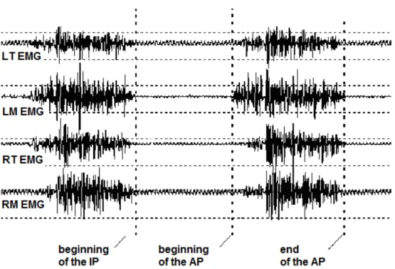
inactive period (IP) and the active period (PA) of each muscle (Figure 1). For this purpose, the irst and last identiied cycles of each attempt were disregarded and the central cycle was selected. It is
a routine calculation faster and more accurate when
Figure 1 – EMG signal of Left Temporal (LT), Left Masseter (LM), Right Temporal (RT) and Right Masseter (RM) muscles for a chewing cycle (Inactive Period (IP) and Active Period (AP).
This study was approved by the Ethics Committee of the institution, under reference number 26/2009.
Participants were characterized using descriptive statistics (mean, standard deviation and 95% coni -dence interval). After checking the normality of the data through the Shapiro-Wilk test, we used the
Spearman correlation coeficient to examine the
relationship between the degree of spasticity, the
level of motor function and topographical classii -cation with parameters of amplitude and duration activation of MA and AT muscles. Correlation values
smaller than 0.20 indicate very low association;
values between 0.20 and 0.39 low association;
values between 0.40 and 0.69 moderate associ-ation; values between 0.70 and 0.89 high associ-ation; and values between 0.90 and 1.0 association too high.
Statistical analysis was performed using the Statistical Package for the Social Sciences (SPSS) version 17.0 for Windows and for all procedures
we adopted the signiicance level of 5% (p <0.05 ),
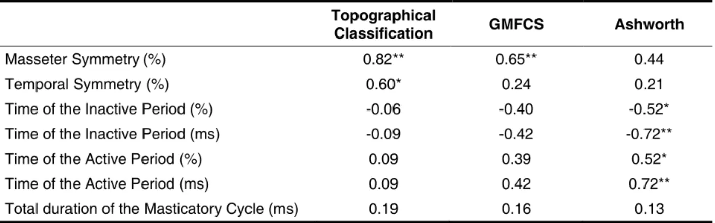
Table 2 shows the values of Spearman
corre-lation coeficient, which was used to examine
the relationship between the degree of spasticity
(MAS), the level of motor function (GMFSC) and topographical classiication with electromyographic
variables.
The topographic classiication was highly
associated with the MA symmetry (p=0.00) and moderately with the AT symmetry (p=0.01). We observed a moderate association between the level of motor function and MA symmetry (p=0.00).
In the analysis of the masticatory cycle time (ms)
was found moderate association between the higher degree of spasticity and the inactive period decrease (p=0.04), as well as with active period
increase (p=0.04). In the analysis of the masticatory
cycle time (%), the greater spasticity was highly associated with inactive period decreased (p=0.00) and active period increase (p=0.00).
RESULTS
Volunteers with spastic CP had a mean age of
9.9 ± 2.0 years, height 1.3 ± 0.2 m and body mass 30.6 ± 8.1 kg. 40.0% (6/15) were male and 60.0% (9/15) were female. Regarding the topographical
classiication 40.0% (6/15) of the volunteers had
hemiparesis, 46.7% (7/15) had diparesis and
13.3% (2/15) showed quadriparesis. In relation to motor impairment assessed by GMFCS, 6.7 (1/15) of subjects were classiied at level I, 46.7% (7/15) at level II, 20.0% (3/15) at the level III and 26.7% (4/15) at level IV. According to the MAS, the mean and standard deviation of the index for the spasticity
degree was 3.8 ± 2.5 points.
Table 1 shows the results of electromyographic variables, MA symmetry, AT symmetry, time of the inactive period, time of the active period and the total duration of the masticatory cycles.
Mean SD 95% CI
Masseter Symmetry(%) 80.62 9.65 75.27 - 85.96
Temporal Symmetry (%) 81.10 7.09 77.17 - 85.03
Time of the Inactive Period (%) 43.45 13.12 36.19 - 50.71
Time of the Inactive Period (ms) 473.25 176.64 375.40 - 571.10
Time of the Active Period (%) 56.55 13.12 49.28 - 63.81
Time of the Active Period (ms) 614.01 250.42 475.30 - 752.70
Total duration of the Masticatory Cycle (ms) 1087.26 269.15 938.20 - 1236.30
Table 1 – Mean values , standard deviations (SD) and conidence intervals of the mean (95% CI) of the
muscle symmetry and masticatory cycle time of volunteers with cerebral palsy (n = 15).
ms= millisecond
Topographical
Classification GMFCS Ashworth
Masseter Symmetry(%) 0.82** 0.65** 0.44
Temporal Symmetry (%) 0.60* 0.24 0.21
Time of the Inactive Period (%) -0.06 -0.40 -0.52*
Time of the Inactive Period (ms) -0.09 -0.42 -0.72**
Time of the Active Period (%) 0.09 0.39 0.52*
Time of the Active Period (ms) 0.09 0.42 0.72**
Total duration of the Masticatory Cycle (ms) 0.19 0.16 0.13
Table 2 – Correlation between the level of Gross Motor Function Classiication System (GMFCS), degree of spasticity (Ashworth) and topographical classiication in relation to the masticatory cycle
time of volunteers with cerebral palsy (n = 15).
The sum of the degrees of muscle tone in the different joints obtained by MAS has been used in clinical practice and research11,23,24. Spastic cerebral
palsy is characterized by increased muscle tone causes alterations in posture and movement and prevents proper motor development.
It was expected that the degree of spasticity
was associated with measures of the amplitude of the electromyographic activity of the masticatory muscles, but was only associated with the time.
The sample size may have inluenced the absence
of some associations. No studies were found that evaluated the association of spasticity with param-eters of the electrical activity of oral motor function of children with CP. There are also a few studies that evaluate the association of spasticity with
other alterations of the stomatognathic system. In
the assessment of oral function of children with CP, Ries and Bérzin (2005) found no association between the severity of spasticity with the severity of temporomandibular dysfunction11.
In this sample the highest degree of spasticity
was associated with a shorter time inactive period and longer active period of the masticatory muscles. The shorter time inactive period and longer active
period shows the greatest dificulty in relaxation of
volunteers more spastic. Although there was no relationship with the total duration of the masticatory cycles, this behavior in the MA and AT muscles activity can cause an overload in the stomatognathic system, which in turn may contribute to altera-tions in oral motor function. The alteraaltera-tions in the parameters of the time periods of the masticatory cycle characterize the muscle activity dysfunction
of spastic CP. Knowledge of the speciic alteration
that causes a movement disorder of masticatory muscles allow a multidisciplinary team to establish more appropriate therapeutic strategies.
CONCLUSION
These results indicate multifactorial relation-ships for alterations in oral motor function in the presence of CP. Most spasticity was associated
with less time to relax and the largest contraction time during the chewing cycle. Volunteers with
hemiparesis compared to those with quadriparesis and diparesis showed a lighter level of motor and functional impairment, yet with greater asymmetry of the muscles AT and MA. The symmetry and the time of activation of AT and MA muscles should be
considered during therapeutic approach for dificulty
chewing in CP.
DISCUSSION
The CP is a disorder characterized by the presence of abnormal muscle tone and lower performance on functional abilities and gross motor function17. In this study, the spasticity, the gross
motor function and the topographical classiication
were associated with parameters of amplitude and activation time of the masticatory muscles, showing aspects of oral motor dysfunction.
The mandibular movement during mastication is considered asymmetric18,19. Normative values of
symmetry of masticatory activity for children were
not found in the scientiic literature. Despite the
small sample size in this study, it was observed in 12 children with typical development (average age
of eight years), a symmetry index of MA muscle
91.5 ± 6.2% and of AT muscle 92.0 ± 8.3%8. In
healthy adults, was considered a normal Symmetry
index of electrical activity was considered normal in
healthy adults of at least 82.0 ± 1.3%20 or 87.1 ±
1.6% for the MA muscle and 88.1 ± 1.4% for the AT muscle21. The present study found values lower
than these parameters, although not be the same population. Children with CP have activity patterns of the muscles involved in chewing more asymmetric compared with the typical development group8.
The symmetry parameters of MA and AT muscles showed, respectively, high and moderate
association with the topographic classiication of CP. In the presence of spastic quadriparesis and
spastic diparesis we found more symmetry in the electrical activity of the masticatory muscles when
compared with spastic hemiparesis. One expla -nation for this result is the fact that these volunteers, although more committed, have a more symmetric
distribution of their alterations. In children with
spastic hemiparesis are observed early asymme-tries of movements and functional abilities due to a unilateral distribution of spasticity22. The functional
asymmetry characteristic of hemiparesis is also observed in oral motor function.
The symmetry of the MA was moderately
associated with the GMFCS. Thus, the higher
the symmetry of the electrical activity of the MA muscles in CP volunteers, the greater their gross motor function limitations and functional abilities. This result, seemingly contradictory, may be related to greater asymmetry of volunteers with spastic hemiparesis and a lighter level of motor and
functional impairment observed with the GMFCS. In
REFERENCES
1. Bax M, Goldstein M, Rosenbaum P, Leviton
A, Paneth N, Dan B, Jacobsson B, Damiano D.
Proposed deinition and classiication of cerebral
palsy. Dev Med Child Neurol. 2005;47:571-6. 2. Mancini MC, Fiúza PM, Rebelo JM, Magalhães
LC, Coelho ZAC, Paixão ML et al. Comparação
do desempenho de atividades funcionais em
crianças com desenvolvimento normal e crianças
com paralisia cerebral. Arq Neuropsiquiatr. 2002;60(2-B):446-52.
3. Mayston MJ. People With Cerebral Palsy: Effects of and Perspectives for Therapy. Neural Plast. 2001;8(1-2):51-69.
4. Miscio G, Del Conte C, Pianca D, Colombo R, Panizza M, Schieppati M et al. Botulinum toxin in post-stroke patients: stiffness modiications and
clinical implications. J Neurol. 2004;251(2):189-96. 5. Priori A, Cogiamanian F, Mrakic-Sposta S. Pathophysiology of spasticity. Neurol Sci. 2006;27(4):S307-S9.
6. Giubbina CA, Assencio-Ferreira VJ. A deglutição
na Paralisia Cerebral. Rev CEFAC. 2002;4:29-34.
7. Vivone GP, Tavares MMM, Bartolomeu RS, Nemr K, Chiappetta ALML. Análise da Consistência Alimentar e Tempo de Deglutição em Crianças com Paralisia Cerebral Tetraplégica Espástica. Rev
CEFAC. 2007;9(4):504-11.
8. Ries LGK, Bérzin F. Ativação Assimétrica dos Músculos Temporal e Masseter em Crianças com
Paralisia Cerebral. Fisioter Mov. 2009;22(1):45-52. 9. Palisano R, Rosenbaum P, Walter S, Russell D,
Wood E, Galuppi B. Developmental and reliability
of a system to classify gross motorfunction in
children with cerebral palsy. Dev Med Child Neurol. 1997;39:214-23.
10. Bohannon RW, Smith MB. Inter reliability of a modiied Ashworth scale of muscle spasticity. Phys
Ther. 1987;67(2):206-7.
11. Ries LGK, Bérzin F. Signs and symptoms
of temporomandibular disorders in children with cerebral palsy. Rev Bras Fisioter. 2005;9:341-6.
12. Sommerich CM, Joines SMB, Hermans V, Moon
SD. Use of surface electromyography to estimate
neck muscle activity. J Electromyogr Kinesiol.
2000;10:377-98.
13. Abbink JH, van der Bilt A, van der Glas HW.
Detection of onset and termination of muscle activity in surface electromyograms. J Oral Rehabil. 1998;25:365-9.
14. Ferrario VF, Sforza C, Colombo A, Ciusa V.
An electromyographic investigation of masticatory muscles symmetry in normo-occlusion subjects. J Oral Rehabil. 2000;27:33-40.
15. Ferrario VF, Tartaglia GM, Galletta A, Grassi GP, Sforza C. The Inluence of Occlusion on Jaw and Neck Muscle Activity: a Surface EMG
Study in Healthy Young Adults. J Oral Rehabil. 2006;33:341-8.
16. Ries LGK, Alves MC, Berzin F. Asymmetric
Activation of Temporalis, Masseter, and Sternocleidomastoid Muscles in Temporomandibular Disorder Patients. Cranio 2008;26(1):59-64.
17. Assumpção MS de, Piucco EC, Corrêa ECR, Ries LGK. Coativação, espasticidade, desempenho
motor e funcional na paralisia cerebral. Motriz. 2011;17(4):650-9.
18. Green JR, Moore CA, Ruark JL, Rodda PR, Morvée WT, Vanwitzenburg MJ. Development
of chewing in children from 12 to 48 months:
RESUMO
Objetivo: veriicar a existência de associação entre o grau de espasticidade, o nível de função motora ampla e a classiicação topográica de voluntários com PC com parâmetros de amplitude e tempo da ativação eletromiográica dos músculos temporal (TA) e masseter (MA). Métodos: a atividade
muscu-lar durante a tarefa de mastigação foi avaliada em quinze voluntários com PC. Os instrumentos clíni
-cos utilizados foram: a Escala Modiicada de Ashworth para espasticidade, o Sistema de Classiicação de Função Motora Ampla (GMFCS) para função motora ampla. Foram analisados os parâmetros de simetria muscular e tempo do período ativo e inativo do ciclo mastigatório. Resultados: durante o
ciclo mastigatório observou-se associação entre o nível de função motora ampla e a simetria do MA, entre o maior grau de espasticidade e a diminuição do período inativo e aumento do período ativo e entre a classiicação topográica e a simetria do MA e do TA. Conclusão: a simetria e o tempo da
atividade dos músculos TA e MA devem ser considerados durante a terapia da função motora oral
na PC.
indices in healthy Brazilian young adults and data reproducibility. J Oral Rehabil. 2009;36:577-83. 22. Jones MW, Morgan E, Shelton JE, Thorogood
C. Cerebral Palsy: Introduction and Diagnosis (Part I). J Pediatr Health Care. 2007;21(3):146-52.
23. Bhakta BB, Cozens JA, Chamberlain MA, Bamford JM. Quantifying associated reactions in the paretic arm in stroke and their relationship to spasticity. Clin Rehabil. 2001;15(2):195-206.
24. Blackburn M, van Vliet P, Mocket SP. Reliability of Mensurements Obtained With the Modiied Ashworth Scale in the Lower Extremities of People
With Stroke. Phys Ther. 2002;82(1):25-34.
longitudinal study of EMG patterns. J Neurophysiol.
1997;77(5):2704-16.
19. Widmalm SE, Lee Y-S, McKay D. Clinical Use
of Qualitative Electromyography in the Evaluation
of Jaw Muscle Function: A Practitioner’s Guide.
Cranio. 2007;25(1):63-73.
20. Ferrario VF, Sforza C, Miani Jr A, D’Addona
A, Barbini E. Electromyographic activity of human masticatory in normal young people. statistical evaluation of reference values for clinical applications. J Oral Rehabil. 1993;20:271-80.
21. Felicio CM, Sidequersky FV, Tartaglia GM,
Sforza C. Electromyographic standardized
Received on: January 18, 2012 Accepted on: April 29, 2012
Mailing address:
Lilian Gerdi Kittel Ries
Rua Pascoal Simone, 358 – Coqueiros Florianópolis – SC
CEP: 88080-350