www.reumatologia.com.br
REVISTA BRASILEIRA DE
REUMATOLOGIA
R E V B R A S R E U M A T O L . 2 0 1 3 ;5 4 ( 1 ): 7 5 – 7 6
Letter to the editor
Sonographic appearance of pseudopodagra in Behçet’s disease
Aspecto ultrassonográfi co de pseudopodagra na doença de Behçet
Dear Editor,
Arthritis of the i rst metatarsophalangeal (MTP) joint of a di-fferent cause than gout is identii ed by the term pseudopoda-gra. Although the majority causes of pseudopodagra are de-posits of other micro crystals, there has been other rare cause, such as Behcet’s disease.1,2 We present the case of a patient with a pseudopodagra due to Behcet’s disease.
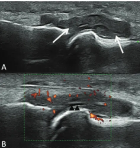
A 43-year-old female patient suffering from Behcet’s dis-ease for 10 years presented to the emergency department (ED) with discomfort in her right MTP joint. The onset was char-acterized by acute pain, swelling, erythema, tenderness, and limitation of movement of the i rst MTP joint of the right foot. The patient denied any fever, chills, trauma, urethral symp-toms, conjunctivitis, or history of gout. She had a past several attacks affecting oligoarticular involving knees and ankles. There was no family history of rheumatic disease. On physi-cal examination, she was afebrile and all vital signs were normal. The area overlying her i rst MTP joint was red, hot, swollen, and exquisitely tender to touch and to any move-ment of the great toe. The skin was not disrupted, and no lym-phangitis or adenopathy was present. All other joints were normal, as was the remainder of the physical examination. Patient refused arthrocentesis procedure. We preferred ultra-sound imaging as a i rst line imaging technique instead of magnetic resonance imaging in order to save time. Grayscale ultrasound examination of dorsal aspect of 1st MTP showed marked synovial thickening (arrows) and minimal synovial l uid (arrowhead) (Fig. 1A). There was no gouty tophus at the MTP joint. On power doppler imaging increased color signals are seen within the hyperthrophic synovium consistent with hyperemia (Fig. 1B). On left side, minimal concentric prolifera-tion of synovial lining cell tissue and synovial l uid (arrow-head) was seen (Fig. 2A). There were no color signals within the synovium on the 1st MTP of the left foot (Fig. 2B). Pre-liminary diagnosis was pseudopodagra rather than metatar-sophalangeal arthritis because signii cant extra-articular soft tissue changes were evident. Soft tissue changes can occur
Fig. 1 – A, Grayscale ultrasound examination of dorsal aspect. B, Power doppler imaging increased color signals.
Fig. 2 – A, Minimal concentric proliferation of synovial lining cell tissue and synovial l uid. B, There were no color signals within the synovium.
R E V B R A S R E U M A T O L . 2 0 1 3 ;5 4 ( 1 ): 7 5 – 7 6
76
with rheumatological diseases including familial Mediterra-nean fever (FMF)'s joint attack, acute rheumatic fever (ARF), calcium pyrophosphate dihydrate (CPPD) crystal deposition disease and gout. Rheumatoid arthritis also commonly affects the MTP joints, although this is characterized with a symmet-ric polyarthritis, making it a rare differential diagnosis. In our patient FMF and ARF were not considered as a relevant dif-ferential diagnosis because those diseases were inconsistent with clinical and laboratory i ndings. Sonographic features of CPPD deposits depend on the amount and distribution, vary-ing from homogeneously punctate pattern or sharply deined hyperechoic bands within the articular cartilage or loating in synovial luid to rounded or amorphous-shaped hyper-echoic areas in ibrocartilage.3 In the gout, monosodium urate crystals tend to result in hyperechoic enhancement on the supericial margin of hyaline cartilage. The most frequent ultrasonographic characteristic of gouty tophi is hyperecho-genicity.4 In addition, tophi are generally heterogeneous, with poorly dei ned contours, multiple grouped and surrounded by an anechoic halo.5,6 However, these sonographic i ndings were not seen in our patient.
Musculoskeletal ultrasound has signiicant potential use-fulness for the diagnosis, severity assessment, decision to treat, and treatment efi cacy assessment of patients with un-known etiology arthritis.7 The ultrasound examination as a fast, reproducible diagnostic method has now become part of the routine diagnosis in rheumatological disorders.
Conl icts of interest
The authors declare no conl icts of interest.
R E F E R E N C E S
1. Benamour S.Pseudopodagra in Behçet's disease. Rev Rhum
Engl Ed 1995; 62(2):153-4.
2. Giacomello A, Sorgi ML, Zoppini A. Pseudopodagra in
Behçet's syndrome.Arthritis Rheum 1981; 24(5):750-1.
3. Lin YY, Wang TG, Li KJ, Lew HL.Imaging Characteristics
of Calcium Pyrophosphate Dihydrate Crystal Deposition Disease. Am J Phys Med Rehabil 2012; Http://dx.doi.org/ 10.1097/PHM.0b013e31825566aa.
4. Fernandes EA, Lopes MG, Mitraud SA, Ferrari AJ, Fernandes AR. Ultrasound characteristics of gouty tophi in the
olecranon bursa and evaluation of their reproducibility. Eur J Radiol 2012; 81(2):317-23.
5. de Ávila Fernandes E, Kubota ES, Sandim GB, Mitraud SA, Ferrari AJ, Fernandes AR. Ultrasound features of tophi in chronic tophaceous gout.
Skeletal Radiol 2011; 40(3):309-15.
6. de Ávila Fernandes E, Sandim GB, Mitraud SA, Kubota ES, Ferrari AJ, Fernandes AR. Sonographic description and classii cation of tendinous involvement in relation to tophi in chronic tophaceous gout. Insights Imaging 2010; 1(3):143-8. 7. Howard RG, Pillinger MH, Gyftopoulos S, Thiele
RG, Swearingen CJ, Samuels J. Reproducibility of
musculoskeletal ultrasound for determining monosodium urate deposition: concordance between readers. Arthritis Care Res (Hoboken) 2011; 63(10):1456-62.
Fuat Ozkan*, Gozde Yildirim Cetin, Mehmet Sayarlioglu
Kahramanmaras Sutcu Imam University, Faculty of Medicine, Department of Radiology, Kahramanmaras, Turkey
*Corresponding author.