155
Radiol Bras. 2012 Mai/Jun;45(3):155–159
Redundant nerve roots of the cauda equina: review
of the literature
*
Raízes nervosas redundantes da cauda equina: revisão da literatura
Marcello Henrique Nogueira-Barbosa1, Leonor Garbin Savarese2, Carlos Fernando Pereira da Silva Herrero3, Helton Luiz Aparecido Defino4
In imaging diagnosis, redundant nerve roots of the cauda equina are characterized by the presence of elongated, enlarged and tortuous nerve roots in close relationship with a high-grade lumbar spinal canal stenosis. This is not an independent entity, but it is believed to be a consequence of the chronic compression at the level of the lumbar canal stenosis and thus may be part of the natural history of lumbar spinal stenosis. The present paper is aimed at reviewing the histopathological, electrophysiological and imaging findings, particularly at magnetic resonance imaging, as well as the clinical meaning of this entity. As the current assessment of canal stenosis and root compression is preferably performed by means of magnetic resonance imaging, this is the imaging method by which the condition is identified. The recognition of redundant nerve roots at magnetic resonance imaging is important, particularly to avoid misdiagnosing other conditions such as intradural arteriovenous malformations. The literature approaching the clinical relevance of the presence of redundant nerve roots is controversial. There are articles suggesting that the pathological changes of the nerve roots are irreversible at the moment of diagnosis and therefore neurological symptoms are less likely to improve with surgical decompression, but such concept is not a consensus.
Keywords: Cauda equina; Spinal stenosis; Spine; Magnetic resonance imaging.
A redundância das raízes nervosas da cauda equina é caracterizada, no diagnóstico por imagem, pela presença de raízes nervosas alongadas, espessadas e tortuosas junto a uma área de estenose do canal vertebral lombar. Não é uma entidade independente, mas acredita-se que ocorra como o resultado da compressão crônica ao nível da este-nose do canal lombar e que, portanto, pode fazer parte da evolução natural da esteeste-nose. O objetivo deste trabalho é revisar a histopatologia, a eletrofisiologia, as características de imagem, especialmente na ressonância magnética, e o significado clínico desta entidade. Como a avaliação atual da estenose de canal e das compressões radiculares é realizada preferencialmente por meio da ressonância magnética, é nesse método de imagem que a redundância das raízes nervosas da cauda equina será identificada. O reconhecimento desta entidade nos exames de ressonância magnética é importante, principalmente para evitar equívocos que poderiam levar ao diagnóstico de outras afecções, particularmente de malformações arteriovenosas intradurais. A literatura é controversa a respeito da importância clí-nica da presença de redundância das raízes nervosas da cauda equina. Há artigos que sugerem que as alterações patológicas da raiz nervosa são irreversíveis no momento do diagnóstico e que os sintomas neurológicos não são mais suscetíveis de melhora com a descompressão cirúrgica, porém este conceito não é um consenso.
Unitermos: Cauda equina; Estenose espinhal; Coluna vertebral; Ressonância magnética.
Abstract
Resumo
* Study developed at the Service of Radiodiagnosis of Centro de Ciências das Imagens e Física Médica (CCIFM), Hospital das Clínicas da Faculdade de Medicina de Ribeirão Preto da Univer-sidade de São Paulo (HC-FMRPUSP), Ribeirão Preto, SP, Brazil. 1. PhD, Professor at Centro de Ciências das Imagens e Física Médica (CCIFM), Faculdade de Medicina de Ribeirão Preto da Universidade de São Paulo (FMRPUSP), Ribeirão Preto, SP, Brazil. 2. Graduate Student of Medicine, Faculdade de Medicina de Ribeirão Preto da Universidade de São Paulo (FMRPUSP), Ribei-rão Preto, SP, Brazil.
3. Master, MD, Physician Assistant at the Spine Surgery Sec-tion, Division of Orthopedics, Hospital das Clínicas da Faculdade de Medicina de Ribeirão Preto da Universidade de São Paulo (HC-FMRPUSP), Ribeirão Preto, SP, Brazil.
4. PhD, Professor, Department of Biomechanics, Medicine and Rehabilitation of the Locomotor System at Faculdade de Medi-cina de Ribeirão Preto da Universidade de São Paulo (FMRPUSP), Ribeirão Preto, SP, Brazil.
Nogueira-Barbosa MH, Savarese LG, Herrero CFPS, Defino HLA. Redundant nerve roots of the cauda equina: review of the literature. Radiol Bras. 2012 Mai/Jun;45(3):155–159.
stenosis(1–11). Initially, RNR of the cauda
equina was described as being identified at myelography as serpiginous filling de-fects associated with partial or total block-age preventing the transit of contrast mate-rial(1–9). Most recently imaging findings of
RNR of the cauda equina were also de-scribed for magnetic resonance imaging (MRI)(10–12). The prevalence of this
syn-drome may reach 33.8–42% of patients with lumbar canal stenosis(8,10,13) and has
been detected in 8.2% of elderly cadavers in Japan(8).
INTRODUCTION
Redundant nerve roots (RNR) of the cauda equina is characterized by the pres-ence of elongated, enlarged and tortuous nerve roots in the subarachnoid space of the lumbar column adjacent to a site of canal
Mailing Address: Leonor Garbin Savarese. Faculdade de Me-dicina de Ribeirão Preto – Universidade de São Paulo. Avenida Bandeirantes, 3900, Campus Universitário. Ribeirão Preto, SP, Brazil, 14049-090. E-mail: [email protected]
Some studies attribute the first descrip-tion of RNR of the cauda equina to Ver-biest(11–13). Such studies specifically
men-tion an article where Verbiest has described a series of surgically confirmed cases of lumbar spinal stenosis(14) but, as the
origi-nal text was evaluated, the authors of the present review concluded that Verbiest described elongated nerve roots in another series of cases about rare presentations of cauda equina compression(15). However,
the term “RNR of the cauda equina” was first utilized by Cressman & Pawl(1).
The literature is controversial on the clinical relevance of the presence of redun-dant roots of the cauda equina. There are articles suggesting that the degenerative pathological changes in the affected nerve roots are irreversible and, therefore, the neurological symptoms would be less likely to improve with surgical decompres-sion, but consensus is still to be reached on such concept.
Redundancy of the nerve roots is prob-ably the pathological result of a chronic compressive force at the level of the site of spinal canal constriction(9).
The present article is aimed at review-ing the findreview-ings described on publications related to RNR of the cauda equina, with emphasis on imaging findings, and particu-larly at MRI. Additionally, studies on the clinical significance of this entity were re-viewed.
HISTOPATHOLOGICAL STUDY
Suzuki et al. have analyzed clinical as-pects, radiological studies and electro-physiology studies of patients with and without RNR of the cauda equina and in-vestigated anatomical and histopathologi-cal findings of redundant and non-redun-dant nerve roots in cadavers(8,9). The main
pathological findings in patients with RNR of the cauda equina were derangement and reduction in the number of nervous fibers, demyelination, besides endoneural fibrosis and Schwann cells proliferation(8,9). In their
investigation, those authors have not dem-onstrated any significant change in the an-terior horn and in the anterolateral columns of the spinal cord. Low degree of nerve cells loss and spinal ganglion fibrosis were observed, but such changes were equally
present in non-redundant roots and were, therefore, considered as changes related to aging.
The topographic distribution in ob-served in cases of redundant nerve roots was 33.3% in S1, 33.3% at S2, 16% at L5 and 17.3% below the S2 roots(8). The
ana-tomical study developed by Suzuki et al. has demonstrated that all redundant nerve roots pass through the spinal canal constric-tion. No redundancy was observed in roots which did not pass by the site of canal con-striction(8). Demyelination and axonal loss
was considered as being a consequence of the continuous mechanical compression of nerve roots confined to the stenosed spinal canal(8).
ELECTROPHYSIOLOGY
The electrophysiological study of re-dundant nerve roots has demonstrated tem-poral dispersion of the action potential and delay of the velocity of the sensitive ner-vous conduction, although the difference in the velocity of sensitive nervous conduc-tion has not been statistically significant(8).
The study authors have suggested that such results reflected the reduction and degen-eration of the nerve fibers(8). They also have
not found any significant difference in the electrophysiological manifestation be-tween ventral and dorsal redundant nerve roots.
Measurements of the redundant and non-redundant nerve roots conduction ve-locity were performed during surgery(13). In
that study, the velocity of conduction of re-dundant nerve roots was approximately one half the normal value and, in general, the redundant nerve roots presented multiphase action potentials temporarily dispersed as compared with the normal roots.
Only one case report approached the evaluation of electromyographic changes related to RNR of the cauda equina in the postoperative period(16). Such report has
documented preoperative partial denerva-tion of the left medial segment of the gas-trocnemius muscle and, in spite of the sig-nificant pain relief experienced by the pa-tient after laminectomy, electromyographic changes in the gastrocnemius muscle were still present over the three-month and six-month follow-up.
INTRAOPERATIVE FINDINGS
Several authors have described cases of elongated and tortuous nerve roots after opening the dura mater, confirming the preoperative imaging findings(1,2,4–6,11,13).
Spontaneous extrusion of elongated roots usually occurs during the surgical opening of the dura mater(2,6,7,11,13). The opening of
the dura mater may be performed to rule out the hypothesis of arteriovenous malforma-tion(7) or simply because surgeons have
decided that such a procedure should be a part of the decompression(6,13). Also, there
are reports on the indication of duroplasty in association with decompressive laminec-tomy was indicated for the relief of nerve root compression(11,13).
MYELOGRAPHY
In the literature, the diagnosis of RNR of the cauda equina was first described in studies approaching myelographic im-ages(1–9). At the time, myelography was the
only imaging method available to docu-ment the anatomy and pathology of nerve roots of the cauda equina in vivo. Currently, myelography has been widely replaced by MRI and its utilization has remained re-stricted to the rare cases where MRI is con-traindicated or for those cases where post-operative metal artifacts are enough to se-verely degrade MRI images.
The myelographic pattern described in redundant nerve roots of the cauda equina corresponds to serpiginous intradural fill-ing defects associated with a partial or com-plete blockage of the transit of contrast material and, therefore, spinal canal steno-sis(1–7). Variations in the positioning of the
patients may influence the presentation of RNR of the cauda equina at the images. The orthostatic position yields a better vi-sualization of redundant roots(1,7,8,10).
Serpiginous filling defects may also be identified by myelography in the presence of dural or intradural arteriovenous malfor-mations(17), thus constituting an important
differential diagnosis. Less frequently, a plexiform neurofibroma or a neurinoma may cause thickening and redundancy of nerve roots(18–20). Other diseases can cause
demyelinat-ing polyneuropathy and some hereditary neuropathies; but association between such entities and serpiginous nerve roots of cauda equina was not reported.
MAGNETIC RESONANCE IMAGING
The characteristics of RNR of the cauda equina at MRI images have already been described by different authors(10–12).
Thick-ened, elongated and tortuous or serpiginous nerve roots adjacent to a site of lumbar vertebral canal stenosis represent typical
findings. In the literature, signal intensity abnormalities nerve roots have not been reported in association with RNR of the cauda equina. Figures 1 and 2 illustrate the typical MRI findings in cases of redundant nerve roots.
At MRI, the main differential diagnosis must be done with dural arteriovenous malformations or arteriovenous fistulas. In such conditions, MRI usually shows intra-dural serpiginous vessels and ectasia of the coronal venous plexus (Figure 3). How-ever, the clinical presentation of vascular
malformations is different from that of ca-nal stenosis, and there are MRI findings that help in the differentiation between these two conditions. RNR are typically associated with vertebral canal stenosis, and clinically the patient presents neuro-genic claudication. At the images, arterio-venous malformations are frequently asso-ciated with great draining vessels, and may
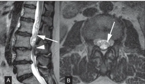
Figure 1. A 61-year-old woman with neurogenic claudication. Preoperative MRI images obtained for plan-ning of surgical decompression. A: Sagittal MRI T2-weighted image demonstrates degenerative changes of the spine with disc herniation and vertebral canal stenosis at L3-4 and L4-5 levels (arrowhead). Elon-gated and tortuous nerve roots of the cauda equina can be identified (arrow). B: Axial MRI T2-weighted image obtained at a level slightly above the L3-4 stenosis, also presenting tortuous nerve roots (arrow).
Figure 3. Surgically confirmed spinal arteriovenous fistula. A,B: Sagittal MRI T2-weighted image showing dilated serpiginous ves-sels (arrow). Presence of in-creased signal intensity in the spinal cord substance (arrow-head) associated with medullary ischemia secondary to steal phenomenon. C: Coronal MRI T2-weighted image also demon-strating dilated serpiginous ves-sels in the venous plexus (ar-row).
clinically present with signs of myelopathy, subarachnoid hemorrhage or medullary is-chemia(7,17). Dural arteriovenous fistula is
usually associated with abnormalities in signal intensity of the spinal cord on MRI T2-weighted sequences(17). Another
impor-tant imaging finding in the diagnosis of arteriovenous fistula is the exaggerated enhancement of the coronal venous plexus after gadolinium injection in 88% of the patients with such condition(17).
Ono et al. have selected 44 patients with L4-5 spondylolisthesis submitted to de-tailed pre- and postoperative clinical evalu-ation, all of them presenting vertebral ca-nal stenosis and complete blockage of the contrast material transit at preoperative myelography(10). All the patients in that
study were preoperatively submitted to myelography and MRI, and for the purpose of statistical analysis, the patients were di-vided into three groups, as follows: group A, patients with RNR of the cauda equina found both at myelography and MRI (16 patients); group B, patients with RNR of the cauda equina found only at myelogra-phy (14 patients); and group C, where none of the two studies demonstrated RNR of the cauda equina (14 patients). Statistically significant difference in relation to clinical symptoms was found between groups A and C. The patients group where MRI dem-onstrated RNR of the cauda equina (group A) presented more severe clinical symp-toms than group C, both pre- and postop-eratively. Ono et al. have identified that the group of patients in whom MRI could dem-onstrate RNR of the cauda equina pre-sented worse clinical symptoms and, there-fore, the identification of such finding by MRI would tend to have clinical signifi-cance(10). Then, the same authors
specu-lated that the redundancy would have been underestimated at MRI in the less severe cases, where the redundancy was only dem-onstrated at myelography, probably be-cause of the fact that, at MRI, the lumbar spine image acquisition is routinely per-formed with the patient in a neutral posi-tioning, while the myelography study was performed with dynamic evaluation in the orthostatic position.
In another study, the relative length of the redundant nerve roots was measured on sagittal MR images and presented
statisti-cal relationship with clinistatisti-cal improvement within the group of patients with RNR(12).
The authors standardized the measurement of the relative length of the nerve roots on the most central sagittal image of the lum-bar column. The relative length was ob-tained by measuring the distance from the maximum stenosis level to the most distant point where the presence of redundant roots could be identified, and by dividing such obtained value by the height of the verte-bral body located above the stenosis level. The greater the relative length, the better the postoperative outcomes, a result that, at a first analysis, seems to be unexpected, leading the study authors to raise the hy-pothesis that such result was related to a greater accommodation capability of the longer redundant roots during flexion and extension of the spinal column, as related to effects of traction forces(12). The present
study authors did not find any other study investigating such type of imaging finding in relation to postsurgical outcomes.
DISCUSSION
RNR of the cauda equina is a relatively common MRI finding in the lumbar verte-bral column in cases where canal stenosis is present. The clinical condition of patients with RNR of the cauda equina is related to the natural history of the lumbar spinal canal stenosis. One believes that chronic lumbar stenosis is the primary cause of symptoms. The results from the study de-veloped by Suzuki et al. suggest that pa-tients with RNR of the cauda equina present more advanced ages, longer symp-toms duration and greater severity of neu-rological signs and symptoms than patients presenting canal stenosis without nerve root redundancy(8). Min et al. also have
found that the group of patients with RNR presented more advanced age, They have not found any statistically significant dif-ference between groups of patient with and without RNR of the cauda equina in rela-tion to symptom durarela-tion, and observed a non-statistically significant tendency for better postoperative outcomes in the group of patients without RNR of the cauda equina(12).
The literature is controversial about the implication of the presence of RNR in the
indication for decompressive surgery for spinal canal stenosis. Some investigations suggest that such abnormalities of the nerve roots are irreversible, and, therefore, the neurological symptoms are not susceptible to improvement after surgical decompres-sion(8,9). Ono et al. have found that cases of
RNR of the cauda equina diagnosed by means of MRI were more severe, and that the presence of such change may negatively affect the surgical outcomes(10). In two
other studies, the postoperative improve-ment was not statistically different in pa-tients with and without RNR of the cauda equina(12,13), but in one of those studies a
tendency towards worse results for patients with RNR of the cauda equina was identi-fied, similarly to the results reported by Ono et al., as mentioned in the previous para-graph(12). Anyway, the complete regression
of stenosis symptoms after surgical decom-pression is rare in patients with typical RNR, and they frequently continue com-plaining of dysesthesia and paresthesia(13).
CONCLUSION
At MRI, RNR are often associated with degenerative stenosis of the lumbar verte-bral canal and therefore the recognition of such entity is important to avoid equivocal diagnosis and mainly false-positive results for arteriovenous malformations. The clini-cal significance of such abnormality of the cauda equina in the progression of lumbar canal stenosis is still controversial, but there are indications in the literature sug-gesting that its identification at MRI may indicate a tendency towards worse postop-erative results. Therefore the authors of the present review suggest that radiologists should look for RNR of the cauda equina at MRI and, if applicable, describe such finding in their reports.
REFERENCES
1. Cressman MR, Pawl RP. Serpentine myelo-graphic defect caused by a redundant nerve root. Case report. J Neurosurg. 1968;28:391–3. 2. Fox JL. Redundant nerve roots in the cauda
equina. Case report. J Neurosurg. 1969;30:74–5. 3. Ehni G, Moiel RH, Bragg TG. The “redundant” or “knotted” nerve root: a clue to spondylotic cauda equina radiculopathy. Case report. J Neurosurg. 1970;32:252–4.
5. Duncan AW, Kido DK. Serpentine cauda equina nerve roots. Radiology. 1981;139:109–11. 6. de Tribolet N, Campiche R. Redundant nerve
roots of the cauda equina. A rare disease? Eur Neurol. 1982;21:169–74.
7. Hacker DA, Latchaw RE, Yock DH Jr, et al. Re-dundant lumbar nerve root syndrome: myelo-graphic features. Radiology. 1982;143:457–61. 8. Suzuki K, Ishida Y, Ohmori K, et al. Redundant
nerve roots of the cauda equina: clinical aspects and consideration of pathogenesis. Neurosurgery. 1989;24:521–8.
9. Suzuki K, Takatsu T, Inoue H, et al. Redundant nerve roots of the cauda equina caused by lum-bar spinal canal stenosis. Spine. 1992;17:1337– 42.
10. Ono A, Suetsuna F, Irie T, et al. Clinical signifi-cance of the redundant nerve roots of the cauda equina documented on magnetic resonance
im-aging. J Neurosurg Spine. 2007;7:27–32. 11. Hakan T, Celiko™lu E, Aydoseli A, et al. The
re-dundant nerve root syndrome of the cauda equina. Turk Neurosurg. 2008;18:204–6.
12. Min JH, Jang JS, Lee SH. Clinical significance of redundant nerve roots of the cauda equina in lumbar spinal stenosis. Clin Neurol Neurosurg. 2008;110:14–8.
13. Tsuji H, Tamaki T, Itoh T, et al. Redundant nerve roots in patients with degenerative lumbar spinal stenosis. Spine. 1985;10:72–82.
14. Verbiest H. A radicular syndrome from develop-mental narrowing of the lumbar vertebral canal. J Bone Joint Surg Br. 1954;36-B:230–7. 15. Verbiest H. Sur certaines formes rares de
com-pression de la queue de cheval. In: Hommage à Clovis Vincent. Paris: Maloine; 1949. p. 161–74. 16. Renstein L, Twardzik FG, Russo GL, et al. Elec-tromyographic abnormalities in redundant nerve
root syndrome of the cauda equina. Arch Phys Med Rehabil. 1984;65:270–2.
17. Gilbertson JR, Miller GM, Goldman MS, et al. Spinal dural arteriovenous fistulas: MR and my-elographic findings. AJNR Am J Neuroradiol. 1995;16:2049–57.
18. Friedrich H, Gilsbach J, Mennel HD, et al. Knot-ted neurinoma or plexiform neurofibroma in the cauda equina (author’s transl). Neurochirurgia (Stuttg). 1978;21:135–8.
19. Rengachary SS, McGregor DH, Watanabe I, et al. Suggested pathological basis of “redundant nerve root syndrome” of the cauda equina. Neurosur-gery. 1980;7:400–11.