Changes in endogenous tissue
glutathione level in relation to
murine ascites tumor growth and
the anticancer activity of cisplatin
Cell and Tumor Biology Laboratory, Department of Zoology, School of Life Sciences, North-Eastern Hill University, Shillong, India D. Khynriam and
S.B. Prasad
Abstract
Changes in glutathione levels were determined in tissues of 11- to 12-week-old Swiss albino mice at different stages of Daltons lymphoma tumor growth and following cisplatin (8 mg/kg body weight, ip) treatment for 24-96 h, keeping 4-5 animals in each experimental group. Glutathione levels increased in spleen of tumor-bearing com-pared to normal mice (9.95 ± 0.14 vs 7.86 ± 1.64 µmol/g wet weight, P£0.05) but decreased in blood (0.64 ± 0.10 vs 0.85 ± 0.09 mg/ml) and testes (9.28 ± 0.15 vs 10.16 ± 0.28 µmol/g wet weight, P£0.05). Daltons lymphoma cells showed an increase in glutathione concen-tration (4.43 ± 0.26 µmol/g wet weight) as compared to splenocytes, their normal counterpart (3.62 ± 0.41 µmol/g wet weight). With the progression of tumor in mice, glutathione levels decreased significant-ly in testes (~10%) and bone marrow cells (~13%) while they in-creased in Daltons lymphoma cells (28-46%) and spleen (15-27%). Glutathione levels in kidney, Daltons lymphoma cells and bone marrow cells (8.50 ± 1.22, 4.43 ± 0.26 and 3.28 ± 0.17 µmol/g wet weight, respectively) decreased significantly (6.04 ± 0.42, 3.51 ± 0.32 and 2.17 ± 0.14 µmol/g wet weight, P£0.05) after in vivo cisplatin treatment for 24 h. Along with a decrease in glutathione level, the glutathione-S-transferase (GST) activity also decreased by 60% in tumor cells after cisplatin treatment. The elevated drug uptake by the tumor cells under the conditions of reduced glutathione concentration and GST activity after treatment could be an important contributory factor to cisplatins anticancer activity leading to tumor regression. Furthermore, lower doses of cisplatin in combination with buthionine sulfoximine (an inhibitor of glutathione synthesis) may be useful in cancer chemotherapy with decreased toxicity in the host.
Correspondence
S.B. Prasad
Department of Zoology School of Life Sciences North-Eastern Hill University Shillong 793022
India
Fax: +91-364-55-0076/55-0108 E-mail: [email protected] or [email protected] Research supported by the University Grants Commission, New Delhi (COSIST, DRS program) and by North-Eastern Hill University, Shillong, India.
Received December 10, 2001 Accepted September 13, 2002
Key words
·Cisplatin
·Dalton’s lymphoma ·Glutathione
·Glutathione-S-transferase ·Chemotherapy
Introduction
Cis-diamminedichloroplatinum (II), com-monly known as cisplatin, is a widely used anticancer drug against several animal (1,2) and human malignancies (3). Many of its
tissue calcium and potassium concentrations (7), various enzymes such as 5'-nucleoti-dase, arginase, cathepsins and lactate dehy-drogenase (8), and mitochondria (9), proper-ties that have led to the proposal of the involvement of cisplatin in multistep and multilevel actions on the tumor cell/host re-lationship during cisplatin-mediated chemo-therapy (8,9). However, the therapeutic effi-cacy of the drug is limited due to various dose-limiting side effects mainly involving nephrotoxicity (10), hematotoxicity (11), muta-genicity (12,13) and the development of ac-quired resistance (14). The changes in gluta-thione levels have been shown to be of im-portance in the occurrence of these side ef-fects (11-15). L-buthionine-(S,R)-sulfoximine (BSO) is an inhibitor of g-glutamylcysteine synthetase and has often been used to deplete cellular reduced glutathione (16). In vitro stud-ies have demonstrated that cancer cells resis-tant to alkylating agents including cisplatin can be made sensitive to these drugs through BSO-induced glutathione depletion (17).
Glutathione, an endogenous intracellular thiol-containing tripeptide (L-g -glutamyl-L-cysteinyl-glycine), is an important antioxi-dant and has been the focus of interest in cancer chemotherapy (18). Under normal physiological conditions mammalian cells maintain more than 98% of glutathione in the reduced form (GSH) at intracellular con-centration of 0.5 to 10 mM (19). GSH plays a crucial role in numerous biochemical pro-cesses including diverse bioreductive reac-tions, transport, protection against harmful free radicals and xenobiotics, and detoxifi-cation of metals and electrophiles (19). GSH has been implicated in the metabolism of cisplatin (20) and other metals causing alter-ations in the rate of metal uptake and elimi-nation. The reactive thiol group of GSH is also responsible for many other functions, i.e., i) maintenance of protein structure and function by reducing the disulfide linkages of proteins, ii) regulation of protein synthe-sis and degradation, iii) maintenance of
im-mune function, iv) leukotriene and prosta-glandin metabolism, v) modulation of mi-crotubule-related processes, vi) bile forma-tion, and vii) DNA synthesis (19).
Therefore, it was of interest to determine GSH levels in various tissues during ascites Daltons lymphoma growth in vivo and cis-platin treatment. Platinum uptake and gluta-thione-S-transferase (GST; EC 2.5.1.18) ac-tivity were also measured in Daltons lym-phoma cells collected from mice under dif-ferent experimental conditions in an attempt to identify the mechanism of GSH changes and its significance in cisplatin-mediated cancer chemotherapy.
Material and Methods
Chemicals
GSH, BSO, 5,5'-dithiobis-2-nitrobenzoic acid (DTNB), and 1-chloro-2,4-dinitroben-zene (CDNB) were purchased from Sigma (St. Louis, MO, USA). Cisplatin was ob-tained from Biochem Pharmaceutical Indus-tries, Mumbai, India. Other chemicals used in the experiments were of analytical grade. The solutions were prepared in twice glass-distilled water.
Tumor maintenance
Inbred Swiss albino mice were main-tained in the laboratory with free access to commercially available food pellets and wa-ter. For each experimental determination, 4-5 mice aged 10-12 weeks were used. Ascites Daltons lymphoma tumor was maintained
in vivo by intraperitoneal (ip)
transplanta-tion of 1 x 10⁷ tumor cells per animal (0.25
Cisplatin treatment and tissue collection
On the 5th, 10th and 15th day of tumor growth, tumor-bearing mice were killed by cervical dislocation, and liver, kidney, spleen, blood, ascites tumor, testes, humerus and femur bones were collected. Bone marrow cells were prepared from humerus and femur by flushing in PBS with a hypodermic sy-ringe and by centrifugation (3000 g, 10 min) to collect the bone marrow cell pellet. As-cites tumor was also centrifuged (3000 g, 10 min) to separate the Daltons lymphoma cell pellet and the ascites supernatant. Accord-ing to the dose and treatment schedule used earlier by us (11), a single dose of cisplatin (8 mg/kg body weight, ip) was administered to tumor-bearing mice on the 10thday post-tumor transplantation and after 24, 48, 72 and 96 h of treatment tissues were collected and used for GSH determinations. GSH was also determined in the same tissues of con-trol (untreated tumor-bearing mice injected with normal saline only) and normal (un-treated, non-tumor-bearing) mice. Spleno-cytes were prepared from the spleens of normal mice as described earlier (6) and used as the normal counterpart of Daltons lymphoma cells in GSH determinations.
Blood glutathione
Blood GSH was determined by the method of Beutler et al. (21). Virtually all non-protein sulfhydryl of red blood cells is in the form of GSH. DTNB is a disulfide compound which is readily reduced by sulf-hydryl compounds forming a highly colored yellow anion.
Briefly, 0.9 ml water and 1.5 ml precipi-tating solution (1.67 g glacial metaphospho-ric acid, 0.20 g Na2EDTA, 30 g NaCl, 100 ml
water) were added immediately to 100 µl of blood and mixed well. After 5 min of incuba-tion at room temperature the reacincuba-tion mix-ture was centrifuged (3000 g at 4ºC, 15 min) and 2.0 ml of 0.3 mol/l phosphate solution
and 250 µl DTNB solution (200 mg in 100 ml of 1% sodium citrate solution) were added to 500 µl of the clear supernatant. A blank was prepared with 1 ml phosphate solution, 1 ml water, 0.5 ml precipitating solution, and 250 µl DTNB solution. Both the blank and sample reaction mixtures were read against water at 412 nm in a Beckman DU-640 spectrophotometer. GSH concentration was calculated on the basis of a millimolar ex-tinction coefficient of 13.6 and a molecular weight of 307.
Glutathione in other tissues
Total GSH was estimated in various tis-sues by the method of Sedlak and Lindsay (22). Briefly, 5% tissue homogenates were prepared in 20 mM EDTA, pH 4.7, and 100 µl of the homogenate or pure GSH was added to 0.2 M Tris-EDTA buffer (1.0 ml, pH 8.2) and 20 mM EDTA, pH 4.7 (0.9 ml) followed by 20 µl of Ellmans reagent (10 mmol/l DTNB in methanol). After 30 min of incubation at room temperature, absorbance was read at 412 nm in a Beckman DU-640 spectrophotometer. Samples were centri-fuged before the absorbance of the superna-tants was measured.
Glutathione-S-transferase assay
GST activity was assayed in Daltons lymphoma cells of tumor-bearing mice un-der different experimental conditions. As-cites tumor was centrifuged at 3000 g at 4ºC for 10 min and the tumor cell pellet was collected. A 10% homogenate of liver and Daltons lymphoma cells in 0.1 M sodium phosphate-1 mM Na2EDTA buffer, pH 6.5,
was obtained with a motor-driven Teflon-pestle homogenizer at 4ºC. The tissue homo-genates were centrifuged at 27,000 g at 4ºC for 20 min and the respective supernatants were collected as the enzyme source.
1.0 ml contained 850 µl of 0.1 M sodium phosphate-1 mM Na2EDTA buffer, pH 6.5,
50 µl of 20 mM GSH in deionized water, and 50 µl of 20 mM CDNB in 95% ethanol. The reaction mixture, maintained at 30ºC, was started by adding 50 µl enzyme and the increase in absorbance at 340 nm was moni-tored for 3 min with a Beckman DU-640 spectrophotometer. The enzyme activity was calculated using the extinction coefficient (E340 =9.6 mM-1 cm-1). One unit of enzyme
activity was defined as the amount of en-zyme that catalyzed the conjugation of 1 µmol CDNB per minute.
The same supernatants used for enzyme assays were also used for protein determina-tion by the method of Lowry et al. (24) using bovine serum albumin as standard. The spe-cific activity of the enzyme is reported per mg protein.
Buthionine sulfoximine and cisplatin treatment
To standardize the time of maximum BSO-induced depletion, BSO at the dose of 5 mM/kg body weight (11) was injected ip
into a group of mice on the 10th day post-tumor transplantation. Different tissues were then collected and used for GSH determina-tions at 2-h intervals after treatment. Since the maximum GSH depletion (43-45%) in-duced by BSO in Daltons lymphoma cells was observed 8 h after treatment, in the experiments involving BSO plus cisplatin treatments BSO was given 8 h prior to cis-platin treatment.
Determination of platinum uptake by Dalton’s lymphoma cells
The Daltons lymphoma cell pellet used for GSH estimation was also processed for platinum uptake analysis. The pellet (0.5 g) was digested with 5.0 ml nitric acid and a few drops of hydrogen peroxide in a clean conical flask with gentle heating to near
dryness. Perchloric acid (5.0 ml) was added to the digests and again heated to near dry-ness to remove excess nitric acid. This last stage was repeated until a clear solution resulted. The digests were finally dissolved in 5 ml of 5% nitric acid (distilled water and nitric acid at the ratio of 95:5 ml), and the filtrate was stored in polypropylene bottles for platinum analysis using a plasma lab ICP-OES emission spectrometer operated at 700 volts PMT and 214,438 nm wavelength after calibrating the instrument with the ap-propriate standard solutions.
Macrophage-Dalton’s lymphoma cell interactions in vitro
Peritoneal exudate cells (PEC) from nor-mal mice were harvested as described by Dileepan et al. (25). Four days prior to PEC collection, mice were injected ip with 2 ml of 4% thioglycolate and cells were collected by lavage of the peritoneal cavity with 5 ml serum-free 199 medium containing 100 U/ml penicillin. PEC were centrifuged at 400 g for 10 min and washed twice with 199 medium. The cells were resuspended in com-plete 199 medium supplemented with 10% fetal bovine serum at a concentration of 1 x 10⁵
/ml on a 24-well culture dish (Genetix, New Delhi, India) and incubated at 37ºC in an incubator in the presence of 5% CO2-95%
air. After 2 h, the culture plates were shaken and washed with warm medium (37ºC) to remove non-adherent cells. Fresh medium was added and the mixture was incubated overnight. Different groups of tumor-bear-ing mice were treated with cisplatin (4 and 8 mg/kg body weight) and with BSO (8 h prior to cisplatin treatment) plus cisplatin. Twenty-four hours after cisplatin treatment, Daltons lymphoma cells were aspirated from the peri-toneal cavity, washed with 199 medium, resuspended in complete medium at a con-centration of 1 x 10⁴
cells/ml and used as target cells for macrophages.
with warm medium and target Daltons lym-phoma cells (treated and control) were added at an effector:target ratio of 10:1 and further incubated for 24 h. After incubation, the supernatants were discarded, fresh medium was added and the number of macrophage-Daltons lymphoma interacting cells was scored and analyzed under different treat-ment conditions.
Host survival
Tumor-transplanted animals were ran-domly divided into four groups consisting of 10 mice per group. On the 8th day post-tumor transplantation, mice in groups I and II received cisplatin at the dose of 4 and 8 mg/kg body weight, respectively. Mice in group III were treated with BSO (5 mM/kg body weight 8 h prior to cisplatin treatment) plus cisplatin (4 mg/kg body weight). The mice in group IV received the same volume of normal saline and were used as control. The deaths, if any, of the hosts in different groups were recorded daily and the survival pattern of the hosts was determined for dif-ferent groups. The antitumor efficacy of the drug in different groups is reported as percent increase in life span calculated as [(T-C/C)] x 100, where T and C are the mean survival days of treated and untreated con-trol mice, respectively.
Results
GSH levels during tumor growth and cisplatin treatment
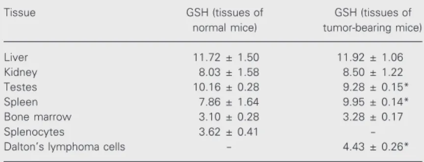
GSH levels did not change much in liver and kidney at different stages of tumor growth. However, a significant decrease in GSH levels was noted in testes (~10%) and bone marrow cells (~13%), while an in-crease was observed in spleen (15-27%) and Daltons lymphoma cells (28-46%) during 5 to 15 days of tumor growth (Figure 1).
Cisplatin treatment of tumor-bearing mice for 24-96 h showed a significant (P£0.05) decrease of GSH level in kidney (18-29%), Daltons lymphoma cells (14-21%), spleen (12-18%), bone marrow cells (20-34%) (Table 1) and blood (26-37%) for one or more observation times (Table 2).
Comparison of GSH levels in tissue of normal and tumor-bearing mice revealed an increase in spleen, but a decrease in testes (Table 3) and blood (Table 2) of tumor-bearing mice. A 22% increase in GSH con-tent was observed in Daltons lymphoma cells as compared to splenocytes which were used as their normal controls (Table 3).
Glutathione-S-transferase activity
GST specific activity (units/mg protein)
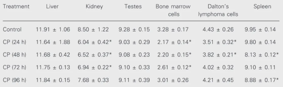
Table 1. Changes in total GSH content in the tissues of tumor-bearing mice after cisplatin treatment in vivo.
Treatment Liver Kidney Testes Bone marrow Dalton’s Spleen
cells lymphoma cells
Control 11.91 ± 1.06 8.50 ± 1.22 9.28 ± 0.15 3.28 ± 0.17 4.43 ± 0.26 9.95 ± 0.14
CP (24 h) 11.64 ± 1.88 6.04 ± 0.42* 9.03 ± 0.29 2.17 ± 0.14* 3.51 ± 0.32* 9.80 ± 0.14
CP (48 h) 11.68 ± 0.42 6.52 ± 0.37* 9.08 ± 0.23 2.20 ± 0.15* 3.82 ± 0.21* 8.13 ± 0.12*
CP (72 h) 11.75 ± 0.13 6.94 ± 0.22* 9.10 ± 0.33 2.61 ± 0.12* 4.02 ± 0.32 9.10 ± 0.11
CP (96 h) 11.84 ± 0.15 7.68 ± 0.33 9.11 ± 0.39 3.01 ± 0.26 4.21 ± 0.45 8.88 ± 0.17*
Table 2. Changes in blood GSH levels of tumor-bearing mice after cis-platin treatment.
Treatment GSH
Normal mice 0.85 ± 0.09
Tumor-bearing mice (control) 0.64 ± 0.10**
Cisplatin (24 h) 0.57 ± 0.02
Cisplatin (48 h) 0.47 ± 0.06*
Cisplatin (72 h) 0.40 ± 0.01*
Cisplatin (96 h) 0.42 ± 0.08*
Normal mice = mice without tumor or treatment. GSH content is re-ported as mg/ml. Data are rere-ported as means ± SD (N = 4-5).
*P<0.05 compared to control; **P<0.05 compared to normal mice (Stu-dent t-test).
Table 3. GSH levels in the tissues of normal and tumor-bearing mice on the 10th day following tumor transplantation.
Tissue GSH (tissues of GSH (tissues of
normal mice) tumor-bearing mice)
Liver 11.72 ± 1.50 11.92 ± 1.06
Kidney 8.03 ± 1.58 8.50 ± 1.22
Testes 10.16 ± 0.28 9.28 ± 0.15*
Spleen 7.86 ± 1.64 9.95 ± 0.14*
Bone marrow 3.10 ± 0.28 3.28 ± 0.17
Splenocytes 3.62 ± 0.41
-Dalton’s lymphoma cells - 4.43 ± 0.26*
Splenocytes from normal mice were used as the normal control of Dalton’s lym-phoma cells. GSH content is reported as µmol/g wet weight. Data are reported as means ± SD (N = 4-5).
*P<0.05 compared to the respective tissue from normal animal (Student t-test).
in Daltons lymphoma cells (1.28 ± 0.19) decreased to 0.43 ± 0.05, 0.55 ± 0.04, 0.25 ± 0.03, and 0.26 ± 0.04 at 24, 48, 72 and 96 h, respectively, of cisplatin treatment, correspond-ing to a ~60-80% reduction in GST activity.
BSO treatment
Treatment of mice with BSO, an inhibi-tor of GSH synthesis, decreased GSH levels in all tissues, with a gradual recovery at 10-12 h of treatment. In Daltons lymphoma cells maximum (>40%) depletion in GSH concentration was noted at 8 h of BSO treat-ment (Figure 2).
Platinum uptake by Dalton’s lymphoma cells
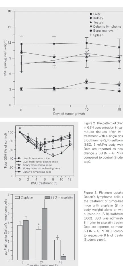
Platinum uptake by Daltons lymphoma cells at 8 h of cisplatin treatment was 3.6 µg platinum/g Daltons lymphoma cells, later decreasing from 24 to 48 h (Figure 3). How-ever, when cisplatin was given to the mice in combination with BSO, platinum uptake by cells was found to be higher than after cis-platin treatment alone at 8 and 24 h of treat-ment (Figure 3), followed by a decrease at 48 h of treatment.
In vitro macrophage-Dalton’s lymphoma cell interactions
In in vitro studies on
macrophage-Daltons lymphoma cell interactions, one or two Daltons lymphoma cells connecting to macrophages through cytoplasmic bridges were counted as interacting cells. These in-teractions were much lower (~9%) in the control and increased significantly (P£0.05) in cisplatin-treated groups in a dose-depend-ent manner (~28-55%). However, as com-pared to the mice treated with cisplatin (4 mg/kg body weight) alone, combined treat-ment with BSO plus cisplatin (4 mg/kg body weight) caused a significant (P£0.05) in-crease in the interactions of Daltons lym-phoma cells with macrophages.
Table 4. Antitumor activity of cisplatin and L-buthionine-(S,R)-sulfoximine (BSO) plus cisplatin against murine ascites Dalton’s lymphoma.
Treatment Survival days I.L.S. (%)
Control 19.00 ± 2.5
-Cisplatin (4 mg) 29.25 ± 3.0* 53.95
Cisplatin (8 mg) >60* >216
BSO + cisplatin (4 mg) 42.50 ± 1.0* 123.68
BSO was administered ip 8 h prior to cisplatin treatment on the 8th day post-tumor transplantation. I.L.S. (%) = percent increase in the life span was calculated as described in Methods. Data are reported as means ± SD (3-4 experimental determina-tions for 10 mice per group).
Figure 1. Changes in GSH levels in the tissues of mice at different stages of tumor growth in vivo. Results are reported as means ± SD (N = 4-5). *P£0.05 compared to day 5 for Dalton’s lymphoma cells, and to day 0 for other tissues (Student t-test).
µg Platinum/g Dalton’s lymphoma cells
7
6
5
4
3
2
1
0
*
*
*
8 24 48
Cisplatin treatment (h)
Cisplatin BSO + cisplatin
Figure 3. Platinum uptake by Dalton’s lymphoma cells after the treatment of tumor-bearing mice with cisplatin (8 mg/kg body weight) alone or with L-buthionine-(S,R)-sulfoximine (BSO). BSO was administered 8 h prior to cisplatin treatment. Data are reported as means ± SD (N = 4). *P£0.05 compared to respective 8 h of treatment (Student t-test).
Host survival patterns
Following tumor transplantation, an early sign of tumor development was noted from the 3rd-4th day onwards. Tumor-inoculated animals survived for 19 ± 2.5 days. In the group of mice treated with a therapeutic dose of cisplatin (8 mg/kg body weight) about 80% of the animals survived for more than 60 days and were tumor free.However, com-parison of treatment of tumor-bearing mice with a subtherapeutic dose of cisplatin (4 mg/kg body weight) alone and its combina-tion with BSO showed that combined treat-ment with BSO and a subtherapeutic dose of cisplatin resulted in a further increase in survival time up to 40 days (Figure 4). Thus, the percent increase in life span was about 53% in mice receiving a subtherapeutic dose of cisplatin and doubled to ~123% after combination chemotherapy with BSO and a subtherapeutic dose of cisplatin. However, 8 mg cisplatin treatment provided an even longer survival time than BSO plus 4 mg cisplatin (Table 4).
Discussion
Glutathione, the most abundant thiol in the cell, is maintained in reduced form (GSH) by NADPH-dependent glutathione reductase (19). The present results showed that GSH concentrations differ widely among differ-ent tissues in the mouse. The highest con-centration, found in the liver, was approxi-mately four times that observed in bone mar-row cells which showed the lowest concen-tration (Figure 1). GSH concenconcen-tration did not change significantly in liver and kidney during in vivo tumor growth in mice, but
GSH (µmol/g wet weight)
18
+ 15
12
9
6
3
0
+ +
+
+ *
* *
*
*
* *
0 5 10 15
Days of tumor growth
Liver Kidney Testes
Dalton’s lymphoma Bone marrow Spleen
*
Figure 2. The pattern of changes in GSH concentration in various mouse tissues after in vivo
treatment with a single dose of L-buthionine-(S,R)-sulfoximine (BSO, 5 mM/kg body weight). Data are reported as percent change ± SD (N = 4). *P<0.05 compared to control (Student t -test).
Total GSH (% of control)
100
80
60
40
20
0
0 2 4 6 8 10 12
BSO treatment (h)
Liver from normal mice Liver from tumor-bearing mice Kidney from normal mice Kidney from tumor-bearing mice Dalton’s lymphoma cells
* **
* ***
* * * **
* *
* *
*
decreased in testes and bone marrow and increased in spleen and Daltons lymphoma cells (Figure 1). The observed GSH increase in spleen and Daltons lymphoma cells was maximum on day 10 of tumor growth, slightly decreasing thereafter over the next 4-5 days when tumor growth was probably reduced. In Ehrlich ascites tumor cells, maximal GSH concentration was observed by about the 7th day of tumor growth, followed by a signifi-cant decrease on the 14th day of tumor growth, which was correlated with a de-crease in cell proliferation and in the rate of GSH synthesis (26).
Cancer cells can generate large amounts of hydrogen peroxide which may contribute to their ability to mutate and damage normal tissues, and, moreover, facilitate tumor growth and invasion (27). It has been sug-gested that persistent oxidative stress in tu-mor cells could partly explain some impor-tant characteristics of cancer, such as acti-vated proto-oncogenes, genomic instability, drug resistance, invasion and metastasis (28), and the resistance of many cells against oxi-dative stress is often associated with high intracellular levels of GSH (29). The ob-served increase of GSH mainly in tumor cells suggests its involvement in facilitating the proliferation and metabolism of tumor
cells in the host and agrees with the report that elevation of intracellular GSH in tumor cells is associated with mitogenic stimula-tion (30) and that GSH controls the onset of tumor cell proliferation by regulating pro-tein kinase C activity and intracellular pH (31). A decrease in the rate of cancer cell proliferation has also been correlated with a decrease in GSH level in tumor cells (26). Thus, the variation of GSH concentration in the Daltons lymphoma cells and other tis-sues with tumor growth in the host may reflect alterations in the antioxidant machin-ery accompanied by changes in the rate of proliferation of Daltons lymphoma cells in the host.
As compared to liver, kidney and bone marrow of normal animals, GSH levels in-creased slightly in the respective tissues of tumor-bearing animals (Table 3). However, significant changes were noted in other tis-sues, i.e., an increase in spleen and Daltons lymphoma cells and a decrease in testes (Table 3) and blood (Table 2). Some amino acid precursors for glutathione synthesis, particularly glutamine, serve as important respiratory fuel for tumor cells (26) and tu-mor progression has been associated with an avid consumption of host glutamine by tu-mor cells causing a decrease in internal GSH concentration (32). The increase in GSH level in Daltons lymphoma cells with tumor growth may also reflect increased uptake of essential amino acids from plasma with a consequent depletion of blood precursors for GSH synthesis, thereby reducing blood GSH concentration in tumor-bearing ani-mals (Table 2). The GSH content of Daltons lymphoma cells was also higher than that of splenocytes (the normal counterpart of Daltons lymphoma cells) (Table 3). As GSH is known to play a role in detoxifying many reactive metabolites (33), its increased lev-els in Daltons lymphoma cells and tissues of tumor-bearing mice could also represent a protective mechanism in response to various toxic radicals.
0 4 8 12 16 20 24 28 32 36 40 44 48 Days after tumor transplantation
% Survival
100
Control
Cisplatin (4 mg/kg body weight) Cisplatin (8 mg/kg body weight) BSO + cisplatin (4 mg/kg body weight) 80
60
40
20
0 Figure 4. Mean survival pattern
Cisplatin treatment of mice caused a de-crease of GSH in Daltons lymphoma cells, kidney, bone marrow, spleen (Table 1) and blood (Table 2). The decrease in blood GSH levels in tumor-bearing mice and after cis-platin treatment may result in decreased an-tioxidant capacity. It has been observed that changes in glutathione levels in blood and development of various types of hematotox-icity in the host are inversely related in cis-platin-mediated chemotherapy (11). Evi-dence of low blood GSH has been reported in a variety of diseases including cancer (34). Similarly, the decrease in GSH levels in kidney, bone marrow and spleen after cisplatin treatment of the host may contri-bute to nephrotoxicity, mutagenicity and immune response, respectively. Indeed, when GSH levels were increased in the hosts, the nephrotoxic as well as mutagenic effects of cisplatin were found to be decreased (Khynriam D and Prasad SB, unpublished results).
In an attempt to understand the relation-ship between cisplatin-mediated cytotoxic-ity and decreased GSH levels in the hosts, particularly in Daltons lymphoma cells, GST activity was assayed in Daltons lymphoma cells. GST represents an integral part of the detoxification system and protects cells against oxidative and chemical-induced tox-icity and stress by catalyzing the S-conjuga-tion between the thiol group of GSH and the electrophilic moiety of toxic substrates in-cluding cisplatin (35). Cisplatin-GSH com-plexes have been proposed to be ejected from the cells in an ATP-dependent fashion by the glutathione S-conjugate (GS-X) ex-port pump (36), thereby preventing the drug from reaching the critical DNA. GST activ-ity decreased by 60-80% after cisplatin treat-ment. Thus, the decreased activity of GST as well as GSH concentration (Table 1) in Daltons lymphoma cells after cisplatin treat-ment suggest the possibility of a reduced conjugation of GSH with cisplatin because it is known that cisplatin-GSH conjugates can
be formed directly or be catalyzed by GST (36). This may suggest reduced elimination of the drug through export pumps and avail-ability of more drug in tumor cells causing cytotoxic effects. Modulation of GST activ-ity has been shown to affect the sensitivactiv-ity of tumor cell lines to alkylating agents. Inhibi-tion of GST by either ethacrynic acid or piriprost enhanced alkylator cytotoxicity to both rat and human cancer cell lines (37). Cisplatin also behaves like an alkylating agent, and therefore a similar modulation mechanism might be involved. Both GSH depletion by BSO and GST inhibition have increased the tumoricidal activity of mel-phalan, a proteolytic alkylating drug (38), supporting the present view of involvement of cisplatin-mediated decrease of GSH and inhibition of GST in its anticancer activity. This is further strengthened by the results of the experiments involving combined treat-ment with BSO, a GSH depleting agent, with cisplatin.
been suggested to have better therapeutic efficacy than cisplatin alone, and decreased side effects in the host (39). Here, combined treatment with BSO and a subtherapeutic dose of cisplatin showed that, as compared to cisplatin alone, macrophage-Daltons lym-phoma cell interactions were significantly increased by about 37% in the BSO plus cisplatin-treated groups. This clearly sug-gests that the therapeutic response to a sub-therapeutic dose of cisplatin could be en-hanced when used in combination with BSO, which itself is a nontoxic agent. As the vari-ous side effects developed in the hosts are dose dependent, it may be suggested that, compared to higher doses of cisplatin, the modulation of GSH levels in combination with low doses of cisplatin should be very useful in decreasing the toxicity, with en-hanced cytotoxicity in the host. This sugges-tion is supported by the results of host sur-vival patterns under different treatment con-ditions. The percent increase in life span was about 53% in mice receiving a subtherapeu-tic dose of cisplatin alone and was doubled (~123%) after combination chemotherapy with BSO and a subtherapeutic dose of cis-platin (Figure 4, Table 4).
The enhanced drug uptake by Daltons lymphoma cells under the conditions of re-duced GSH levels and GST activity in Daltons lymphoma cells should be an
im-portant determining factor in the cytotoxic effects of cisplatin against murine ascites Daltons lymphoma. Tumor cells are defi-cient in the ability to repair DNA after reac-tion with cisplatin (40). Although some re-covery of GSH in Daltons lymphoma cells was noted later at 72-96 h following cis-platin treatment (Table 1) along with elimi-nation of the drug from the cells, a decrease in GSH levels and availability of more drug during the initial stage of treatment may be expected to give rise to a variety of meta-bolic dysfunctions directly or indirectly re-lated to cisplatin cytotoxicity which may be partially repaired or retained within the Daltons lymphoma cells, leading to tumor regression.
The data presented here suggest that changes in GSH levels in Daltons lymphoma cells and other tissues during tumor growth and after cisplatin treatment could be an important factor contributing to tumor pro-gression and cisplatin-mediated anticancer activity.
Acknowledgments
We thank the Head, Regional Sophisti-cated Instrumentation Center, North-East-ern Hill University, Shillong, for help with the platinum determinations.
References
1. Rosenberg B (1985). Fundamental studies with cisplatin. Cancer, 55: 2303-2316.
2. Prasad SB & Giri A (1994). Antitumour effect of cisplatin against murine ascites Dalton’s lymphoma. Indian Journal of Experimental Biology, 32: 155-162.
3. Go RS & Adjei AA (1999). Review of the comparative pharmacology and clinical activity of cisplatin and carboplatin. Journal of Clinical Oncology, 17: 409-422.
4. Chu G (1994). Cellular responses to cisplatin. Journal of Biological Chemistry, 269: 787-790.
5. Collins JL & Kao M (1989). The anticancer drug cisplatin increases the naturally occurring cell-mediated lysis of tumor cells. Cancer Immunology, Immunotherapy, 29:17-22.
6. Prasad SB & Sodhi A (1981). Effect of cis-dichlorodiammineplatinum (II) on the agglutinability of tumor and normal cells with concanavalin
A and wheat germ agglutinin. Chemico-Biological Interactions, 36: 355-367.
7. Prasad SB & Giri A (1999). Cisplatin-induced changes in tissue calcium and potassium concentrations in tumour-bearing mice.
Medical Science Research, 27: 459-462.
8. Prasad SB, Giri A, Khynriam D, Kharbangar A, Nicol BM & Lotha C (1999). Cisplatin-mediated enzymatic changes in mice bearing as-cites Dalton’s lymphoma. Medical Science Research, 27: 723-730. 9. Kharbangar A, Khynriam D & Prasad SB (2000). Effect of cisplatin on
mitochondrial protein, glutathione, and succinate dehydrogenase in Dalton lymphoma-bearing mice. Cell Biology and Toxicology, 16: 363-373.
10. Krakoff IH (1979). Nephrotoxicity of cis-dichlorodiammineplatinum.
Cancer Treatment Reports, 63: 1523-1525.
glutathi-one levels after cisplatin treatment of tumor-bearing mice. Cell Biology and Toxicology,17: 357-370.
12. Giri A, Khynriam D & Prasad SB (1998). Vitamin C mediated protec-tion on cisplatin-induced mutagenicity in mice. Mutation Research, 421: 139-148.
13. Prasad SB & Khynriam D (2002). Mutagenicity and endogenous glutathione levels in tumor-bearing mice after cisplatin treatment. In: Khassanova L, Collery P, Maymard I, Khassanova Z & Etienne JC (Editors), Metal Ions in Biology and Medicine.Vol. 7. John Libbey Eurotext, Paris, France, 580-585.
14. Kartalou M & Essigmann JM (2001). Mechanisms of resistance to cisplatin. Mutation Research, 478: 23-48.
15. Giri A, Khynriam D & Prasad SB (1998). Use of vitamin C against cisplatin induced mutagenicity and nephrotoxicity. In: Sharan RN (Editor), Trends in Radiation and Cancer Biology. Forschungszentrum Jülich GmbH, Jülich, Germany, 166-176.
16. Bailey HH (1998). L-S,R-buthionine sulfoximine: historical develop-ment and clinical issues. Chemico-Biological Interactions, 111-112: 239-254.
17. Prasad SB, Khokhar AR & Siddik ZH (1994). Potentiation by L-buthionine sulfoximine (BSO) of cytotoxicity of platinum(IV) com-plexes in sensitive and resistant human ovarian carcinoma cells. In: Collery P, Poirier LA, Littlefield NA & Etienne JC (Editors), Metal Ions in Biology and Medicine. Vol. 3.John Libbey Eurotext, Paris, France, 365-370.
18. Arrick BA & Nathan CF (1984). Glutathione metabolism as a determi-nant of therapeutic efficacy: a review. Cancer Research,44: 4224-4232.
19. Wang W & Ballatori N (1998). Endogenous glutathione conjugates: occurrence and biological functions. Pharmacological Reviews,50: 335-355.
20. Suzuki CAM & Cherian MG (1990). The interaction of cis- diam-minedichloroplatinum with metallothionein and glutathione in rat liver and kidney. Toxicology,64: 113-127.
21. Beutler E, Dunn O & Kelly BM (1963). Improved method for the determination of blood glutathione. Journal of Laboratory and Clini-cal Medicine, 65: 882-888.
22. Sedlak J & Lindsay RH (1968). Estimation of total, protein-bound and nonprotein sulfhydryl groups in tissues with Ellman’s reagent. Ana-lytical Biochemistry, 25: 192-205.
23. Habig WH, Pabot MJ & Jarkoby WB (1974). Glutathione-S-trans-ferase. The first enzymatic step in mercapturic acid formation. Jour-nal of Biological Chemistry,249: 7130-7139.
24. Lowry OH, Rosebrough NJ, Farr AL & Randall RJ (1951). Protein measurement with the Folin phenol reagent. Journal of Biological Chemistry, 193:265-275.
25. Dileepan KN, Lorsbach RB & Stechschutte DJ (1993). Mast cell granules inhibit macrophage-mediated lysis of mastocytoma cells (P815) and nitric oxide production. Journal of Leukocyte Biology, 53: 446-453.
26. Estrela JM, Sternandez R, Terradez P, Asensi M, Puertes IR & Vina J (1992). Regulation of glutathione metabolism in Ehrlich ascites
tu-mour cells. Biochemical Journal, 286: 257-262.
27. Szatrowski TP & Nathan CF (1991). Production of large amounts of hydrogen peroxide by human tumor cells. Cancer Research, 51: 794-798.
28. Toyokuni S, Okamoto K, Yodoi J & Hiai H (1995). Persistent oxidative stress in cancer. FEBS Letters, 358: 1-3.
29. Estrela JM, Obrador E, Navarro J, Lasso De La Vega MC & Pellicer JA (1995). Elimination of Ehrlich tumours by ATP-induced growth inhibition, glutathione depletion and X-rays. Nature Medicine,1: 84-88.
30. Shaw JP & Chou IN (1986). Elevation of intracellular glutathione content associated with mitogenic stimulation of quiescent fibro-blasts. Journal of Cellular Physiology, 129: 193-198.
31. Terradez P, Asensi M, Lasso De La Vega MC, Puertes IR, Vina J & Estrela JM (1993). Depletion of tumour glutathione in vivo by buthionine sulfoximine: modulation by the rate of cellular prolifera-tion and inhibiprolifera-tion of cancer growth. Biochemical Journal, 292: 477-483.
32. Amores-Sanchez MI & Medina MA (1999). Glutamine as a precursor of glutathione and oxidative stress. Molecular Genetics and Metabo-lism, 67: 100-105.
33. Hinchman CA & Ballotori N (1994). Glutathione conjugation and conversion to mercapturic acid can occur as an intrahepatic process.
Journal of Toxicology and Environmental Health, 41: 387-409. 34. DellaRovere F, Granata A, Saija A, Broccio M, Tomaino A, Zirilli A, De
Caridi G & Broccio G (2000). -SH groups and glutathione in cancer patient's blood. Anticancer Research, 20: 1595-1598.
35. Welters MJP, Fichtinger-Schepman AMJ, Baan RA, Flens MJ, Scheper RJ & Braakhuis BJM (1998). Role of glutathione, glutathi-one S-transferases and multidrug resistance-related proteins in cis-platin sensitivity of head and neck cancer cell lines. British Journal of Cancer, 77: 556-561.
36. Ishikawa T & Ali-Osman F (1993). Glutathione-associated
cis-diamminedichloroplatinum(II) metabolism and ATP-dependent efflux from leukemia cells: molecular characterization of glutathione plati-num complex and its biological significance. Journal of Biological Chemistry,268: 20116-20125.
37. Hansson J, Berhane K, Castro VM, Jungnelius U, Mannervik B & Ringborg U (1991). Sensitization to human melanoma cells to the cytotoxic effect of melphalan by the glutathione transferase inhibi-tor, ethacrynic acid. Cancer Research,51: 94-98.
38. Canada AT, Herman L, Kidd K, Robertson C & Trump D (1993). Glutathione depletion increases the cytotoxicity of melphalan to the androgen-sensitive prostate cancer cell line, PC-3. Cancer Chemo-therapy and Pharmacology, 32:73-77.
39. Prasad SB, Giri A & Arjun J (1992). Use of subtherapeutical dose of cisplatin and vitamin C against murine ascites Dalton’s lymphoma.
Polish Journal of Pharmacology and Pharmacy, 44: 383-391. 40. Szymkowski DE, Yarema K, Essigmann JM, Lippard SJ & Wood RD