The role of chemokines and chemokine
receptors in eosinophil activation
during inflammatory allergic reactions
1Departamento de Ciências Básicas, Faculdade de Odontologia de Araçatuba,
Universidade Estadual Paulista, Araçatuba, SP, Brasil
2Department of Pathology, University of Michigan Medical School, Ann Arbor,
MI, USA S.H.P. Oliveira1
and N.W. Lukacs2
Abstract
Chemokines are important chemotactic cytokines that play a funda-mental role in the trafficking of leukocytes to sites of inflammation. They are also potent cell-activating factors, inducing cytokine and histamine release and free radical production, a fact that makes them particularly important in the pathogenesis of allergic inflammation. The action of chemokines is regulated at the level of agonist produc-tion and processing as well as at the level of receptor expression and coupling. Therefore, an analysis of the ligands must necessarily consider receptors. Eosinophils are target cells involved in the allergic inflammatory response since they are able to release a wide variety of mediators including CC and CXC chemokines and express their receptors. These mediators could damage the airway epithelial cells and might be important to stimulate other cells inducing an amplifica-tion of the allergic response. This review focuses on recently emerging data pertaining to the importance of chemokines and chemokine receptors in promoting eosinophil activation and migration during the allergic inflammatory process. The analysis of the function of eosino-phils and their chemokine receptors during allergic inflammation might be a good approach to understanding the determinants of asthma severity and to developing novel therapies.
Correspondence
S.H.P. Oliveira
Departamento de Ciências Básicas Faculdade de Odontologia de Araçatuba
Rua José Bonifácio, 1193 16015-050 Araçatuba, SP Brasil
Fax: +55-18-624-4890 E-mail: shpoliv@foa.unesp.br Publication supported by FAPESP.
Received May 8, 2003 Accepted July 4, 2003
Key words
•Chemokines •Chemokine receptors •Allergic inflammation •Eosinophil
Chemokines are a large family of cyto-kines that play a highly important role in orchestrating the exquisitely organized and regulated movement of cells to specific loca-tions within the body. There are large reper-toires of chemokine receptors expressed in different cell types. In addition, chemokines promote leukocyte migration and are potent cell activators (1,2). After binding to their receptors on neutrophils, eosinophils, baso-phils, mast cells and other cells, chemokines elicit granule exocytosis, oxidative burst with
antago-nists represents an important approach to controlling these specific pathologies.
Chemokines are 8-12-kDa heparin-bind-ing proteins that are rich in basic amino acids and contain conserved cysteine motifs. The latter are involved in the formation of essen-tial disulfide bonds located between the first and third and the second and fourth cys-teines in the vicinity of the N-terminus of the protein, thereby defining four structural mo-tifs: CXC, CC, C and CX3C (8). The chemo-kine nomenclature has been in place since 1996 and has been officially endorsed by the Nomenclature Committee of the International Union of Pharmacological Reviews (2000, 52: 145-176). In an attempt to clarify the complex nomenclature associated with
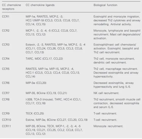
chemokines, Drs. Yoshie and Zlotnik have devised a systematic nomenclature parallel-ing that of the receptor nomenclature system (8). The chemokine Nomenclature Sub-Com-mittee of the Nomenclature ComSub-Com-mittee of the International Union of Immunological Societies has considered this system and recommended its adoption by IUIS/WHO (Tables 1 and 2).
The chemokine receptors exert most of their biological effects by binding to a large family of Gi-protein-coupled seven-trans-membrane receptors leading to activation of multiple intracellular signaling pathways (9). Redundancy exists in the signaling of chemo-kines through chemokine receptors because many chemokines bind to more than one
Table 1. CC receptor family, CC chemokine ligands and biological function.
CC chemokine CC chemokine ligands Biological function receptors
CCR1 MIP-1α, RANTES, MCP-2, -3, Eosinophil and monocyte migration, HCC-1/MIP-1δ (CCL3, CCL5, CCL8, CCL7, decreased Th2 cytokines and airway CCL14, CCL15) remodelling. Antiviral activity.
CCR2 MCP-1, -2, -3, -4, -5 (CCL2, CCL8, CCL7, Monocyte, lymphocyte and basophil CCL13, CCL12) recruitment. Mast cell degranulation/
activation.
CCR3 Eotaxin, -2, -3, RANTES, MIP-1α, MCP-2, -3, -4 Eosinophil/mast cell chemotaxis/ (CCL11, CCL24, CCL26, CCL5, CCL3, CCL8, activation. Eosinophil, basophil and CCL7, CCL13) Th2 cell recruitment.
CCR4 TARC, MDC (CCL17, CCL22) Th2 cell, monocyte recruitment, dendritic cell recruitment.
CCR5 RANTES, MIP-1α, MIP-1ß, MCP-2, -4, Th2 cell, macrophage recruitment. HCC-1 (CCL5, CCL3, CCL4, CCL8, CCL13, Decreased eosinophilia and airway
CCL14) hyperreactivity.
CCR6 MIP-3α (CCL20) Decreased eosinophilia, airway hyperreactivity and lung IL-5.
CCR7 MIP-3ß, 6Ckine (CCL19, CCL21) NK cell recruitment.
CCR8 I-309, TCA-3 (mouse), TARC, HCC-4 (CCL1, Th2 recruitment, smooth muscle cell CCL17, CCL16) contraction, decreased eosinophilia
and serum IL-5.
CCR9 TECK (CCL25) T-cell recruitment.
CCR10 Eskine, MIP-3α, 6Ckine (CCL27, CCL20, CCL19) T-cell recruitment.
CCR11 MIP-3ß, 6Ckine, TECK, MCP-1, -2, -3, -4, -5 Monocyte recruitment. (CCL19, CCL21, CCL25, CCL2, CCL8, CCL7,
chemokine receptor, and many chemokine receptors bind more than one chemokine (Tables 1 and 2).
The G-protein receptor ligation involves activation of the heterotrimeric Gα/ßγ com-plex that is responsible, when activated, for different cell functions. For example, the stimulation of chemotaxis by a chemokine requires functional coupling of the receptor to Gαi, since migration is completely inhib-ited by treatment of the cells with pertussis toxin. However, Gαi itself appears not to be necessary for cell migration. The essential step is the release of the heterotrimeric G-protein ßγ subunits from Gαi and the Gi -protein-coupled receptor (10). It was shown that only ßγ subunits released from Gi -coupled receptors, but not those released from Gs- or Gq-coupled receptors, could mediate cell migration (11).
The G-protein effector activation often modifies the concentrations of a second mes-senger and involves the signal transduction machinery. Most chemokines share the
abil-ity to bind to chemokine receptors that trig-ger these downstream cascades, rapidly acti-vating phosphoinositide-specific phospholi-pase C-ß2 (PLC-ß2) and PLC-ß3 isoenzymes, which lead to inositol-1,4,5-triphosphate for-mation and to a transient rise in the concen-tration of intracellular free calcium (Ca2+). This pathway has been widely used to test the responsiveness of chemokine receptors to different chemokines (12). In neutrophils of mice that lack the genes encoding PLC-ß2 and PLC-ß3, the chemokine-induced calcium elevation is fully suppressed, which sup-ports the conclusion that PLC-ß2 and PLC-ß3 are the sole PLC isoforms that are activated by chemokines in immune cells (13).
Several other chemokines have been shown to inhibit adenylate cyclase and to activate mitogen/extracellular signal-regu-lated kinase (MEK)-1 and/or extracellular signal-regulated kinase (ERK)-1/2. In addi-tion, these responses stimulate tyrosine phos-phorylation of focal adhesion complex com-ponents and activate nuclear factor-κB
(NF-Table 2. CXC, CX3C and XC receptors, their chemokine ligands, and biological function.
CX chemokine CXC chemokine ligands Biological function receptors
CXCR1 IL-8, ENA-78, GCP-2 (CXCL8, Neutrophil chemotaxis and functional CXCL5, CXCL6) modulation.
CXCR2 IL-8, NAP-2, ENA-78, GROα, GROß, Neutrophil chemotaxis and functional GROγ (CXCL8, CXCL7, CXCL5, CXCL1, modulation.
CXCL2, CXCL3)
CXCR3 Mig, IP-10, I-TAC (CXCL9, Attraction of Th1 cells, activated eosinophils; CXCL10, CXCL11) allograft rejection.
CXCR4 SDF-1 (CXCL12) Reduction of tissue eosinophil recruitment and airway hyperreactivity, B cell responses, stem cell homing.
CXCR5 BCA-1 (CXCL13) Formation of B cell compartment; effector of T-cell generation. Activated CD4+ T-cell.
CXCR6 CXCL16 Activated T-cell.
CX3CR1 Fractalkine, neurotactin Microglia expression, renal inflammation, vascular injury.
XCR1 Lymphotactin α Fungal infection, mast cell response.
κB) as well as signal transducer and activa-tor of transcription 1 (STAT1) and STAT3 (14,15). Thus, chemokines can couple to distinct signaling pathways that have been demonstrated to mediate not only migration, but also cell growth and transcriptional acti-vation.
One particular signaling pathway, namely that controlled by lipid kinase phosphoinosi-tide 3-kinase (PI3K), has been the focus of much attention with respect to its activation by chemokine receptors and the role it plays in regulating cell migration. PI3Ks are a family of proteins that catalyze the phospho-rylation of the 3-OH position of the inositol head group of phosphoinositide lipids, phos-phatidylinositol (PtdIns), phosphatidylino-sitol (4) phosphate [PtdIns(4)P] and phos-phatidylinositol (4,5) bisphosphate [PtdIns (4,5)P2]. This results in the formation of PtdIns(3)P, PtdIns(3,4)P2 and PtdIns(3,4,5)P3, respectively, collectively termed 3'-phospho-inositide lipids (16). PI3K is considered to be one of the direct substrates of heterotri-meric G-protein ßγ subunit activation that generate lipid second messenger molecules, resulting in the activation of multiple intra-cellular signaling cascades. These events regulate a broad array of cellular responses, including survival, activation, differentiation and proliferation, and are now recognized as playing a key role in a number of physiologi-cal and pathophysiologiphysiologi-cal processes in the lung (17).
PI3K has been shown to be a key regula-tor of both neutrophil recruitment and acti-vation. In mice lacking the catalytic subunit of the myeloid restricted PI3K-γ, neutrophil migration to the inflamed peritoneum was severely compromised (18,19). The role of PI3K in eosinophil degranulation is not known, but these enzymes are required for activation of the eosinophil NADPH oxidase complex (20).
In eosinophils, eotaxin (CCL11), a CC chemokine that binds CCR3, induces de-granulation and chemotaxis through the
acti-vation of ERK-2 (a downstream molecule from p42/44 mitogen-activated protein ki-nases, MAPK) and p38 MAPK (21). How-ever, p38 MAPK plays a greater role than ERK-2 in eosinophil differentiation and mac-rophage inflammatory protein 1α (MIP-1α, CCL3) production by eosinophils (22). In-terestingly, MIP-1ß (CCL4) and T-cell acti-vation gene 3 (TCA-3, CCL1) (CCR5 and CCR8 ligands) induce eosinophil chemo-taxis by a Gαi-independent mechanism, be-cause the migration was not inhibited by treatment of the cells with pertussis toxin (Oliveira SHP, unpublished data). Thus, dif-ferent pathways may be used by eosinophils to carry out their functions.
Chemokines, eosinophils and asthma
Bronchial asthma is a chronic inflamma-tory disorder characterized by airway in-flammation and infiltration by eosinophils, neutrophils and T lymphocytes (23-25). Fur-thermore, there is a mass of evidence show-ing that the severity of the disease depends on eosinophil accumulation and activation within the airways (25-27). Eosinophils con-tain a number of products that when released directly cause mucosal injury and contribute to the disturbances in lung physiology. These products include major basic protein, eosin-ophil cationic protein, eosineosin-ophil-derived neurotoxin, and eosinophil peroxidase, in addition to oxygen and nitrogen metabolites. In addition, eosinophils generate lipid me-diators, including platelet-activating factor, leukotrienes B4 and C4, cytokines and chemo-kines that are able to activate eosinophils, other leukocytes, and structural cells. In this cascade of events, structural cells release more chemotactic factors such as chemokines and leukotriene B4 that create a more intense in-flammatory response (28-30).
molecules and local generation of chemotac-tic agents that direct cell migration from the vascular compartment into the inflamed air-ways (1).
For many years, research has been fo-cussed on the mechanisms of activation and recruitment of eosinophils into the airways in asthmatic patients or in animals used as experimental models. Recently, chemokines have been implicated in contributing to al-lergic disorders, such as asthma (31-33). These chemokines seem to be related to the severity of asthmatic inflammation and reac-tive airway response.
Chemokines, such as RANTES (CCL5, regulated on activation, normal T-cell ex-pressed and secreted), MIP-1α (CCL3) and monocyte chemoattractant protein 5 (MCP-5, CCL12) are upregulated early after aller-gen challenge, but one cannot easily demon-strate a correlation between this upregula-tion and the recruitment of defined leuko-cyte subsets (34). In contrast, the kinetics of production of eotaxin (CCL11), MCP-1 (CCL2), monocyte-derived chemokine (MDC, CCL22) and thymus- and activation-regulated chemokine (TARC, CCL17) shows a good correlation with the recruitment of specific leukocyte subsets expressing the re-ceptors for these chemokines (35-37).
In studies using animal models of aller-gic airway inflammation, the neutralization
of either MIP-1α (CCL3) or RANTES
(CCL5), but not MCP-1 (CCL2), signifi-cantly reduced the intensity of the eosinophil recruitment to the lung and airway during the allergic airway response. In contrast, neu-tralization of MCP-1 (CCL2) significantly reduced total leukocyte migration. Further examination of the effect of MCP-1 (CCL2) depletion indicated that both CD4+ and CD8+ lymphocyte subsets were decreased. Deple-tion of MCP-1 (CCL2) significantly reduced the airway hyperreactivity to near control levels, whereas depletion of MIP-1α (CCL3) or RANTES (CCL5) did not affect the inten-sity of airway hyperreactivity. These data
suggest that multiple CC chemokines are involved in the recruitment of the particular leukocyte populations during the allergic process (38).
Eotaxin (CCL11), the first CC chemo-kine showing the ability to selectively recruit eosinophils, has drawn most attention from researchers studying allergic responses. Eo-taxin (CCL11) was first discovered in the bronchoalveolar lavage fluid of guinea pigs after an allergen challenge (39). It has since been cloned in humans and mice, and ap-pears to have similar functions, although a paucity of data still exists with respect to inflammatory disorders. Eotaxin (CCL11), eotaxin 2 (CCL24), RANTES (CCL5), MCP-3 (CCL7), -4 (CCL1MCP-3) and CCRMCP-3 expression were observed in bronchial biopsies from atopic and nonatopic asthmatics (40). In ad-dition, an increased bone marrow pool of CCR3(+) mature and immature eosinophils has been described in subjects with asthma (41). Thus, it appears that CCR3 is substan-tially involved in the recruitment of inflam-matory cells in the allergic response. Con-firming this issue, the pretreatment of eosino-phils from allergic and eosinophilic donors with a monoclonal antibody to CCR3 blocked chemotaxis and calcium flux induced by all CCR3 ligands (42). These results demon-strated the importance of CCR3 for eosino-phil responses.
Table 3. CC chemokine receptor expression in leukocytes and structural cells.
CCR1 CCR2 CCR3 CCR4 CCR5 CCR6 CCR7 CCR8 CCR9 CCR10 CCR11
Eosinophils + + + - + + - + - -
-Neutrophils + + + - - -
-Mast cells + + + + + + + - - -
-Macrophages + + - - + - - - + -
-Lymphocytes + + + + + + + + + + +
Fibroblasts - + + - - - +
-Epithelial cells - - + - - + - - - +
-Endothelial cells - + + + - - - +
-Airway smooth - + - - -
-muscle cells
thought to be specific to eosinophils, but has subsequently been detected on Th2 cell sub-sets, as well as basophils, mast cells, neural tissue and airway epithelia. It appears that CCR3 is important for the basal trafficking of eosinophils to the intestinal mucosa but not to the lung. Furthermore, CCR3 may be involved in mast cell homing to epithelial tissues (44,45) as well as leukotriene C4 production by eosinophils and basophils (46). Other chemokine receptors vary consid-erably in their expression patterns between leukocyte subsets (Tables 3 and 4). CCR1, CCR5 and CXCR3 are preferentially ex-pressed in Th1 cells, whereas CCR3, CCR4 and CCR8 seem to be typical of Th2 cells. Likewise, mast cells also express a number of chemokine receptors including, CCR1, CCR2, CCR3, CCR4, CCR5, CCR6, CX3CR1 and XCR1 (47-49). These obser-vations highlight the complexity and diffi-culty researchers have faced in the func-tional characterization of chemokines and their receptors.
When examining responses in various cell types, the complexity of the chemokine system becomes readily evident. There are experimental data demonstrating that eotaxin (CCL11) is the most potent eosinophil che-motactic factor. However, additional studies have now demonstrated that other non-CCR3-binding chemokines, including MDC (CCL22), MIP-1ß (CCL4), TCA-3 (CCL1), C10, and TARC (CCL17), can also induce
eosinophil activation as well as eosinophil migration. In studies from our laboratories, we have observed that eosinophils elicited from the peritoneum of sensitized mice dis-play additional chemotactic responses to spe-cific CC chemokines, which are absent from peripheral blood eosinophils. We demon-strated that CCR5 and CCR8 ligands MIP-1ß (CCL4) and TCA-3 (CCL1) were able to induce elicited eosinophils to migrate using in vitro chemotactic assays. Furthermore, the
pretreated IL-4- or -TNF-α-elicited eosino-phils show upregulation of the expression and function of CCR5 and CCR8 (50) (Tables 5 and 6). We also observed that these chemo-kines were able to induce a dramatic eosino-phil peroxidase release, suggesting that MIP-1ß (CCL4) and TCA-3 (CCL1) are potent eosinophil activators (Table 6). These stud-ies have been supported by data in CCR8-/-mice, where the most significant alterations were decreased eosinophil accumulation and release of eosinophil peroxidase into the bronchoalveolar lavage fluid (51).
eosinophils to migrate, release eosinophil cationic protein and induce NF-AT complex nuclear translocation (52).
The functional role of CXCR4 expres-sion in eosinophils was observed to be
in-ducible and SDF-1α (CXCL12) elicited
strong migration comparable to that induced by eotaxin (CCL11). Th2 cytokines such as IL-4 and IL-5 drastically inhibited the ex-pression of CXCR4 (53). These studies pro-vide useful insights into novel mechanisms of action of the CXC chemokines in the pathophysiology of allergic inflammation, including initiation, progression and termi-nation of the eosinophilic processes.
In support for a role for CXCR4, a CXCR4 antagonist (AMD3100) was observed to at-tenuate allergic lung inflammation and air-way hyperreactivity in mice. AMD3100-treated animals had reduced airway hyperre-activity, peribronchial eosinophilia and over-all inflammatory responses, as well as re-duced IL-4 and IL-5 levels (54).
The CXCR3-selective chemokine ligands also may represent an important therapeutic approach to control the allergic processes, since the CXC ligands, IP-10 (CXCL10), Mig (CXCL9) and I-TAC (CXCL11) can act as antagonists of CCR3 and thereby inhibit the infiltration of Th2 cells, in addition to their agonist effects on CXCR3, which lead to attraction of Th1 cells (55). Therefore, although most of the research in allergic asthma has concentrated on the CC family of chemokines, the CXC family also appears to have a distinct function in the overall devel-opment and severity of disease.
Conclusion
Chemokines play an important role in the allergic inflammatory process through the initiation of leukocyte recruitment, activa-tion and regulaactiva-tion of disease severity. Chemokines are very promiscuous and bind to multiple receptors. Therefore, determin-ing the precise function of the individual
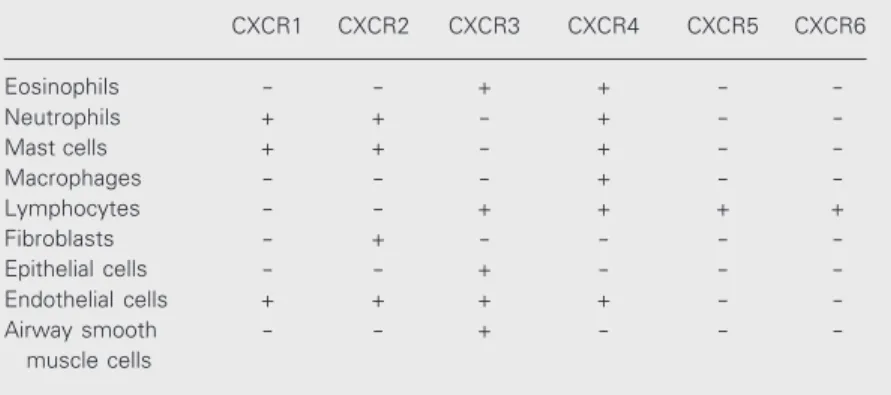
Table 4. CXC chemokine receptor expression in leukocytes and structural cells.
CXCR1 CXCR2 CXCR3 CXCR4 CXCR5 CXCR6
Eosinophils - - + + -
-Neutrophils + + - + -
-Mast cells + + - + -
-Macrophages - - - + -
-Lymphocytes - - + + + +
Fibroblasts - + - - -
-Epithelial cells - - + - -
-Endothelial cells + + + + -
-Airway smooth - - + - -
-muscle cells
Table 6. CC chemokine receptor functional activity (chemotactic activity, EPO release and Ca2+ flux activity) on eosinophils (Eos) stimulated with TNF-α or IL-4.
Stimulus Chemotactic activity EPO release Ca2+ flux
Eos+Medium Eos+IL-4 Eos+TNF-α Eos+Medium Eos+Medium
MIP-1α (CCR1) ++ - +++ + ++
MCP-1 (CCR2) - - - -
-Eotaxin (CCR3) +++ +++ +++ ++ +++
MDC (CCR4) - - - +++
-MIP-1ß (CCR5) - ++ ++ +++
-TCA-3 (CCR8) - ++ ++ +++
-Results are reported as the following parameters: - = no chemotactic activity, EPO release or Ca2+ flux; + = weak chemotactic activity, EPO release or Ca2+ flux; ++ = intermediate chemotactic activity, EPO release or Ca2+ flux; +++ = strong chemotactic activity, EPO release or Ca2+ flux, when compared with control (nonstimulated eosino-phils). EPO = eosinophil peroxidase.
Table 5. CC chemokine receptor expression on eosinophils stimulated with TNF-α or IL-4.
Stimulus mRNA receptor expression
CCR1 CCR2 CCR3 CCR4 CRR5 CCR8
Medium (control) ++ - +++ - -
-+ TNF-α +++ - +++ - ++ ++
+ IL-4 + - +++ - ++ ++
References
1. Luster AD (1998). Chemokines-chemotactic cytokines that mediate inflammation. New England Journal of Medicine, 338: 436-445. 2. Lukacs NW, Oliveira SHP & Hogaboam CM (1999). Chemokines and
asthma: redundancy of function or a coordinated effort? Journal of Clinical Investigation, 104: 995-999.
3. Rollins BJ (1997). Chemokines. Blood, 90: 909-928.
4. Furie MB & Randolph GJ (1995). Chemokines and tissue injury.
American Journal of Pathology, 146: 1287-1301.
5. Baggiolini M & Dahinden CA (1994). CC chemokines in allergic inflammation. Immunology Today, 15: 127-133.
6. Strieter RM, Standiford TJ, Huffnagle GB, Colletti LM, Lukacs NW & Kunkel SL (1996). “The good, the bad, and the ugly.” The role of chemokines in models of human disease. Journal of Immunology, 156: 3583-3586.
7. Christopherson 2nd K & Hromas R (2001). Chemokine regulation of normal and pathologic immune responses. Stem Cells, 19: 388-396. 8. Zlotnik A & Yoshie O (2000). Chemokines: a new classification
system and their role in immunity. Immunity, 12: 121-127. 9. Thelen M (2001). Dancing to the tune of chemokines. Nature
Immu-nology, 2: 129-134.
10. Neptune ER & Bourne HR (1997). Receptors induce chemotaxis by releasing the betagamma subunit of Gi, not by activating Gq or Gs.
Proceedings of the National Academy of Sciences, USA, 94: 14489-14494.
11. Neptune ER, Liri T & Bourne HR (1999). Galphai is not required for chemotaxis mediated by Gi-coupled receptors. Journal of Biological Chemistry, 274: 2824-2828.
12. Baggiolini M, Dewald B & Moser B (1997). Human chemokines. An update. Annual Review of Immunology, 15: 675-705.
13. Li Z, Jiang H, Xie W, Zhang Z, Smrcka AV & Wu D (2000). Roles of PLC-beta2 and -beta3 and PI3Kgamma in chemoattractant-mediated signal transduction. Science, 287: 1046-1049.
14. Ward SG & Westwick J (1998). Chemokines: understanding their role in T lymphocyte biology. Biochemical Journal, 333: 457-470. 15. Ward SG, Bacon KB & Westwick J (1998). Chemokines and T
lymphocytes: more than an attraction. Immunity, 8: 1-11.
16. Curnock AP, Logan MK & Ward SG (2002). Chemokine signaling: pivoting around multiple phosphoinositide 3-kinases. Immunology, 105: 125-136.
17. Krymskaya VP, Penn RB, Orsini MJ, Scott PH, Plevin RJ, Walker TR, Eszterhas AJ, Amrani Y, Chilvers ER & Panettieri Jr RA (1999). Phosphatidylinositol 3-kinase mediates mitogen-induced human air-way smooth muscle cell proliferation. American Journal of Physiolo-gy, 277 (1 Part 1): L65-L78.
18. Sasaki T, Irie-Sasaki J, Horie Y et al. (2000). Colorectal carcinomas in mice lacking the catalytic subunit of PI(3)Kgamma. Nature, 406: 897-902.
19. Hirsch E, Wymann MP, Patrucco E, Tolosano E, Bulgarelli-Leva G,
Marengo S, Rocchi M & Altruda F (2000). Analysis of the murine phosphoinositide 3-kinase gamma gene. Gene, 256: 69-81. 20. Hofmann C, Dichmann S, Zimpfer U, Czech W, Herouy Y, Wagner E
& Norgauer J (2000). Metabolism and function of 3-D-phosphory-lated phosphoinositides in C5a-stimu3-D-phosphory-lated eosinophils. Biochemical and Biophysical Research Communications, 269: 816-821. 21. Kampen GT, Stafford S, Adachi T, Jinquan T, Quan S, Grant JA, Skov
PS, Poulsen LK & Alam R (2000). Eotaxin induces degranulation and chemotaxis of eosinophils through the activation of ERK2 and p38 mitogen-activated protein kinases. Blood, 95: 1911-1917.
22. Adachi T, Choudhury BK, Stafford S, Sur S & Alam R (2000). The differential role of extracellular signal-regulated kinases and p38 mitogen-activated protein kinase in eosinophil functions. Journal of Immunology, 165: 2198-2204.
23. Djukanovic R, Wilson JW, Britten KM, Wilson SJ, Walls AF, Roche WR, Howarth PH & Holgate ST (1990). Quantitation of mast cells and eosinophils in the bronchial mucosa of symptomatic atopic asthmatics and healthy control subjects using immunohistochemis-try. American Review of Respiratory Disease, 142: 863-871. 24. Bradley BL, Azzawi M, Jacobson M, Assoufi B, Collins JV, Irani AM,
Schwartz LB, Durham SR, Jeffery PK & Kay AB (1991). Eosinophils, T-lymphocytes, mast cells, neutrophils, and macrophages in bron-chial biopsy specimens from atopic subjects with asthma: compari-son with biopsy specimens from atopic subjects without asthma and normal control subjects and relationship to bronchial hyperre-sponsiveness. Journal of Allergy and Clinical Immunology, 88: 661-674.
25. Kay AB & Corrigan CJ (1992). Asthma. Eosinophils and neutrophils.
British Medical Bulletin, 48: 51-64.
26. Kay AB, Barata L, Meng Q, Durham SR & Ying S (1997). Eosinophils and eosinophil-associated cytokines in allergic inflammation. Inter-national Archives of Allergy and Immunology, 113: 196-199. 27. Kitayama J, Mackay CR, Ponath PD & Springer TA (1998). The C-C
chemokine receptor CCR3 participates in stimulation of eosinophil arrest on inflammatory endothelium in shear flow. Journal of Clinical Investigation, 101: 2017-2024.
28. Smith RS, Smith TJ, Blieden TM & Phipps RP (1997). Fibroblasts as sentinel cells. Synthesis of chemokines and regulation of inflamma-tion. American Journal of Pathology, 151: 317-322.
29. Hogaboam CM, Smith RE & Kunkel SL (1998). Dynamic interactions between lung fibroblasts and leukocytes: implications for fibrotic lung disease. Proceedings of the Association of American Physi-cians, 110: 313-320.
30. Chung KF (2000). Airway smooth muscle cells: contributing to and regulating airway mucosal inflammation? European Respiratory Jour-nal, 15: 961-968.
31. Kita H & Gleich GJ (1996). Chemokines active on eosinophils: poten-tial roles in allergic inflammation. Journal of Experimental Medicine,
chemokines and receptors will be essential to the definition of the correct targets. The analysis of the function of eosinophils and their chemokine receptors during inflamma-tion continues to be a good approach to understanding the determinants of asthma
183: 2421-2426.
32. Lukacs NW, Standiford TJ, Chensue SW, Kunkel RG, Strieter RM & Kunkel SL (1996). C-C chemokine-induced eosinophil chemotaxis during allergic airway inflammation. Journal of Leukocyte Biology, 60: 573-578.
33. Griffiths-Johnson DA, Collins PD, Jose PJ & Williams TJ (1997). Animal models of asthma: role of chemokines. Methods in Enzymol-ogy, 288: 241-266.
34. Gutierrez-Ramos JC, Lloyd C, Kapsenberg ML, Gonzalo JA & Coyle AJ (2000). Non-redundant functional groups of chemokines operate in a coordinate manner during the inflammatory response in the lung. Immunological Reviews, 177: 31-42.
35. Gonzalo JA, Lloyd CM, Kremer L, Finger E, Martinez-A C, Siegelman MH, Cybulsky M & Gutierrez-Ramos JC (1996). Eosinophil recruit-ment to the lung in a murine model of allergic inflammation. The role of T cells, chemokines, and adhesion receptors. Journal of Clinical Investigation, 98: 2332-2345.
36. Gonzalo JA, Lloyd CM, Wen D et al. (1998). The coordinated action of CC chemokines in the lung orchestrates allergic inflammation and airway hyperresponsiveness. Journal of Experimental Medicine, 188: 157-167.
37. Gonzalo JA, Pan Y, Lloyd CM et al. (1999). Mouse monocyte-derived chemokine is involved in airway hyperreactivity and lung inflamma-tion. Journal of Immunology, 163: 403-411.
38. Lukacs NW, Strieter RM, Warmington K, Lincoln P, Chensue SW & Kunkel SL (1997). Differential recruitment of leukocyte populations and alteration of airway hyperreactivity by C-C family chemokines in allergic airway inflammation. Journal of Immunology, 158: 4398-4404.
39. Jose PJ, Griffiths-Johnson DA, Collins PD, Walsh DT, Moqbel R, Totty NF, Truong O, Hsuan JJ & Williams TJ (1997). Eotaxin: a potent eosinophil chemoattractant cytokine detected in a guinea pig model of allergic airway inflammation. Journal of Experimental Medi-cine, 179: 881-887.
40. Ying S, Meng Q, Zeibecoglou K, Robinson DS, Macfarlane A, Humbert M & Kay AB (1999). Eosinophil chemotactic chemokines (eotaxin, eotaxin-2, RANTES, monocyte chemoattractant protein-3 (MCP-3), and MCP-4), and C-C chemokine receptor 3 expression in bronchial biopsies from atopic and nonatopic (intrinsic) asthmatics.
Journal of Immunology, 163: 6321-6329.
41. Zeibecoglou K, Ying S, Yamada T, North J, Burman J, Bungre J, Meng Q, Kay AB & Robinson DS (1999). Increased mature and immature CCR3 messenger RNA+ eosinophils in bone marrow from patients with atopic asthma compared with atopic and nonatopic control subjects. Journal of Allergy and Clinical Immunology, 103 (1 Part 1): 99-106.
42. Heath H, Qin S, Rao P, Wu L, Larosa G, Kassam N, Ponath PD & Mackay CR (1997). Chemokine receptor usage by human eosino-phils. The importance of CCR3 demonstrated using an antagonistic monoclonal antibody. Journal of Clinical Investigation, 99: 178-184. 43. Nagase H, Kudo K, Izumi S, Ohta K, Kobayashi N, Yamaguchi M,
Matsushima K, Morita Y, Yamamoto K & Hirai K (2001). Chemokine receptor expression profile of eosinophils at inflamed tissue sites: De-creased CCR3 and inDe-creased CXCR4 expression by lung eosinophils.
Journal of Allergy and Clinical Immunology, 108: 563-569.
44. Humbles AA, Lu B, Friend DS, Okinaga S, Lora J, Al-Garawi A, Martin TR, Gerard NP & Gerard C (2002). The murine CCR3 receptor regulates both the role of eosinophils and mast cells in allergen-induced airway inflam-mation and hyperresponsiveness. Proceedings of the National Academy of Sciences, USA, 99: 1479-1484.
45. Ma W, Bryce PJ, Humbles AA, Laouini D, Yalcindag A, Alenius H, Friend DS, Oettgen HC, Gerard C & Geha RS (2002). CCR3 is essential for skin eosinophilia and airway hyperresponsiveness in a murine model of aller-gic skin inflammation. Journal of Clinical Investigation, 109: 621-628. 46. Bandeira-Melo C, Phoofolo M & Weller PF (2001). Extranuclear lipid
bodies, elicited by CCR3-mediated signaling pathways, are the sites of chemokine-enhanced leukotriene C4 production in eosinophils and ba-sophils. Journal of Biological Chemistry, 276: 22779-22787.
47. Bonecchi R, Bianchi G, Bordignon PP et al. (1998). Differential expres-sion of chemokine receptors and chemotactic responsiveness of type 1 T helper cells (Th1s) and Th2s. Journal of Experimental Medicine, 187: 129-134.
48. Sallusto F, Mackay CR & Lanzavecchia A (1997). Selective expression of the eotaxin receptor CCR3 by human T helper 2 cells. Science, 277: 2005-2007.
49. Oliveira SH & Lukacs NW (2001). Stem cell factor and IgE-stimulated murine mast cells produce chemokines (CCL2, CCL17, CCL22) and express chemokine receptors. Inflammation Research, 50: 168-174. 50. Oliveira SH, Lira S, Martinez-A C, Wiekowski M, Sullivan L & Lukacs NW
(2002). Increased responsiveness of murine eosinophils to MIP-1beta (CCL4) and TCA-3 (CCL1) is mediated by their specific receptors, CCR5 and CCR8. Journal of Leukocyte Biology, 71: 1019-1025.
51. Chensue SW, Lukacs NW, Yang T-Y et al. (2001). Aberrant in vivo T helper type 2 cell response and impaired eosinophil recruitment in CC chemokine receptor 8 knockout mice. Journal of Experimental Medi-cine, 193: 573-584.
52. Jinquan T, Jing C, Jacobi HH et al. (2000). CXCR3 expression and activation of eosinophils: role of IFN-gamma-inducible protein-10 and monokine induced by IFN-gamma. Journal of Immunology, 165: 1548-1556.
53. Nagase H, Miyamasu M, Yamaguchi M, Fujisawa T, Ohta K, Yamamoto K, Morita Y & Hirai K (2000). Expression of CXCR4 in eosinophils: functional analyses and cytokine-mediated regulation. Journal of Immu-nology, 164: 5935-5943.
54. Lukacs NW, Berlin A, Schols D, Skerlj RT & Bridger GJ (2002). AMD3100, a CXCR4 antagonist, attenuates allergic lung inflammation and airway hyperreactivity. American Journal of Pathology, 160: 1353-1360. 55. Loetscher P, Pellegrino A, Gong JH, Mattioli I, Loetscher M, Bardi G,
Baggiolini M & Clark-Lewis I (2001). The ligands of CXC chemokine receptor 3, I-TAC, Mig, and IP10, are natural antagonists for CCR3.