Tyrosol Suppresses Allergic Inflammation by
Inhibiting the Activation of Phosphoinositide
3-Kinase in Mast Cells
In-Gyu Je1☯, Duk-Sil Kim2☯, Sung-Wan Kim2, Soyoung Lee1, Hyun-Shik Lee3, Eui Kyun Park4, Dongwoo Khang5, Sang-Hyun Kim1*
1Department of Pharmacology, School of Medicine, Kyungpook National University, Daegu 700–422, Republic of Korea,2Department of Thoracic and Cardiovascular Surgery, CHA Gumi Medical Center, CHA University, Gumi 730–040, Republic of Korea,3School of Life Sciences, Kyungpook National University, Daegu 702–701, Republic of Korea,4Department of Oral Pathology and Regenerative Medicine, School of Dentistry, Kyungpook National University, Daegu 700–412, Republic of Korea,5Department of Molecular Medicine, School of Medicine, Gachon University, Incheon 406–840, Republic of Korea
☯These authors contributed equally to this work.
Abstract
Allergic diseases such as atopic dermatitis, rhinitis, asthma, and anaphylaxis are attractive research areas. Tyrosol (2-(4-hydroxyphenyl)ethanol) is a polyphenolic compound with di-verse biological activities. In this study, we investigated whether tyrosol has anti-allergic in-flammatory effects. Ovalbumin-induced active systemic anaphylaxis and immunoglobulin E-mediated passive cutaneous anaphylaxis models were used for the immediate-type aller-gic responses. Oral administration of tyrosol reduced the alleraller-gic symptoms of hypothermia and pigmentation in both animal models. Mast cells that secrete allergic mediators are key regulators on allergic inflammation. Tyrosol dose-dependently decreased mast cell degran-ulation and expression of inflammatory cytokines. Intracellular calcium levels and activation of inhibitor ofκB kinase (IKK) regulate cytokine expression and degranulation. Tyrosol blocked calcium influx and phosphorylation of the IKK complex. To define the molecular tar-get for tyrosol, various signaling proteins involved in mast cell activation such as Lyn, Syk, phosphoinositide 3-kinase (PI3K), and Akt were examined. Our results showed that PI3K could be a molecular target for tyrosol in mast cells. Taken together, these findings indicated that tyrosol has anti-allergic inflammatory effects by inhibiting the degranulation of mast cells and expression of inflammatory cytokines; these effects are mediated via PI3K. There-fore, we expect tyrosol become a potential therapeutic candidate for allergic inflammatory disorders.
Introduction
There are a range of allergic disorders including atopic dermatitis, allergic rhinitis, asthma, food allergy, and anaphylaxis. Mast cells are known to play key roles in these diseases through a11111
OPEN ACCESS
Citation:Je I-G, Kim D-S, Kim S-W, Lee S, Lee H-S, Park EK, et al. (2015) Tyrosol Suppresses Allergic Inflammation by Inhibiting the Activation of Phosphoinositide 3-Kinase in Mast Cells. PLoS ONE 10(6): e0129829. doi:10.1371/journal.pone.0129829
Academic Editor:Kottarappat N Dileepan, University of Kansas Medical Center, UNITED STATES
Received:March 18, 2015
Accepted:May 12, 2015
Published:June 11, 2015
Copyright:© 2015 Je et al. This is an open access article distributed under the terms of theCreative Commons Attribution License, which permits unrestricted use, distribution, and reproduction in any medium, provided the original author and source are credited.
Data Availability Statement:All relevant data are within the paper and its Supporting Information files.
Funding:This work was supported by the National Research Foundation of Korea grant funded by the Korea government (2014R1A5A2009242 and 2013R1A1A3009525). The funder had no role in study design, data collection and analysis, decision to publish, or preparation of the manuscript.
the production and secretion of allergic mediators; histamine, chemokines, cytokines, and growth factors [1]. Type 2 helper T (Th2) cells differentiated by stimulation of antigen-presenting cells activate B cells to produce immunoglobulin E (IgE), which binds to high affini-ty IgE receptor (FcεRI) on the surface of mast cells [2]. FcεRI-mediated mast cell activation is triggered by antigen-IgE cross-linking and leads to the degranulation and expression of inflam-matory cytokines [3]. Mast cell signaling has been investigated thoroughly. Activation of Lyn and Syk causes phosphorylation of phosphoinositide 3-kinase (PI3K), which stimulates Akt and phospholipase C (PLC)γ[4]. Phosphorylation of the inhibitor ofκB (IκB) kinase (IKK) complex by Akt and protein kinase C (PKC) results in activation of nuclear factor (NF)-κB and synaptosomal-associated protein (SNAP)23. In addition, PLCγcatalyzes the production of ino-sitol 1,4,5-trisphosphate (IP3), which binds to IP3receptors on the surface of the endoplasmic
reticulum (ER). It causes release of calcium stored in the ER into the cytoplasm. Subsequently, the loss of calcium in the ER triggers a sudden increase of calcium influx from outside of the cell [5]. As a result, the expression and release of allergic molecules are enhanced by NF-κB, SNAP23, and increased intracellular calcium.
Histamine is the most important molecule in the acute allergy manifesting edema, warmth, and erythema by causing vasodilation, increasing vascular permeability, and leukocyte recruit-ment [6]. Inflammatory cytokines such as tumor necrosis factor (TNF)-α, interleukin (IL)-1β, and IL-4 lead to the chronic allergic phase by enhancing T cell activation or B cell survival [7]. Rat basophilic leukemia (RBL)-2H3 cells are suitable forin vitrostudies of mast cell-mediated allergic inflammation involving the degranulation and expression of inflammatory cytokines [8]. Ovalbumin (OVA)-induced active systemic anaphylaxis (ASA) and IgE-mediated passive cutaneous anaphylaxis (PCA) are well-characterized animal models for immediate-type hyper-sensitivity [9,10].
Tyrosol (2-(4-hydroxyphenyl)ethanol), a polyphenolic compound contained in olive oil, has been reported to possess a wide range of biological activities including oxidative, anti-apoptotic, and anti-inflammatory effects [11–13]. The present study compared the activities of tyrosol with gallic acid (3,4,5-trihydroxybenzoic acid) and dexamethasone already known for anti-allergic inflammatory properties [14,15]. In this study, we aimed to assess the effects of tyrosol on allergic inflammation using animal models for immediate-type hypersensitivity. In addition, anti-allergic effects related to the inhibition of mast cell degranulation and inflamma-tory cytokine expression was investigated using mast cells.
Materials and Methods
Animals
Male Imprinting Control Region (ICR) mice (aged 6 weeks) and Sprague-Dawley (SD) rats (aged 10 weeks) were purchased from the Dae-Han Biolink (Daejeon, Korea). Throughout the study, five animals per cage were housed in a room with laminar air flow, a temperature of 22 ± 2°C, and relative humidity of 55 ± 5%. Animal care and treatments were carried out in ac-cordance with the guidelines established by the Public Health Service Policy on the Humane Care and Use of Laboratory Animals and were approved by the Institutional Animal Care and Use Committee of Kyungpook National University.
Reagents and cell culture
Dulbecco’s modified Eagle’s medium andα-minimum essential medium (GIBCO, Grand Is-land, NY) respectively supplemented with 10% heat-inactivated fetal bovine serum (FBS), 100 U/ml penicillin G, 100μg/ml streptomycin, and 250 ng/ml amphotericin B. RBL-2H3 cells
were used throughout the study at a passage number ranging from 4 to 8.
Preparation of RPMCs
Peritoneal cells were isolated from SD rats as previously described [10]. In brief, the rats were anesthetized with CO2and injected with 20 ml of Tyrode’s buffer A (137 mM NaCl, 5.6 mM
glucose, 12 mM NaHCO3, 2.7 mM KCl, 0.3 mM NaH2PO4, and 0.1% gelatin) into the
peritone-al cavity, followed gentle massage of the abdomen for approximately 90 s. The peritoneperitone-al cavity was carefully opened, and the fluid containing the peritoneal cells was aspirated using a Pasteur pipette. The cells were collected after centrifugation at 150gfor 10 min at room temperature and then resuspended in 1 ml of Tyrode’s buffer A. This suspension was layered on 2 ml of 0.235 g/ml Histodenz solution and centrifuged at 400gfor 15 min at room temperature in order to separate the mast cells from other major components of rat peritoneal cells, i.e., mac-rophages and small lymphocytes. The cells at the buffer-Histodenz interface were discarded, and the cells in the pellet were washed and resuspended. Mast cell preparations had a purity of approximately 95%, based on toluidine blue staining. More than 97% of the cells were viable, based on trypan blue staining.
OVA-induced ASA
Mice (n= 10/group) were sensitized with the OVA mixture (100μg of OVA and 2 mg of alum
adjuvant in 200μl of phosphate-buffered saline [PBS]) by intraperitoneal injection on day 0
and day 7. Drugs including tyrosol, gallic acid, and dexamethasone were orally administered 3 times at doses of 0.1–10 mg/kg body weight (BW) (once every 2 days after the second sensitiza-tion). On day 14, 200μg of OVA was intraperitoneally injected, and then the rectal temperature
was measured every 10 min for 1 h. After 1 h, blood was obtained from the abdominal artery of each mouse for the measurement of serum histamine, OVA-specific IgE, and IL-4 levels.
IgE-mediated PCA
An IgE-mediated PCA model was established as described previously [16]. To induce the PCA reaction, the skin on the ears of the mice (n= 5/group) was sensitized with an intradermal in-jection of anti-DNP IgE (0.5μg/site) for 48 h. Drugs including tyrosol, gallic acid, and
dexa-methasone were orally administered at doses of 0.1–10 mg/kg BW 2 h before intravenous injection of DNP-HSA (1 mg/mouse) and 4% Evans blue (1:1) mixture. Thirty minutes later, the mice were euthanized, and both ears were collected to measure dye pigmentation. The amount of dye was determined colorimetrically after extraction with 0.5 ml of 1 M KOH and 4.5 ml of an acetone and phosphoric acid (5:13) mixture. The absorbance of the extract was measured using a spectrophotometer at a wavelength of 620 nm.
Determination of mast cell degranulation
however, these were challenged with DNP-HSA for only 30 min. The cells were separated from the media by centrifugation at 150gfor 5 min at 4°C. To separate histamine from serum and media, 0.1 N HCl and 60% perchloric acid were added. After centrifugation, the supernatant fluid transferred to effendorf tube containing 5 N NaOH, 5 M NaCl, andn -buta-nol and vortexed. The organic phase was gathered, shaken with 0.1 N HCl andn-haptane, and then centrifuged. The histamine in the aqueous phase is assayed using theo -phthaldial-dehyde spectrofluorometric procedure as previously described [17]. Fluorescent intensity was detected using a fluorescent plate reader (Molecular Devices) at an excitation wavelength of 360 nm and an emission wavelength of 440 nm. Release ofβ-hexosaminidase is also widely used as a marker for mast cell degranulation [16]. After incubation of media withβ -hexosa-minidase substrate buffer (100 mM sodium citrate, 1 mM 4-nitrophenylN -acetyl-β-D-galac-tosaminide, pH 4.5) for 1 h at 37°C, the reaction was terminated using stop solution (0.1 M Na2CO3and NaHCO3), and the absorbance was measured using a spectrophotometer at a
wavelength of 405 nm.
Determination of cell viability
Cell viability was determined by colorimetric analysis using 3-(4,5-dimethylthiazol-2-yl)-2,5-diphenyltetrazolium bromide (MTT) [18]. Water-soluble MTT is converted into water-in-soluble formazan by mitochondrial dehydrogenase. RBL-2H3 cells (3 × 10⁴/well in 96-well plates) were pretreated with various concentrations of tyrosol for 24 h and incubated with 1 mg/ml MTT at 37°C. After 2 h, the formazan crystals were dissolved with DMSO, and then the absorbance was measured using a spectrophotometer (Molecular Devices, Sunnyvale, CA) at a wavelength of 570 nm. The absorbance of the formazan formed in untreated control cells was considered to represent 100% viability.
RNA extraction and real-time PCR
Prior to isolation of total cellular RNA, RBL-2H3 cells (5 × 10⁵/well in 12-well plates) were sensitized with anti-DNP IgE (50 ng/ml). After incubating overnight, the cells were pre-treated with or without drugs including tyrosol, gallic acid, and dexamethasone for 1 h and challenged with DNP-HSA (100 ng/ml) for 1 h. RNAiso Plus reagent (Takarabio Bio Inc., Shiga, Japan) was used to extract total RNA, in accordance with the manufacturer’s protocol. Complementary DNA (cDNA) was synthesized from 2μg of total RNA using the Maxime
RT-Pre Mix Kit (iNtRON Biotechnology, Daejeon, Korea). Quantitative real-time PCR was carried out using the Thermal Cycler Dice TP850 (Takarabio Bio Inc.) according to the man-ufacturer’s protocol. The total 25μl reaction mixture was composed as follows: 1.5μl of
cDNA (150 ng), 1μl of each of the forward and reverse primers (0.4μM), 12.5μl of SYBR
Premix Ex Taq (Takarabio Bio Inc.), and 9μl of dH2O. The conditions used for PCR were
similar to those described previously [8].
Enzyme-linked immunosorbent assay (ELISA)
The levels of inflammatory cytokines in media from RBL-2H3 cells challenged with DNP-HSA and serum were measured by ELISA [10]. ELISA was performed on a 96-well Nunc immuno plate using ELISA kits (BD Biosciences, San Diego, CA) according to the manufacturer’s proto-col. Before the detection of OVA-specific IgE, the immune plate was coated with 20μg of OVA
Determination of the intracellular calcium level
Intracellular calcium was measured using the fluorescent indicator Fluo-3/AM (Invitrogen, Carlsbad, CA) [10]. RBL-2H3 cells (3 × 10⁴/well in 96-well plates) were sensitized with anti-DNP IgE (50 ng/ml). After incubating overnight, the cells were preincubated with Fluo-3/ AM for 1 h at 37°C and washed with Tyrode’s buffer B (137 mM NaCl, 5.5 mM glucose, 12 mM NaHCO3, 2.7 mM KCl, 0.2 mM NaH2PO4, 1 mM MgCl2, and 1.8 mM CaCl2) to remove
the dye from the cell surface. The cells were pretreated with or without tyrosol for 1 h and then challenged with DNP-HSA (100 ng/ml). BAPTA-AM (Calbiochem, La jolla, CA), a cal-cium chelator, was used as a positive control. Fluorescent intensity was detected using a fluo-rescent plate reader at an excitation wavelength of 485 nm and an emission wavelength of 510 nm. The intracellular calcium level in untreated control cells was assigned a relative ab-sorbance value of 1.
Western blot
Nuclear and cytosolic proteins were extracted as previously described [16]. Before protein extraction, RBL-2H3 cells (2 × 10⁶/well in 6-well plates) were sensitized with anti-DNP IgE (50 ng/ml). After incubating overnight, the cells were pretreated with or without tyrosol for 1 h and challenged with DNP-HSA (100 ng/ml). PP2 (Calbiochem), an inhibitor of Src fami-ly kinases, was used as a positive control. After suspension in 100μl of cell lysis buffer A
(0.5% Triton X-100, 150 mM NaCl, 10 mM HEPES, 1 mM EDTA/Na3VO4, 0.5 mM PMSF/
DTT, and 5μg/ml leupeptin/aprotinin), the cells were vortexed, incubated for 5 min on ice,
and centrifuged at 400gfor 5 min at 4°C. The supernatant was collected and used as the cy-tosolic protein extract. The pellets were washed 3 times with 1 ml of PBS and then suspended in 25μl of cell lysis buffer B (25% glycerol, 420 mM NaCl, 20 mM HEPES, 1.2 mM MgCl2,
0.2 mM EDTA, 1 mM Na3VO4, 0.5 mM PMSF/DTT, and 5μg/ml leupeptin/aprotinin),
vor-texed, sonicated for 30 s, incubated for 20 min on ice, and centrifuged at 15,000gfor 15 min at 4°C. The supernatant was collected and used as the nuclear protein extract. Proteins were separated using 8–12% sodium dodecyl sulfate-polyacrylamide gel electrophoresis and transferred to a nitrocellulose membrane. Immunodetection was carried out using a chemi-luminescent substrate (Thermo Scientific). The following antibodies were purchased from Santa Cruz Biotech (Santa Cruz, CA); phospho-IKK (sc-21661, Ser176, goat polyclonal, 1:1000), IKK (sc-7607, rabbit polyclonal, 1:1000), NF-κB (sc-109, rabbit polyclonal, 1:1000), IκBα(sc-371, rabbit polyclonal, 1:1000), actin (sc-8432, mouse monoclonal, 1:1000). The following antibodies were purchased from Cell signaling Technology (Beverly, MA); phos-pho-Lyn (#2731, Tyr507, rabbit polyclonal, 1:1000), phospho-Syk (#2711, Tyr525/526, rabbit polyclonal, 1:1000), phospho-PI3K (#4228, Tyr458, rabbit polyclonal, 1:1000), phospho-Akt (#9271, Ser473, rabbit polyclonal, 1:1000), Lyn (#2732, rabbit polyclonal, 1:1000), Syk (#2712, rabbit polyclonal, 1:1000), PI3K (#4292, rabbit polyclonal, 1:1000), Akt (#9272, rabbit poly-clonal, 1:1000).
Statistical analysis
Results
Effects of tyrosol on systemic and local anaphylaxis
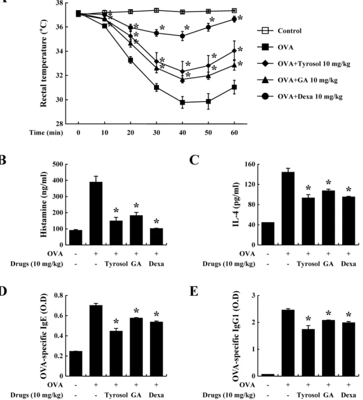
The OVA-induced systemic anaphylaxis model is appropriate for the investigation of the anti-allergic inflammatory effects of drug candidates [9]. After challenges of OVA, mice repetitively sensitized with OVA and alum adjuvant developed anaphylaxis and were monitored for 1 h. The rectal temperature of the mice decreased over 40–50 min; administration of tyrosol allevi-ated this hypothermia, which was associallevi-ated with the serum histamine level (Fig 1A). The serum histamine level increased by about 4 times and was also diminished by tyrosol (Fig 1B). In addition, tyrosol attenuated the serum IL-4 level (Fig 1C). IL-4 is a representative Th2 cyto-kine, that plays important role in inducing IgE synthesis [19]. Binding of IgE and IgG1 with FcεRI is necessary for the activation of mast cells and allergic responses, especially anaphylaxis [20]. We measured OVA-specific IgE and IgG1 levels, which were considerably increased by sensitizations of OVA (Fig1Dand1E). Tyrosol showed more effective inhibition in the genera-tion of both immunoglobulins than did gallic acid and dexamethasone.
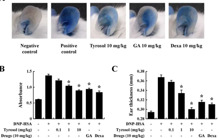
PCA is another widely used animal model for immediate-type allergic reactions [16]. After challenges of antigen, a blue spot developed at the sensitized site because of the increased vas-cular permeability caused by histamine released from mast cells. Tyrosol decreased the size and color of the blue spot in a dose-dependent manner (Fig2Aand2B). Increased vascular perme-ability induced ear swelling, which was also reduced by tyrosol (Fig 2C).
Effects of tyrosol on the mast cell degranulation and inflammatory
cytokine expression
Mast cells are major producers of histamine, a key molecule in allergic responses [6]. Therefore, inhibition of mast cell degranulation is a useful therapeutic target for the treatment of allergic symptoms. To examine the influence of tyrosol on the degranulation of mast cells, we mea-sured histamine release in RBL-2H3 cells and RPMCs. Histamine was rapidly released after challenges of antigen, however this release was dose-dependently hindered by tyrosol in both mast cells (Fig3Aand3B). Tyrosol showed a similar or superior inhibitory effect in compari-son with gallic acid and dexamethacompari-sone although at 10 times lower concentration. Theβ -hex-osaminidase assay commonly used to evaluate mast cell degranulation was carried out [16]. Suppressive effect of tyrosol presented an equal aspect in both secretion of histamine andβ -hexosaminidase (Fig 3C). MTT assays were performed to investigate the cytotoxicity of tyrosol. Tyrosol did not manifest any significant cytotoxicity following 24 h exposure to concentration of up to 100μM (Fig 3D). These results indicated that tyrosol reduces mast cell degranulation
in the absence of cytotoxicity.
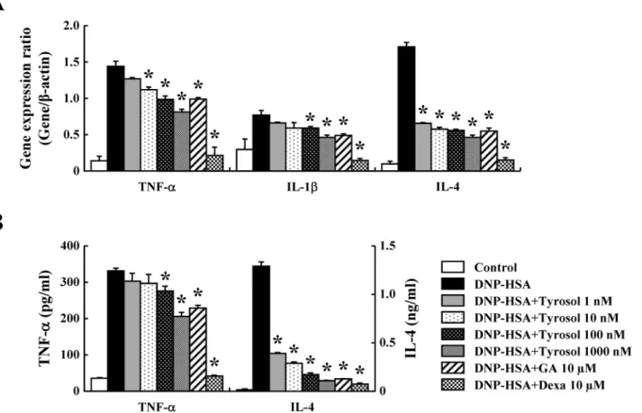
Inflammatory cytokines are known to mediate inflammation by enhancing recruitment and activation of immune cells [21]. To assure the effects of tyrosol on the expression of inflamma-tory cytokines such as TNF-α, IL-1β, and IL-4 in RBL-2H3 cells, real-time PCR and ELISAs were carried out. The expression of these cytokines was elevated by activation of FcεRI, while it was suppressed by tyrosol dose-dependently; these effects were observed at the mRNA and protein levels (Fig4Aand4B).
Effects of tyrosol on intracellular calcium and activation of NF-
κ
B in mast
cells
Fig 1. Effects of tyrosol on ovalbumin-induced active systemic anaphylaxis.Induction of systemic anaphylaxis and oral administration of drugs including tyrosol, GA, and Dexa were described in the Materials and methods section. (A) Rectal temperature was measured every 10 min for 1 h. Blood was obtained from the abdominal artery of each mouse for the measurement of serum histamine, OVA-specific IgE, and IL-4 levels. (B) Histamine level was assayed using theo-phthaldialdehyde spectrofluorometric procedure. (C-E) IL-4, OVA-specific IgE, and OVA-specific IgG1 levels were measured by ELISA. Each data presented as a graph represents the mean±SE (n= 10 per group).*Significant difference from OVA-challenged group atP<0.05. GA: gallic acid; Dexa: dexamethasone.
calcium efflux from the ER; the inflow of extracellular calcium is enhanced by this ER calcium depletion [5]. In the present study, the intracellular calcium level rapidly increased within a few seconds of exposure to DNP-HSA, however tyrosol reduced this rise of intracellular calci-um level (Fig 5A).
NF-κB is a major transcription factor regulating the expression of inflammatory cytokines. Degradation of IκBαallows the movement of NF-κB into the nucleus. The IKK complex acti-vates NF-κB by phosphorylating IκBα[4]. The IKK complex has 3 subunits;α,β, and regulato-ry subunits. Recent research has reported that the IKKβsubunit activates FcεRI-induced exocytosis. Thus, inhibition of the IKK complex might suppress degranulation and inflamma-tory cytokine expression in mast cells. Our results showed that tyrosol hindered activation of NF-κB and the IKK complex (Fig 5B).
Effects of tyrosol on the activation of signaling proteins in mast cells
The intracellular signaling pathways in mast cells have been determined in detail [4]. To assure the phosphorylation of signaling proteins such as Lyn, Syk, PI3K, and Akt, Western blots were performed. Tyrosol did not affect the activation of Lyn and Syk, on the contrary, it reduced the phosphorylation of PI3K and Akt (Fig 6).
Fig 2. Effects of tyrosol on IgE-mediated passive cutaneous anaphylaxis.Mouse ear skin (n= 5/group) was sensitized with an intradermal injection of anti-DNP IgE (0.5μg/site) for 48 h. Drugs including tyrosol, GA, and Dexa were orally administered at doses of 0.1–10 mg/kg BW 2 h before the intravenous injection of DNP-HSA (1 mg/mouse) and 4% Evans blue (1:1) mixture. Thirty minutes later, the thickness of both ears was measured, and the ears were collected to measure pigmentation. Dye was extracted as described in the Materials and methods section and detected using a spectrophotometer. Each data presented as a graph represents the mean±SE (n= 5 per group).*Significant difference from DNP-HSA challenged group atP<0.05. GA: gallic acid; Dexa: dexamethasone.
To confirm that inhibition of PI3K regulates degranulation and cytokine expression in mast cells, we used NVP-BEZ235, an inhibitor of PI3K/Akt [23]. NVP-BEZ235 certainly worked as a PI3K inhibitor in RBL-2H3 cells (Figure B inS1 File). The release of histamine andβ -hexosa-minidase was suppressed by blocking the activation of PI3K (Figure C and D inS1 File). In ad-dition, FcεRI-induced expression of TNF-αand IL-4 was completely blocked by NVP-BEZ235 (Figure E and F inS1 File). According to these results, it seems reasonable to conclude that the inhibition of PI3K by tyrosol reduces allergic responses by suppressing mast cell degranulation and cytokine expression.
Discussion
Anaphylaxis induced by the rapid release of allergic mediators such as histamine, heparin, and various cytokines can have lethal consequences. OVA-induced ASA and IgE-mediated PCA are suitable animal models for immediate-type hypersensitivity. Mice sensitized by OVA and Fig 3. Effects of tyrosol on mast cell degranulation.(A, B) RBL-2H3 cells (5 × 10⁵/well) and RPMCs (2 × 10⁴/well) were sensitized with anti-DNP IgE (50 ng/ml). After incubating overnight, the cells were pretreated with or without drugs including tyrosol, GA, and Dexa for 1 h and then challenged with DNP-HSA (100 ng/ml). Histamine level was assayed using theo-phthaldialdehyde spectrofluorometric procedure. (C) The level ofβ-hexosaminidase was measured usingβ-hexosaminidase substrate buffer. (D) RBL-2H3 cells (3 × 10⁴/well) were pretreated with or without tyrosol for 24 h and then incubated with 1 mg/ml MTT for 2 h. The absorbance intensity was detected using a spectrophotometer. Each data presented as a graph represents the mean±SE of three
independent experiments.*Significant difference from DNP-HSA challenged group atP<0.05. GA: gallic acid; Dexa: dexamethasone.
alum adjuvant developed hypothermia after challenges of OVA because this produced a sud-den increase in the serum histamine level. IgE and IgG levels significantly contribute to system-ic anaphylaxis in msystem-ice [24]. IL-4, a principal Th2 cytokine, differentiates naive T cells into Th2 cells, which stimulate production of IgE in B cells [2]. It is likely that tyrosol reduces hypother-mia by inhibiting histamine release from mast cells and lessening production of IgE and IgG re-sulted from decreasing IL-4 levels in mice. We observed that tyrosol more effectively hindered production of IgE and IgG1 than gallic acid and dexamethasone. The tyrosol-mediated reduc-tion in pigmentareduc-tion observed in PCA is also considered to arise from its suppression of mast cell degranulation. Histamine released from mast cells enhances vascular permeability. Conse-quently, intravascular circulating Evans blue percolates through the endodermis. These results indicated that tyrosol has therapeutic effects on anaphylactic reaction.
Mast cells are important for the manifestation of IgE-mediated allergic responses by releas-ing various mediators such as histamine, lipid-derived mediators, chemokines, cytokines, and growth factors [1]. Therefore, mast cells are key targets for the development of medicines for allergic disorders. To assure the anti-allergic inflammatory effects of tyrosol on mast cells, RBL-2H3 cells and RPMCs were used in this study. We assessed the release of histamine andβ -hexosaminidase to measure mast cell degranulation. Histamine from mast cells causes typical allergic symptoms such as edema, warmth, and erythema [6]. Thus, attenuating mast cell Fig 4. Effects of tyrosol on inflammatory cytokine expression.(A) RBL-2H3 cells (5 × 10⁵/well) were sensitized with anti-DNP IgE (50 ng/ml). After incubating overnight, the cells were pretreated with or without drugs including tyrosol, GA, and Dexa for 1 h and then challenged with DNP-HSA (100 ng/ml). Extraction and analysis of mRNA were performed as described in the Materials and methods section. The gene expression of inflammatory cytokines was determined by real-time PCR. (B) The secretion of inflammatory cytokines was measured by ELISA. Each data presented as a graph represents the mean±SE of three independent experiments.*Significant difference from DNP-HSA challenged group atP<0.05. GA: gallic acid; Dexa: dexamethasone.
degranulation is a viable therapeutic strategy for the treatment of allergic disorders. Tyrosol dose-dependently decreased degranulation without cytotoxic effects.
Inflammatory cytokines such as TNF-α, IL-1β, and IL-4 play crucial roles in prompting and sustaining chronic allergy. TNF-αpromotes adaptive immunity through the activation of NF-κB and stimulates the migration, maturation, and differentiation of immune cells [25,26]. IL-1βaggravates auto-inflammatory and allergic diseases such as contact hypersensitivity, atopic dermatitis, and bronchial asthma [27]. IL-4 is necessary for allergic responses as it drives the generation of IgE in plasma B cells [19]. Our results showed that expression of inflammatory cytokines was increased after activation of FcεRI and this effect was dose-dependently reduced by tyrosol in RBL-2H3 cells. In the previous research, tyrosol suppressed expression of TNF-α Fig 5. Effects of tyrosol on intracellular calcium and the NF-κB activation in mast cells.(A) RBL-2H3 cells (3 × 10⁴/well) were sensitized with anti-DNP IgE (50 ng/ml). After incubating overnight, the cells were preincubated with Fluo-3/AM for 1 h. The cells were pretreated with or without tyrosol for 1 h and then challenged with DNP-HSA (100 ng/ml). Intracellular calcium was detected every 1 min for 5 min using a fluorescent plate reader. Area under the curve (AUC) was calculated over 5 min. BAPTA, a calcium chelator, was used as a positive control. Each data presented as a graph represents the mean±SE of three independent experiments.*Significant difference from DNP-HSA challenged group atP<0.05. (B) RBL-2H3 cells (2 × 10⁶/well) were sensitized with anti-DNP IgE (50 ng/ml). After incubating overnight, the cells were pretreated with or without tyrosol for 1 h and then challenged with DNP-HSA (100 ng/ml). Activations of IKK complex and NF-κB were assayed by Western blot (N-: nuclear, p-: phosphorylated). The bands of actin and total form were used as a loading control. PP2, a Src family inhibitor, was used as a positive control. The band is a representative of three independent experiments.
in an anoxia-induced EAhy926, human endothelial cell line [28]. Furthermore, production of TNF-α, IL-1β, and IL-6 was attenuated on peripheral blood mononuclear cells and RAW 264.7 cells stimulated with lipopolysaccharide [13,29]. These reports support the present research, which assure that tyrosol reduced cytokine expression. As a result, it is possible that tyrosol-mediated blockade of inflammatory cytokines expression alleviates allergic inflammation and limits progression in chronic allergy.
Calcium is a crucial secondary messenger in mast cell signaling [5]. The intracellular calci-um level regulates exocytosis from mast cells and also expression of inflammatory cytokines [30–32]. In our results, the intracellular calcium level in RBL-2H3 cells stimulated with DNP-IgE was rapidly elevated, while tyrosol blocked calcium influx. Activation of NF-κB is important for the expression of various inflammatory cytokines including TNF-α, IL-1β, and IL-4 [33]. Phosphorylation of IκBαprompts its proteolytic degradation, which allows NF-κB to translocate into the nucleus. FcεRI-stimulated degradation of IκBαand nuclear
Fig 6. Effects of tyrosol on the activation of signaling proteins.RBL-2H3 cells (2 × 10⁶/well) were sensitized with anti-DNP IgE (50 ng/ml). After incubating overnight, the cells were pretreated with or without tyrosol for 1 h and then challenged with DNP-HSA (100 ng/ml). Extraction of protein was performed as described in the Materials and methods section. Activation of signaling proteins was assayed by Western blot (p-: phosphorylated). The band of total form was used as a loading control. PP2, a Src family inhibitor, was used as a positive control. The band is a representative of three independent experiments.
translocation of NF-κB were regulated by tyrosol. The IKK complex induces activation of NF-κB [4]. Tyrosol also reduced phosphorylation of IKK, and this seemed to obstruct the degradation of IκBαand nuclear translocation of NF-κB. A recent study informed the rela-tionship between IKK and mast cell degranulation. IKKβstimulated SNAP-23 which has a critical role in mast cell degranulation and anaphylactic responses [34]. We predicted that suppression of the IKK complex might improve allergic inflammatory reactions by lessening degranulation and the expression of allergic mediators. It is likely that tyrosol-related inhibi-tion of degranulainhibi-tion and cytokine expression is associated with the reducinhibi-tion of intracellular calcium level and IKK activation in mast cells, since NF-κB plays is a major transcription fac-tor regulating expression of inflammafac-tory cytokine.
Signaling pathways of mast cells have been extensively studied [4]. Lyn, Syk, PI3K, and Akt were selected as the representative signaling proteins to anticipate the target of tyrosol. Our re-sults indicated that tyrosol interrupted the protein phosphorylation from PI3K without affect-ing Lyn and Syk. Blockade of PI3K reduces activation of PLCγ, which regulates PKC and the intracellular calcium level [35]. PLCγhydrolyzes phosphatidylinositol 4,5-bisphosphate (PIP2)
to diacylglycerol (DAG) and IP3[36]. DAG is reported to activate PKC, which stimulates the
IKK complex; IP3ultimately triggers extracellular calcium influx. These IKK stimulation and
increased intracellular calcium level provoke the secretion and expression of allergic mediators. The Akt dependent- pathway also activates the IKK complex [4]. PI3K produces phosphatidy-linositol 3,4,5-trisphosphate, an important lipid mediator of Akt activation, from PIP2.
Accord-ingly, it is possible that the suppression of PI3K regulates the IKK complex by interrupting the activation of not only PLCγbut also Akt. These studies are consistent with our expectation that tyrosol reduces allergic inflammation by inhibiting the activation of PI3K. Nevertheless, we only estimate that the target point of tyrosol is between Syk and PI3K. Further studies are re-quired to identify the exact target proteins of tyrosol.
In the present study, we aimed to demonstrate the effects of tyrosol on allergic inflammation using animal models and mast cells. Tyrosol attenuated anaphylactic shock in immediate-type hypersensitivity models, and this effect is related to a decreased release of allergic mediators from mast cells. Secretion and expression of allergic molecules are decreased by tyrosol in a dose-dependent manner, and we anticipate that the anti-allergic inflammatory effects of tyrosol are associated with inhibition of PI3K, which regulates activation of the IKK complex and the intracellular calcium level in mast cells. Further studies are needed in order to determine the exact binding target of tyrosol. Nevertheless, this study suggests that tyrosol is a possible thera-peutic candidate for allergic disorders by inhibiting degranulation and the expression of in-flammatory cytokines in mast cells.
Supporting Information
theo-phthaldialdehyde spectrofluorometric procedure. (Figure D) The level of β-hexosamini-dase was measured usingβ-hexosaminidase substrate buffer. (Figure E and F) The secretion of inflammatory cytokines was measured by ELISA. Each data presented as a graph represents the mean ± SE of three independent experiments.Significant difference from DNP-HSA chal-lenged group atP<0.05. GA: gallic acid; Dexa: dexamethasone.
(TIF)
S2 File. The action schema of tyrosol in mast cells.Tyrosol blocked the IgE-mediated phos-phorylation of PI3K. Blockade of PI3K reduces activation of Akt and downstream IKK com-plex. Decrease of IKK and intracellular calcium results in the reduction of secretion of allergic mediators.
(TIF)
S1 Archive. All the images are the original Western blot data forFig 5B,Fig 6, and Figure B inS1 File.
(ZIP)
Author Contributions
Conceived and designed the experiments: IGJ DSK SHK. Performed the experiments: IGJ SL DSK HSL EKP. Analyzed the data: SWK. Wrote the paper: IGJ DK SHK.
References
1. Kraft S, Kinet JP. New developments in FcepsilonRI regulation, function and inhibition. Nat Rev Immu-nol. 2007; 7: 365–378. PMID:17438574
2. Galli SJ, Tsai M. IgE and mast cells in allergic disease. Nat Med. 2012; 18: 693–704. doi:10.1038/nm.
2755PMID:22561833
3. Beghdadi W, Madjene LC, Benhamou M, Charles N, Gautier G, Launay P, et al. Mast cells as cellular sensors in inflammation and immunity. Front Immunol. 2011; 2: 37. doi:10.3389/fimmu.2011.00037
PMID:22566827
4. Kalesnikoff J, Galli SJ. New developments in mast cell biology. Nat Immunol. 2008; 9: 1215–1223. doi:
10.1038/ni.f.216PMID:18936782
5. Vig M, DeHaven WI, Bird GS, Billingsley JM, Wang H, Rao PE, et al. Defective mast cell effector func-tions in mice lacking the CRACM1 pore subunit of store-operated calcium release-activated calcium channels. Nat Immunol. 2008; 9: 89–96. PMID:18059270
6. Galli SJ, Tsai M, Piliponsky AM. The development of allergic inflammation. Nature. 2008; 454: 445– 454. doi:10.1038/nature07204PMID:18650915
7. Kay AB. Allergy and allergic diseases. First of two parts. N Engl J Med. 2001; 344: 30–37. PMID:
11136958
8. Bae Y, Lee S, Kim SH. Chrysin suppresses mast cell-mediated allergic inflammation: involvement of calcium, caspase-1 and nuclear factor-kappaB. Toxicol Appl Pharmacol. 2011; 254: 56–64. doi:10. 1016/j.taap.2011.04.008PMID:21515303
9. Ribeiro-Filho J, Leite FC, Costa HF, Calheiros AS, Torres RC, de Azevedo CT, et al. Curine inhibits mast cell-dependent responses in mice. J Ethnopharmacol. 2014; 155: 1118–1124. doi:10.1016/j.jep. 2014.06.041PMID:24969825
10. Kim HH, Park SB, Lee S, Kwon TK, Shin TY, Park PH, et al. Inhibitory effect of putranjivain A on allergic inflammation through suppression of mast cell activation. Toxicol Appl Pharmacol. 2014; 274: 455– 461. doi:10.1016/j.taap.2013.12.006PMID:24361550
11. Tuck KL, Hayball PJ. Major phenolic compounds in olive oil: metabolism and health effects. J Nutr Bio-chem. 2002; 13: 636–644. PMID:12550060
12. Lecci RM, Logrieco A, Leone A. Pro-oxidative action of polyphenols as action mechanism for their pro-apoptotic activity. Anticancer Agents Med Chem. 2014; 14: 1363–1375. PMID:25244914
14. Kim SH, Jun CD, Suk K, Choi BJ, Lim H, Park S, et al. Gallic acid inhibits histamine release and pro-in-flammatory cytokine production in mast cells. Toxicol Sci. 2006; 91: 123–131. PMID:16322071
15. Wershil BK, Furuta GT, Lavigne JA, Choudhury AR, Wang ZS, Galli SJ. Dexamethasone or cyclosporin A suppress mast cell-leukocyte cytokine cascades. Multiple mechanisms of inhibition of IgE- and mast cell-dependent cutaneous inflammation in the mouse. J Immunol. 1995; 154: 1391–1398. PMID:
7822805
16. Je IG, Kim HH, Park PH, Kwon TK, Seo SY, Shin TY, et al. SG-HQ2 inhibits mast cell-mediated allergic inflammation through suppression of histamine release and pro-inflammatory cytokines. Exp Biol Med (Maywood). 2015; 240: 631–638. doi:10.1177/1535370214555663PMID:25349218
17. Kim HH, Bae Y, Kim SH. Galangin attenuates mast cell-mediated allergic inflammation. Food Chem Toxicol. 2013; 57: 209–216. doi:10.1016/j.fct.2013.03.015PMID:23535185
18. Choi JK, Oh HM, Lee S, Kwon TK, Shin TY, Rho MC, et al. Salvia plebeia suppresses atopic dermatitis-like skin lesions. Am J Chin Med. 2014; 42: 967–985. doi:10.1142/S0192415X1450061XPMID:
25004886
19. Brandt EB, Sivaprasad U. Th2 Cytokines and Atopic Dermatitis. J Clin Cell Immunol. 2011; 2. PMID:
22924155
20. Finkelman FD, Rothenberg ME, Brandt EB, Morris SC, Strait RT. Molecular mechanisms of anaphylax-is: lessons from studies with murine models. J Allergy Clin Immunol. 2005; 115: 449–457; quiz 458. PMID:15753886
21. Sismanopoulos N, Delivanis DA, Alysandratos KD, Angelidou A, Therianou A, Kalogeromitros D, et al. Mast cells in allergic and inflammatory diseases. Curr Pharm Des. 2012; 18: 2261–2277. PMID:
22390690
22. Baba Y, Nishida K, Fujii Y, Hirano T, Hikida M, Kurosaki T. Essential function for the calcium sensor STIM1 in mast cell activation and anaphylactic responses. Nat Immunol. 2008; 9: 81–88. PMID:
18059272
23. Seo BR, Min KJ, Cho IJ, Kim SC, Kwon TK. Curcumin significantly enhances dual PI3K/Akt and mTOR inhibitor NVP-BEZ235-induced apoptosis in human renal carcinoma Caki cells through down-regula-tion of p53-dependent Bcl-2 expression and inhibidown-regula-tion of Mcl-1 protein stability. PLoS One. 2014; 9: e95588. doi:10.1371/journal.pone.0095588PMID:24743574
24. Ishikawa R, Tsujimura Y, Obata K, Kawano Y, Minegishi Y, Karasuyama H. IgG-mediated systemic anaphylaxis to protein antigen can be induced even under conditions of limited amounts of antibody and antigen. Biochem Biophys Res Commun. 2010; 402: 742–746. doi:10.1016/j.bbrc.2010.10.098
PMID:21029722
25. Li J, Yin Q, Wu H. Structural basis of signal transduction in the TNF receptor superfamily. Adv Immunol. 2013; 119: 135–153. doi:10.1016/B978-0-12-407707-2.00005-9PMID:23886067
26. Galli SJ, Tsai M. Mast cells in allergy and infection: versatile effector and regulatory cells in innate and adaptive immunity. Eur J Immunol. 2010; 40: 1843–1851. doi:10.1002/eji.201040559PMID:
20583030
27. Krause K, Metz M, Makris M, Zuberbier T, Maurer M. The role of interleukin-1 in allergy-related disor-ders. Curr Opin Allergy Clin Immunol. 2012; 12: 477–484. doi:10.1097/ACI.0b013e3283574d0c
PMID:22885885
28. Palmieri D, Aliakbarian B, Casazza AA, Ferrari N, Spinella G, Pane B, et al. Effects of polyphenol ex-tract from olive pomace on anoxia-induced endothelial dysfunction. Microvasc Res. 2012; 83: 281– 289. doi:10.1016/j.mvr.2012.02.010PMID:22386654
29. Lu J, Huang G, Wang Z, Zhuang S, Xu L, Song B, et al. Tyrosol exhibits negative regulatory effects on LPS response and endotoxemia. Food Chem Toxicol. 2013; 62: 172–178. doi:10.1016/j.fct.2013.08. 031PMID:23994089
30. Blank U, Cyprien B, Martin-Verdeaux S, Paumet F, Pombo I, Rivera J, et al. SNAREs and associated regulators in the control of exocytosis in the RBL-2H3 mast cell line. Mol Immunol. 2002; 38: 1341– 1345. PMID:12217405
31. Manikandan J, Kothandaraman N, Hande MP, Pushparaj PN. Deciphering the structure and function of FcepsilonRI/mast cell axis in the regulation of allergy and anaphylaxis: a functional genomics paradigm. Cell Mol Life Sci. 2012; 69: 1917–1929. doi:10.1007/s00018-011-0886-0PMID:22146792
32. Tanaka S, Mikura S, Hashimoto E, Sugimoto Y, Ichikawa A. Ca2+ influx-mediated histamine synthesis and IL-6 release in mast cells activated by monomeric IgE. Eur J Immunol. 2005; 35: 460–468. PMID:
15668925
34. Suzuki K, Verma IM. Phosphorylation of SNAP-23 by IkappaB kinase 2 regulates mast cell degranula-tion. Cell. 2008; 134: 485–495. doi:10.1016/j.cell.2008.05.050PMID:18692471
35. Galli SJ, Kalesnikoff J, Grimbaldeston MA, Piliponsky AM, Williams CM, Tsai M. Mast cells as "tunable" effector and immunoregulatory cells: recent advances. Annu Rev Immunol. 2005; 23: 749–786. PMID:
15771585