online | memorias.ioc.fiocruz.br
Chemokines are a group of chemoattractant proteins that induce not only chemotaxis but also activation of tar-get cells (Ulfman et al. 2001). It has been shown that some chemokines influence inflammatory cell infiltration and the cellularity that occurs in the granulomatous response around Schistosoma mansoni eggs. For instance, the
chemokines macrophage inflammatory protein-1α (MIP-1α/CCL3) and regulated upon activation normal T-cell ex
-pressed and secreted (RANTES/CCL5) are produced in a
model of granulomatous inflammation in mice and ablation
of their activity modulates the size of the lesions (Lukacs et al. 1993, 1994, Chensue et al. 1999, Qiu et al. 2001, Souza et al. 2005). Plasma levels of CCL3 are elevated in chron -ic schistosomiasis patients and correlate with the level of
morbidity (Falcão et al. 2002, Souza et al. 2005); blocking CCL3 activity inhibits the granulomatous response in vitro (Falcão et al. 2002). Booth et al. (2004) recently reported
that periportal fibrosis in males with S. mansoni infection
is associated with low concentrations of CCL5.
Eosinophils represent an important line of defence against parasitic infections (Butterworth 1984). In the
parasitic disease caused by exposure to S. mansoni larvae, blood eosinophilia rises due to an increase in interleukin
(IL)-5, IL-1, IL-3, granulocyte-macrophage colony-stim -ulating factor and granulocyte colony-stim-ulating factor (Clutterbuck et al. 1989, Weller 1992) and this increase is dependent on antigens secreted by eggs through the pores
of the shell (Race et al. 1969, Sher et al. 1990). It is well
established that an important step in the extravasation of eosinophils is their adhesion to the vascular endothelium,
which is mediated by selectins and chemokines (Lampinen et al. 2004). However, the acute eosinophil cellular response
against eggs and its contribution to granuloma formation
is not well studied (Pacheco & Lenzi 1997). Furthermore,
there are no reports in the literature showing alteration in the plasma concentration of chemokines in acutely
in-fected patients. Most of the current knowledge on the role
of chemokines in schistosomiasis is derived from mice data. In contrast to experimentation in mice, there is a ma-jor difficulty faced by those researchers trying to answer
questions in acute schistosomiasis in humans. People liv -ing in schistosome endemic areas, where it is easy to find infected patients, are unlikely to develop signs and
symp-toms related to the acute phase of schistosomiasis (Malho
-tra et al. 1997, King et al. 1998).
In the present study, a group of individuals who have not previously acquired schistosomiasis presented vari-ous characterised clinical symptoms of the early phase of Financial support: UNDP World Bank/WHO SPRTTD, CNPq
(478320/2007-8), FAPEMIG (ATC, OAMF, MMT and RCO are CNPq
fellowships)
+ Corresponding author: andreat@cpqrr.fiocruz.br Received 6 January 2009
Accepted 16 October 2009
Seric chemokines and chemokine receptors in eosinophils during
acute human schistosomiasis mansoni
Denise Silveira-Lemos1,2,3,4, Andréa Teixeira-Carvalho2/+, Olindo Assis Martins-Filho2,
Adriano Luiz Souza-Soares3, Pollyanna Castro-Silva1, Matheus Fernandes Costa-Silva1,2,
Pedro Henrique Gazzinelli Guimarães1, Helena Barbosa Ferraz2, Lúcia Alves Oliveira-Fraga5,
Mauro Martins Teixeira3, Rodrigo Corrêa-Oliveira1
1Laboratório de Imunologia Celular e Molecular 2Laboratório de Biomarcadores de Diagnóstico e Monitoração
3Departamento de Bioquímica e Imunologia, Instituto de Ciências Biológicas, Universidade Federal de Minas Gerais, Belo Horizonte,
MG, Brasil 4Núcleo de Pesquisas em Ciências Biológicas, Laboratório de Imunoparasitologia, Departamento de Ciências Biológicas,
Instituto de Ciências Exatas e Biológicas, Universidade Federal de Ouro Preto, Ouro Preto, MG, Brasil 5Núcleo de Pesquisa em
Imunologia, Faculdade de Ciências da Saúde, Universidade Vale do Rio Doce, Governador Valadares, MG, Brasil
The recruitment of circulating eosinophils by chemokines and chemokine receptors plays an important role in the inflammation process in acute human schistosomiasis. Our main focus has been on the plasma chemokines (CXCL8/ CCL2/CCL3/CCL24) and chemokine receptors (CCR2/CCR3/CCR5/CXCR1/CXCR2/CXCR3/CXCR4) expressed by cir-culating eosinophils from acute Schistosoma mansoni infected patients (ACT). Our studies compared ACT patients and healthy individuals as a control group. Our major findings demonstrated a plethora of chemokine secretion with significantly increased secretion of all chemokines analysed in the ACT group. Although no differences were detected for beta-chemokine receptors (CCR2, CCR3 and CCR5) or alpha-chemokine receptors (CXCR3 and CXCR4), a signifi-cantly lower frequency of CXCR1+ and CXCR2+ eosinophils in the ACT group was observed. The association between chemokines and their chemokine receptors revealed that acutely infected schistosome patients displaying decreased plasma levels of CCL24 are the same patients who presented enhanced secretion of CCL3, as well as increased expres-sion of both the CCR5 and CXCR3 chemokine receptors. These findings suggest that CCL24 may influence the kinetics of chemokines and their receptors and eosinophils recruitment during human acute schistosomiasis mansoni.
S. mansoni infection. This allowed observation of cer -tain immunological alterations in a cohesive group of acutely infected patients. In order to get a better insight into eosinophil participation in acute S. mansoni infec-tion, we analysed the expression of chemokine receptors,
namely, CCR2, CCR3, CCR5, CXCR1, CXCR2, CXCR3 and CXCR4 in the blood eosinophils of acutely infected
patients and non-infected individuals. We also measured
the levels of four different chemokines, CCL2 (MCP-1), CCL3 (MIP-1α), CCL24 (eotaxin-2) and CXCL8 (IL-8),
which span different cellular specificities, in the plasma
of these acute patients. Our findings led us to postulate that CCL24 may influence the kinetics of chemokines and
their receptors and leukocyte recruitment during acute human schistosomiasis mansoni.
PATIENTS, MATERIALS AND METHODS
The study population consisted of two groups of in -dividuals, acute S. mansoni-infected patients (ACT) and
healthy blood donors as a control group (CT). The ACT group consisted of 10 patients (6 males and 4 females), with ages ranging from 14-21 years, who acquired their infection in the county village of São Geraldo da Piedade,
an endemic area for S. mansoni infection, situated next
to the city of Governador Valadares in the state of Minas Gerais (MG), Brazil. In our work, acute infected patients
presented various characterized clinical symptoms asso-ciated with early infection by S. mansoni, such as fever, diarrhoea, headache, nausea, eosinophilia and, after a few weeks, the presence of schistosome eggs in their faeces.
Quantitative parasitological stool examinations and detec -tion of S. mansoni eggs was performed using Kato-Katz (Katz et al. 1972). The ultrasonography analysis did not show echogenicity abnormalities. ACT patients did not
receive any corticosteroids or other immunosuppressant chemotherapy, nor were they treated for S. mansoni infec-tion before blood collecinfec-tion. Upon positive laboratory di-agnosis of schistosomiasis, all patients received treatment with a single dose of praziquantel, which was independent of their participation in this study.
Twenty-seven non-infected individuals (11 males and 16 females) were included as a CT group consisting of volunteer blood donors, aged from 19-30 years and were contacted at the Hemominas Blood Bank Foundation in Belo Horizonte, MG, Brazil. All non-infected volunteers
were included after conclusive negative parasitological diagnosis of S. mansoni infection, in addition to negative serology for Chagas disease, leishmaniasis, human im-munodeficiency virus infection and hepatitis.
The inclusion of all individuals in our investigation
was preceded by their written agreement to participate,
signed by the patient or by the patient’s parents. This study was approved by the Ethical Committees of the Fundação Oswaldo Cruz-Fiocruz, Ministry of Health, Brazil.
White blood cell counts - Hemograms were performed in an automated haematology instrument (Coulter MD18, USA), using whole blood collected in 5 mL vacutainer
tubes containing ethylenediamine tetraacetic acid (EDTA)
as the anticoagulant (Becton Dickinson Biosciences, San Diego, CA, USA). The parameter measured was a differen -tial analysis of leukocyte subsets, including absolute counts of eosinophils, neutrophils, lymphocytes and monocytes.
Quantification of plasma chemokines by ELISA - The
plasma concentrations of chemokines were measured by
sandwich enzyme-linked immunosorbent assay (ELISA), as described by Morita et al. (1999). The ELISA kits for human CCL2 and CCL24 were obtained from R&D Sys
-tems, Minneapolis, MN, USA. The antibody pairs and chemokines standards used in the assay for human CCL3 and CXCL8 were purchased from Pharmingen (San Di
-ego, CA, USA). The specific recommendations of the manufacturers were followed. Human plasma samples
were subjected to acid-salt precipitation to avoid
cross-re-activity in ELISA (Falcão et al. 2002). The concentration of chemokines in plasma was analysed in pg/mL.
Immunophenotypic analysis by flow cytometry - Human monoclonal antibodies (mAbs) for anti-CCR2 (48607.211), anti-CCR3 (61828.111), anti-CCR5 (45502.111), anti-CX
-CR1 (42705.111), anti-CXCR2 (48311.211), anti-CXCR3 (49801.111) and anti-CXCR4 (12G5) were labelled with fluorescein isothiocyanate (FITC) (R&D systems). Anti-mouse IgG1 (679.1Mc7) was used as isotype control and purchased from Becton Dickinson Biosciences Pharmin
-gen (San Diego, CA, USA).
Cell staining procedure - One hundred microlitres of EDTA whole blood was incubated in the presence of 5 µL of undiluted anti-human cell surface molecules mAbs for 30 min in the dark and at room temperature (RT). For
CCR2 analysis, monoclonal antibody labelled with
bi-otin (R&D systems, USA) was diluted 1/5 in phosphate-buffered saline (PBS) with avidin-FITC (Diatec, Oslo, Norway) diluted 1/100 in PBS. In this analysis, the cells were incubated with 50 µL of diluted monoclonal an-L of diluted monoclonal an- of diluted monoclonal
an-tibody in the dark for 30 min at RT. Then, erythrocyte lysis was performed using 2 mL of FACS Lysing Solu-L of FACS Lysing Solu- of FACS Lysing Solu
-tion (Becton Dickinson Biosciences Pharmingen, San Diego, CA, USA), followed by incubation for 10 min at room temperature (RT). The leukocyte suspension was further washed with 2 mL of PBS containing 0.01% so
-dium azide. Prior to flow cytometric acquisition, stained
cells were fixed in 200 µL of FACS fix solution (10 g/L
paraformaldehyde, 10.2 g/L sodium cacodylate, 6.65 g/L sodium chloride) for at least 15 min at 4oC. Data collec-tion from 20,000 events was performed using a FACScan flow cytometer (Becton Dickinson, Mountain View, CA, USA). CELLQUESTTM software provided by the
manu-facturer was used for data acquisition and analysis. Analysis of eosinophils was performed by single
colour immunophenotyping with FL-1/FITC-labelled anti-cell surface marker mAbs. The gating strategy was
based on eosinophil selection by their autofluorescent
properties in a non-related FL-3 channel vs. forward scat
-ter (FSC) graphs, as described by Weil and Chused (1981). Eosinophil subpopulations were identified using FSC vs. FL3 dot plots. A confirmatory graph was also set up to identify the eosinophils on FSC vs. side scatter dot plot
distributions confined to a region of high cell complex-ity. Data are presented as the percentage of eosinophils
expressing a given phenotypic marker, except for CCR3,
which was subjected to a semi-quantitative analysis of a
Statistics - Differences between groups were first
eval-uated by MINITAB 13.20 software (San Diego, CA, USA)
to examine three parameters, independence, normality and variance, of the data sets. Considering their non-parametric
nature, all data sets were analysed using Mann-Whitney’s test, with further analysis by Chi-square and Spearman’s rank correlation tests using the Graphpad PRISM 4.02 soft
-ware (La Jolla, CA, USA). Significance was defined in all cases at p < 0.05.
RESULTS
Acute human schistosomiasis is accompanied by eo-sinophilia and monocytosis - The assessment of hemato -logical parameters was evaluated in Fig. 1. Data analysis
demonstrated that the ACT group displayed a significant
increase in the absolute counts of eosinophils as compared
to the CT group (Fig. 1A). This eosinophilic profile was
accompanied by a significant increase in monocytes (p =
0.003) besides of a decrease in the neutrophils (p = 0.0373) counts in the ACT group as compared to the CT group
(Fig. 1C, D, respectively). No significant differences were observed for lymphocyte absolute values (Fig. 1B).
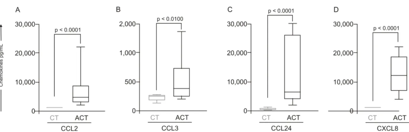
Increased plasma levels of alpha/beta-chemokines are found during acute human schistosomiasis mansoni - The
ex vivo analysis of the alpha and beta-chemokine patterns in acute patients is illustrated in Fig. 2. Interestingly, data analysis demonstrated a plethora of chemokines secretion with a significant increase in secretion of all chemokines
analysed [CCL2 (Fig. 2A), CCL3 (Fig. 2B), CCL24 (Fig. 2C) and CXCL8 (Fig. 2D)] in the ACT group as compared to the CT group. In order to extend our initial findings,
we performed a detailed correlation analysis of circu-lating eosinophil subset counts and chemokine levels.
The results of this investigation revealed a negative cor
-relation between the levels of CCL2 and the number of eosinophils (r = -0.64; p < 0.05) (data not shown). No
additional significant correlations were found with the additional chemokines investigated.
Diminished expression of the alpha-chemokine re-ceptors CXCR1 and CXCR2 is observed during acute human schistosomiasis mansoni - The pattern of alpha
and beta-chemokine receptor expression on eosinophils during acute schistosomiasis mansoni is illustrated in
p < 0.001
CT ACT
15,000
10,000
1,000
0
#
o
f
e
o
s
in
o
p
h
ils
/m
m
3
A
CT ACT
7,500
5,000
2,500
0
#
o
f
ly
m
p
h
o
c
y
te
s/
m
m
3
B
p = 0.003
CT ACT
1,500
1,000
500
0
#
o
f
mon
o
c
y
te
s
/m
m
3
C
p = 0.0373
CT ACT
10,000
5,000
2,500
0
#
o
f
n
e
u
tr
o
p
h
ils
/m
m
3
D
Fig. 1: analysis of leukocytes subsets (A: eosinophils; B: lymphocytes; C: monocytes; D: neutrophils) in the peripheral blood of acute patients (ACT, □ = 10) or control group (CT, □ = 27). The results are expressed in box-plot format. The box stretches from the lower hinge (defined as the 25th percentile) to the upper hinge (the 75th percentile) and therefore contains the middle half of the scores in the distribution. The median is shown as a line across the box. Therefore 1/4 of the distribution is between this line and the top of the box and 1/4 of the distribution is between this line and the bottom of the box. Significant differences (connecting lines) were considered at p < 0.05.
p < 0.0001
CT ACT
30,000
20,000
10,000
0
C
h
e
m
okine
s
p
g
/m
L
A
CCL2
p < 0.0100
CT ACT
2,000
1,000
500
0 B
CCL3
p < 0.0001
CT ACT
30,000
20,000
10,000
0 D
CXCL8 p < 0.0001
CT ACT
30,000
20,000
10,000
0 C
CCL24
Fig. 2: plasmatic levels of alpha and beta-chemokines from acute Schistosoma mansoni infected patients (ACT, □ = 10) and control group
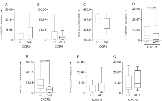
Fig. 3. No differences were detected for beta-chemokine
receptors, including the percentage of CCR2+ or CCR5+ eosinophils and CCR3 eosinophil expression (Fig.
3A-C, respectively) as well as for the alpha-chemokine
re-ceptors CXCR3 and CXCR4 (Fig. 3F, G, respectively).
Interestingly, data analysis demonstrated a shift in the alpha-chemokine receptor profile characterised by a
significantly lower frequency of CXCR1+ and CXCR2+ eosinophils (p = 0.0458 and p = 0.0084, respectively) in the ACT group as compared to the CT group (Fig. 3D, E,
respectively). In addition, a positive correlation between
CCR2 and CCR5 expression and eosinophil absolute counts was observed (r = 0.66, p < 0.05; r = 0.74, p < 0.05, respectively) (data not shown).
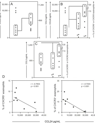
Decreased plasma levels of CCL24 are associated with enhanced secretion of CCL3 and increased expres-sion of CCR5 and CXCR3 in eosinophil subsets during acute S. mansoni infection - The patterns of plasma lev -els of chemokines and chemokine receptor expression are
shown in Fig. 4. Data analysis revealed that ACT patients who displayed decreased plasma levels of CCL24 are the same patients who presented enhanced secretion of CCL3 (Fig. 4A) (p < 0.0001), as well as increased expression of both CCR5 and CXCR3 chemokine receptors (Fig. 4B) (p = 0.002 and p = 0.0337, respectively). In addition, we
performed a further investigation that revealed a direct
association between CCL3 levels and the percentage of
CCR5+ or CXCR3+ eosinophils (Fig. 4C) (p = 0.0001) dur -ing acute human schistosomiasis mansoni.
In order to confirm the initial findings, we
per-formed a correlation between levels of CCL24 and per
-centage of CCR5+ or CXCR3+ eosinophils (Fig. 4D). The results demonstrated the existence of a strong negative
correlation between levels of CCL24 and the frequency of CCR5 or CXCR3 in eosinophils from acute patients (r = -0.7833, p = 0.001; r = -0.7333, p = 0.001, respectively). Together, these findings led us to postulate that CCL24
may influence the kinetics of chemokines and their re-ceptors, as well as leukocyte recruitment during human acute schistosomiasis mansoni.
DISCUSSION
Eosinophilia is a hallmark of the acute phase of schistosomiasis - Eosinophils represent a minor cell type among leukocytes, with basal counts ranging from 400-600 cells/mm3 (Brito-Babapulle 2003). In this study,
acute schistosomiasis patients had a significantly higher
number of eosinophils. Experimental studies have dem
-onstrated that eosinophilia occurs between the 5th-7th
weeks after exposure to the parasite and may be induced
by Th2 cytokines, such as IL-4, IL-10, IL-13, IL-9 and especially IL-5 (Cara et al. 2000). Hoshi et al. (1995)
demonstrated that a transient arrest of rolling
eosino-phils may be induced by chemokine CXCL8 and shifted into firm long-term arrest by CCL24. Eosinophils then
CT ACT 50.00
33.34
16.66
0
A
CCR2
CT ACT 100.00
66.67
33.33
0
B
CCR5
CT ACT 650.0
487.5
325.0
162.5
C
CCR3
p < 0.0485
CT ACT 40.00
26.67
13.33
0
D
CXCR1
%
o
f
C
C
R
+
eo
s
in
o
p
hi
ls
CC
R
3
o
n
e
o
si
n
o
ph
ils
(
FM
C
)
%
o
f
CX
C
R
+
e
o
s
in
op
h
ils
CT ACT 40.00
26.67
13.33
0
G
CXCR4
CT ACT 40.00
26.67
13.33
0
F
CXCR3
CT ACT 40.00
26.67
13.33
0
E
CXCR2
p < 0.0084
%
o
f
C
XC
R
+
e
o
s
in
o
p
h
ils
%
o
f
C
X
C
R
+
e
o
s
in
op
h
ils
Fig. 3: alpha and beta-chemokine receptor expression by peripheral blood eosinophils from Schistosoma mansoni-infected patients (ACT, □ = 10)
and control group (CT, □ = 27). The selection of eosinophils was essentially based on their autofluorescent cells using non-related FL-3 channel versus forward scatter (FSC) and the analysis of chemokine receptor+ eosinophils was carry out by single color immunophenotyping. The results are expressed in box-plot format highlighting the gap of 50% of data set measurement for the percentage of CCR2 (A), CCR5 (B), CXCR1 (D), CXCR2 (E), CXCR3 (F), CXCR4 (G) cells and the fluorescence mean channel (FMC) of CCR3 expression by eosinophils (C). Significant differ
migrate into the tissue in response to chemotactic
fac-tors produced locally at the inflammatory site (Teran et al. 1996). Smithers et al. (1977) demonstrated in ex -perimental models that the cellular infiltrate observed in hepatic granulomas is predominantly formed by eosino-phils and monocytes.
Chemokines in the plasma of acute schistosomiasis patients - The present paper represents the first report
of increased chemokine concentrations in the plasma of
S. mansoni acute-infected patients. All four of the
chemokines measured, CCL2, CCL3, CCL24 and CX
-CL8, were found at higher levels in the plasma of schis -tosomiasis patients in the acute phase of the disease as compared to healthy individuals. Although the presence of these chemokines in plasma is not enough to show a definitive role in the manifestations and general immune response associated with acute schistosomiasis in hu-mans, some evidence is available from animal models.
CCL24 and CXCL8 are generally known for their
chemotactic activity towards eosinophils and neutrophils,
respectively. CCL24 is selectively agonistic towards CCR3 receptors, whose expression on eosinophils (Heath et al. 1997), basophils (Uguccioni et al. 1997) and Th2 lymphocytes (Gerber et al. 1997, Sallusto et al. 1998) is
compatible with a role in allergic disease and the immune response against helminth infections. In this context,
CCL24 could attract eosinophils to inflammatory sites to participate in the initial formation of granulomas. This cell type may comprise up to 40% of the cellular compo
-sition of granulomas. Moreover, an increase of CCL24 could indicate a Th2 type immune response. The role of CXCL8 is more evident in acute inflammation, where
this chemokine attracts neutrophils to inflammatory
sites (Broaddus et al. 1994). Although neutrophils are
not obvious contributors to the formation of schistosome granulomatous, this cell type may have a role in the ini-tial influx of cells or at the interface between innate and acquired immunity, collaborating in the secretion of in-flammatory mediators that will bring other cells types of greater importance. A decreased frequency of circu-lating neutrophils in acute schistosomiasis patients may indicate a migration of this cell type into the
inflam-matory focus. This seems plausible since blood cells
from infected patients release mediators in response to soluble eggs antigen stimulation that are chemotactic to
neutrophils and eosinophils (Dabes et al. 1989). Moreo -ver, schistosome eggs themselves secrete antigens that have direct chemotactic activity on neutrophils and
eosinophils (Owhashi et al. 1985).
The chemokines CCL2 and CCL3 are important me -diators of the influx of monocytes and macrophages, which are relevant cellular types in the granulomatous response around schistosome eggs. In addition, both
macrophages (Koch et al. 1992) and eosinophils (Izumi et al. 1997) are sources of CCL2. Tillie-Leblond et al.
(2000) demonstrated that chemotactic activity from eo-sinophils from bronchial lavage fluid in asthmatic
sub-jects is associated with higher levels of CCL2, CCL3 and CCL5. Moreover, CCL3-deficient schistosome-infected
mice exhibit smaller granulomas compared to wild-type
mice (Souza et al. 2005). In humans, a correlation be
-tween high levels of CCL3 and severity of disease has
been observed during schistosome infection (Falcão et
al. 2002). Thus, CCL2 and CCL3 could promote the
accumulation of macrophages and eosinophils in the early phases of granuloma formation. In our study, both chemokines are increased in plasma from patients with acute S. mansoni infection.
Expression profile of chemokines receptors in eosino-phils - Signals via chemokine receptors play an impor -tant role in the accumulation of eosinophils (Nagase et
al. 2001). Previous data have demonstrated a constitutive and as well as or inducible expression of CCR1, CCR3, CXCR1, CXCR2, CXCR3 and CXCR4 by human circu
-lating eosinophils (Heath et al. 1997). In our study, we observed a decrease in the expression of both CXCR1 and CXCR2 by eosinophils from acute patients compared to
healthy individuals and a positive correlation between the
expression of CCR2 and CCR5 in the ACT group.
1,200
600
20
13
0 0
C
15
10
5
0
0 10,000 20,000 30,000 40,00 10,000 20,000 30,000 40,00 30
20
10
0 r = -0.7833
p = 0.001
r = -0.7333 p = 0.001
0
D
32,000
16,000
20
13
0 0
B
32,000
16,000
1,200
600
0 0
A
% of CXCR5
+ eosinophils
% of CXCR3
+ eosinophils
% of CXCR5
+ or CXCR3 +eosinophils
% of CXCR5
+ or CXCR3 +eosinophils
CCL24 pg/mL
CCL3 pg/mL
CCL24 pg/mL CCL3 pg/mL CCL24 pg/mL
Fig. 4: analysis of major discriminatory immunophenotypes of Schis-tosoma mansoni-infected patients (ACT = 8). A dichotomic pattern of
CCL24 levels divides the individuals in two subgroups: high produc -ers (●) and low producers (○). Analysis of individual data from ACT demonstrates an association (dotted rectangles and lines) between
low levels of CCL24 with high levels of CCL3 (A), high frequency of CCR5+ and high frequency of CXCR3+ eosinophils (B). Analysis of individual data from ACT patients demonstrates an association (dot
-ted rectangles and lines) between the high levels of CCL3 with high frequency of both CCR5+ and CXCR3+ eosinophils (C). Confirmatory correlation analysis validates the negative association between levels
of CCL24 and frequency of CCR5+ or CXCR3+ eosinophils (D). Cor-relation analyses (r and p-values) are shown in the figure (D). Signifi
Schuh et al. (2002), while studying CXCR2 knock -out mice during allergic airway reactions and asthma, demonstrated that this chemokine receptor may have significant role in the development and maintenance of
both diseases. Heath et al. (1997) demonstrated that after 5-7 days culture in vitro with human IL-5, CXCR2 and to a lesser degree CXCR1 were detectable on the sur -face of eosinophils and this expression is in parallel with
the ability of these eosinophils to migrate to CXCL8 in chemotaxis assays. However, there are conflicting reports on the expression of CXCR2 on circulating eo
-sinophils (Petering et al. 1999, Jinquan et al. 2000, Na -gase et al. 2001) and little is known about the expression of chemokine receptors on peripheral blood eosinophils
in patients with acute schistosomiasis. Our data suggest that CXCR1 and CXCR2 are not necessary for the acti
-vation of circulating eosinophils in the ACT group. Influence of CCL24 on chemokines and their recep-tors - The study of the importance of chemokines and
their receptors for development and maintenance of
acute human schistosomiasis has been neglected. Of
particular interest are our data describing novel
associa-tions between CCL24 and chemokines, such as CCL3, as well as chemokine receptors such as CCR5 and CXCR3. Eotaxin-2 (CCL24) has long been known to be a specific agonist for CCR3, attracting and activating eosinophils, basophils and Th2 type T lymphocytes and its expres -sion profile coincides with a potential role in allergic inflammation or parasitic diseases as schistosomiasis
(Petkovic et al. 2004, Silveira-Lemos et al. 2008). Re
-cently, it was reported that eotaxin-3 (CCL26) acts as a natural antagonist on CCR1, CCR2 and CCR5 recep
-tors (Ogilvie et al. 2003, Petkovic et al. 2004) and may
play an unrecognised role in the polarisation of cellular
recruitment by attracting Th2 lymphocytes, eosinophils and basophils via CCR3, while concomitantly blocking the recruitment of Th1 lymphocytes and monocytes via CCR1, CCR2 and CCR5. Our data showed a strong nega
-tive correlation between levels of CCL24 and expression of CCR5 and CXCR3, suggesting that a similar mecha -nism may be occurring during acute schistosomiasis. In this context, the increased responsiveness of eosinophils
from acute patients to CCL3 may be mediated by their specific receptors, such as CCR5. Oliveira et al. (2002)
investigated the regulation of chemokine-mediated re-sponses and receptor expression on eosinophils from
mice and reported that CCL3 and eotaxin-1 (CCL11) in -duced a significant intracellular calcium flux in
antigen-elicited and peripheral blood eosinophils and that CCL2, CCL22, CCL4 and CCL1 did not. Peripheral blood eo
-sinophils migrated toward CCL3 and CCL11 but did not migrate toward CCL2, CCL22, CCL4 or CCL1. These
results suggest that eosinophils may up-regulate and use additional chemokine receptors during the progression of inflammatory and stimulus responses for migration and activation and that the chemokine milieu is very im-portant in this process.
Overall, our data demonstrated that the acute phase
of schistosomiasis is characterised by an eosinophilic profile that shows the complex relationship between
chemokines and their receptors and that it may affect the distribution and recruitment of immune cells towards an inflammatory focus as well as determine the establish-ment of distinct patterns of immunological parameters during human acute schistosomiasis mansoni.
ACKNOWLEDGEMENTS
To the technical staff of the Laboratório de Imunologia Celular e Molecular, Fundação Oswaldo Cruz-Fiocruz, and the Núcleo de Pesquisa em Imunologia, UNIVALE, Brazil, for invaluable assistance during this study, and to the
PDTIS-Fiocruz, for use of its facilities.
REFERENCES
Booth M, Mwatha JK, Joseph S, Jones FM, Kadzo H, Ireri E, Kazibwe F, Kemijumbi J, Kariuki C, Kimani G, Ouma JH, Kabatereine NB, Vennervald BJ, Dunne DW 2004. Periportal fibrosis in hu -man Schistosoma mansoni infection is associated with low IL-10,
low IFN-gamma, high TNF-alpha, or low RANTES, depending
on age and gender. J Immunol 15: 1295-1303.
Brito-Babapulle F 2003. The eosinophilias including the idiopathic
hypereosinophilic syndrome. Br J Haematol 121: 203-223.
Broaddus VC, Boylan AM, Hoeffel JM, Kim KJ, Sadick M, Chunt
-harapai A, Hébert CA 1994. Neutralization of IL-8 inhibits neu -trophil influx in a rabbit model of endotoxin-induced pleurisy.
J Immunol 15: 2960-2967.
Butterworth AE 1984. Cell-mediated damage to helminths. Adv Para-sitol 23: 143-235.
Cara DC, Negrao-Correa D, Teixeira MM 2000. Mechanisms under-Mechanisms under -lying eosinophil trafficking and their relevance in vivo. Histol Histopathol 15: 899-920.
Chensue SW, Warmington KS, Allenspach EJ, Lu B, Gerard C, Kunkel SL, Lukacs NW 1999. Differential expression and cross-regu
-latory function of RANTES during mycobacterial (type 1) and
schistosomal (type 2) antigen-elicited granulomatous inflamma-tion. J Immunol 163: 165-173.
Clutterbuck EJ, Hirst EM, Sanderson CJ 1989. Human interleukin-5 (IL-5) regulates the production of eosinophils in human bone marrow cultures: comparison and interaction with IL-1, IL-3, IL-6 and GMCSF. Blood 73: 1504-1512.
Dabes TM, Garcia AA, Colley DG, Ramalho-Pinto FJ 1989. Lymphokine
production by blood or spleen mononuclear cells from patients with schistosomiasis mansoni. Am J Trop Med Hyg 40: 273-281.
Falcão PL, Correa-Oliveira R, Fraga LA, Talvani A, Proudfoot AE, Wells TN, Williams TJ, Jose PJ, Teixeira MM 2002. Plasma con-Plasma con -centrations and role of macrophage inflammatory protein-1alpha during chronic Schistosoma mansoni infection in humans. J In-fect Dis 186: 1696-1700.
Gerber BO, Zanni MP, Uguccioni M, Loetscher M, Mackay CR, Pichler WJ, Yawalkar N, Baggiolini M, Moser B 1997. Functional expression of the eotaxin receptor CCR3 in T lymphocytes
co-localizing with eosinophils. Curr Biol 7: 836-843.
Heath H, Qin S, Rao P, Wu L, LaRosa G, Kassam N, Ponath PD, Mackay CR 1997. Chemokine receptor usage by human eosinophils. The importance of CCR3 demonstrated using an antagonistic mono -clonal antibody. J Clin Invest 99: 178-184.
Hoshi H, Ohno I, Honma M, Tanno Y, Yamauchi K, Tamura G, Shi
Izumi S, Hirai K, Miyamasu M, Takahashi Y, Misaki Y, Takaishi T, Morita Y, Matsushima K, Ida N, Nakamura H, Kasahara T, Ito K 1997. Expression and regulation of monocyte chemoattractant
protein-1 by human eosinophils. Eur J Immunol 27: 816-824.
Jinquan T, Jing C, Jacobi HH, Reimert CM, Millner A, Quan S, Han
-sen JB, Dissing S, Malling HJ, Skov PS, Poul-sen LK 2000. CX
-CR3 expression and activation of eosinophils: role of IFN-gam -ma-inducible protein-10 and monokine induced by IFN-gamma.
J Immunol 165: 1548-1556.
Katz N, Chaves A, Pellegrino JP 1972. A simple device for quantita-A simple device for quantita-tive thick-smear technique in schistosomiasis mansoni. Rev Inst Med Trop Sao Paulo 14: 397-400.
King CL, Malhotra I, Mungai P, Wamachi A, Kioko J, Ouma JH, Kazura
JW 1998. B cell sensitization to helminthic infection develops in utero in humans. J Immunol 160: 3578-3584.
Koch AE, Kunkel SL, Harlow LA, Johnson B, Evanoff HL, Haines GK, Burdick MD, Pope RM, Strieter RM 1992. Enhanced pro -duction of monocyte chemoattractant protein-1 in rheumatoid arthritis. J Clin Invest90: 772-779.
Lampinen M, Carlson M, Håkansson LD, Venge P 2004. Cytokine-regulated accumulation of eosinophils in inflammatory disease.
Allergy59: 793-805.
Lukacs NW, Chensue SW, Smith RE, Strieter RM, Warmington K, Wilke C, Kunkel SL 1994. Production of monocyte chemoattrac -tant protein-1 and macrophage inflammatory protein-1 alpha by inflammatory granuloma fibroblasts. Am J Pathol 144: 711-718.
Lukacs NW, Kunkel SL, Strieter RM, Warmington K, Chensue SW 1993. The role of macrophage inflammatory protein 1 alpha in
Schistosoma mansoni egg-induced granulomatous inflammation.
J Exp Med 177: 1551-1559.
Malhotra I, Ouma J, Wamachi A, Kioko J, Mungai P, Omollo A, Elson L, Koech D, Kazura JW, King CL 1997. In utero exposure to hel -minth and mycobacterial antigens generates cytokine responses similar to that observed in adults. J Clin Invest 99: 1759-1766.
Morita A, Shimosako K, Kikuoka S, Taniguchi Y, Kitaura M, Sasakura K, Tamaki M, Tsuji T, Teraoka H, Yoshie O, Nakajima T, Hirai K
1999. Development of a sensitive enzyme-linked immunosorbent assay for eotaxin and measurement of its levels in human blood.
J Immunol Methods 226: 159-167.
Nagase H, Kudo K, Izumi S, Ohta K, Kobayashi N, Yamaguchi M, Matsushima K, Morita Y, Yamamoto K, Hirai K 2001. Chemokine
receptor expression profile of eosinophils at inflamed tissue sites:
decreased CCR3 and increased CXCR4 expression by lung eo -sinophils. J Allergy Clin Immunol 108: 563-569.
Ogilvie P, Paoletti S, Clark-Lewis I, Uguccioni M 2003. Eotaxin-3
is a natural antagonist for CCR2 and exerts a repulsive effect on human monocytes. Blood 102: 789-794.
Oliveira SH, Lira S, Martinez-A C, Wiekowski M, Sullivan L, Lukacs
NW 2002. Increased responsiveness of murine eosinophils to
MIP-1beta (CCL4) and TCA-3 (CCL1) is mediated by their spe
-cific receptors, CCR5 and CCR8. J Leukoc Biol 71: 1019-1025.
Owhashi M, Horii Y, Ishii A 1985. Schistosoma japonicum: identi-fication and characterization of neutrophil chemotactic factors from egg antigen. Exp Parasitol 60: 229-238.
Pacheco RG, Lenzi HL 1997. Systemic modulation of peripheral eo -sinophilia (air pouch model) in Schistosoma mansoni infection.
Mem Inst Oswaldo Cruz 92 (Suppl. II): 165-172.
Petering H, Götze O, Kimmig D, Smolarski R, Kapp A, Elsner J 1999. The biologic role of interleukin-8: functional analysis and expression of CXCR1 and CXCR2 on human eosinophils. Blood 93: 694-702.
Petkovic V, Moghini C, Paoletti S, Uguccioni M, Gerber B 2004. Eo
-taxin-3/CCL26 is a natural antagonist for CC chemokine recep
-tors 1 and 5. A human chemokine with a regulatory role. J Biol Chem 279: 23357-23363.
Qiu B, Frait KA, Reich F, Komuniecki E, Chensue SW 2001.
Chemokine expression dynamics in mycobacterial (type-1) and schistosomal (type-2) antigen-elicited pulmonary granuloma for-mation. Am J Pathol 158: 1503-1515.
Race GJ, Michaels RM, Martin JH, Larsh JEJr, Matthews JL 1969.
Schistosoma mansoni eggs: an electron microscopic study of shell pores and microbarbs. Proc Soc Exp Biol Med 130: 990-992.
Sallusto F, Lanzavecchia A, Mackay CR 1998. Chemokines and chemokine receptors in T-cell priming and Th1/Th2-mediated
responses. Immunol Today 19: 568-574.
Schuh JM, Blease K, Hogaboam CM 2002. CXCR2 is necessary for
the development and persistence of chronic fungal asthma in mice. J Immunol 168: 1447-1456.
Sher A, Coffman RL, Hieny S, Scott P, Cheever AW 1990. Interleukin 5 is required for the blood and tissue eosinophilia but not granu -loma formation induced by infection with Schistosoma mansoni. Proc Natl Acad Sci USA 87: 61-65.
Silveira-Lemos D, Teixeira-Carvalho A, Martins-Filho OA, Alves Oliveira LF, Costa-Silva MF, Matoso LF, de Sousa LJ, Gazzinelli A, Corrêa-Oliveira R 2008. Eosinophil activation status, cytokines
and liver fibrosis in Schistosoma mansoni infected patients. Acta Trop108: 150-159.
Smithers SR, McLaren DJ, Ramalho-Pinto FJ 1977. Immunity to schis-Immunity to schis-tosomes: the target. Am J Trop Med Hyg26: 11-19.
Souza AL, Roffê E, Pinho V, Souza DG, Silva AF, Russo RC, Guabi
-raba R, Pereira CA, Carvalho FM, Barsante MM, Correa-Olivei
-ra R, F-raga LA, Negrão-Correa D, Teixei-ra MM 2005. Potential
role of the chemokine macrophage inflammatory protein 1 alpha in human and experimental schistosomiasis. Infect Immun73:
2515-2523.
Teran LM, Noso N, Carroll M, Davies DE, Holgate S, Schröder JM 1996. Eosinophil recruitment following allergen challenge is as-Eosinophil recruitment following allergen challenge is as
-sociated with the release of the chemokine RANTES into asth -matic airways. J Immunol 157: 1806-1812.
Tillie-Leblond I, Hammad H, Desurmont S, Pugin J, Wallaert B, Tonnel AB, Gosset P 2000. CC chemokines and interleukin-5
in bronchial lavage fluid from patients with status asthmaticus.
Potential implication in eosinophil recruitment. Am J Respir Crit Care Med 162: 586-592.
Uguccioni M, Mackay CR, Ochensberger B, Loetscher P, Rhis S, LaRosa GJ, Rao P, Ponath PD, Baggiolini M, Dahinden CA 1997. High expression of the chemokine receptor CCR3 in human blood basophils. Role in activation by eotaxin, MCP-4 and other
chemokines. J Clin Invest 100: 1137-1143.
Ulfman LH, Joosten DP, van der Linden JA, Lammers JW, Zwaginga JJ, Koenderman L 2001. IL-8 induces a transient arrest of roll-IL-8 induces a transient arrest of roll -ing eosinophils on human endothelial cells. J Immunol 166:
588-595.
Weil GJ, Chused TM 1981. Eosinophil autofluorescence and its use
in isolation and analysis of human eosinophils using flow micro-fluorometry. Blood 57: 1099-1104.