CLINICAL SCIENCE
The search for stability: bar displacement in three
series of
pectus excavatum
patients treated with the
Nuss technique
Miguel Lia Tedde,IJose´ Ribas Milanez de Campos,I,IIJoa˜o-Carlos Das-Neves-Pereira,IIIFernando Conrado Abra˜o,IFa´bio Biscegli JateneI
IDepartment of Thoracic Surgery, Heart Institute (InCor) do Hospital das Clı´nicas da Faculdade de Medicina da Universidade de Sa˜o Paulo, Sa˜o Paulo/SP,
Brazil.IIHospital Israelita Albert Einstein, Sa˜o Paulo/SP, Brazil.IIIHoˆpital Europe´en Georges Pompidou - Chirurgie Thoracique/ Paris/FR.
OBJECTIVES:To compare bar displacement and complication rates in three retrospective series of patients operated on by the same surgical team.
METHOD:A retrospective medical chart analysis of the three patient series was performed. In the first series, the original, unmodified Nuss technique was performed. In the second, we used the ‘‘third point fixation’’ technique, and in the last series, the correction was performed with modifications to the stabilizer and stabilizer position.
RESULTS: There were no deaths in any of the series. Minor complications occurred in six (4.9%) patients: pneumothorax with spontaneous resolution (2), suture site infection (2), and bar displacement without the reoperation need (2). Major complications were observed in eight (6.5%) patients: pleural effusion requiring drainage (1), foreign body reaction to the bar (1), pneumonia and shock septic (1), cardiac perforation (1), skin erosion/seroma (1), and displacement that necessitated a second operation to remove the bar within the 30 days of implantation (3). All major complications occurred in the first and second series.
CONCLUSION:The elimination of fixation wires, the use of shorter bars and redesigned stabilizers placed in a more medial position results in a better outcome forpectus excavatumpatients treated with the Nuss technique. With bar displacement and instability no longer significant postoperative risks, the Nuss technique should be considered among the available options for the surgical correction ofpectus excavatumin pediatric patients.
KEYWORDS: Pectus excavatum; Nuss technique;Pectusbar; Bar displacement; Bar instability.
Tedde ML, Campos JRM, Pereira JCN, Abra˜o FC, Jatene FB. The search for stability: bar displacement in three series ofpectus excavatumpatients treated with the Nuss technique. Clinics. 2011;66(10):1743-1746.
Received for publication onJune 5, 2011;First review completed onJune 9, 2011;Accepted for publication onJune 30, 2011 E-mail: [email protected]
Tel.: 55 11 2661-5708
INTRODUCTION
Pectus excavatum (PE) is the most common pediatric congenital anomaly of the chest wall and is observed mostly in male patients.1,2 The systemic effects of the deformity range from otherwise asymptomatic presentation to exercise intolerance that necessitates surgical treatment.3The surgi-cal treatment options include chondrosternal resection, sternal osteotomy and elevation, sternal turnover, and other modifications introduced since the late 1950s.
A minimally invasive operation using a pectus bar with interesting results was described by Nuss et al.4This procedure improves the anatomic, aesthetic, and func-tional results without an unaesthetic anterior chest wall
incision. However, early and late complications of the Nuss method have been reported in the literature, includ-ing pneumothorax, pleural effusion, pain, pericardial effusion, cardiac perforation, wound infection, and bar displacement.2
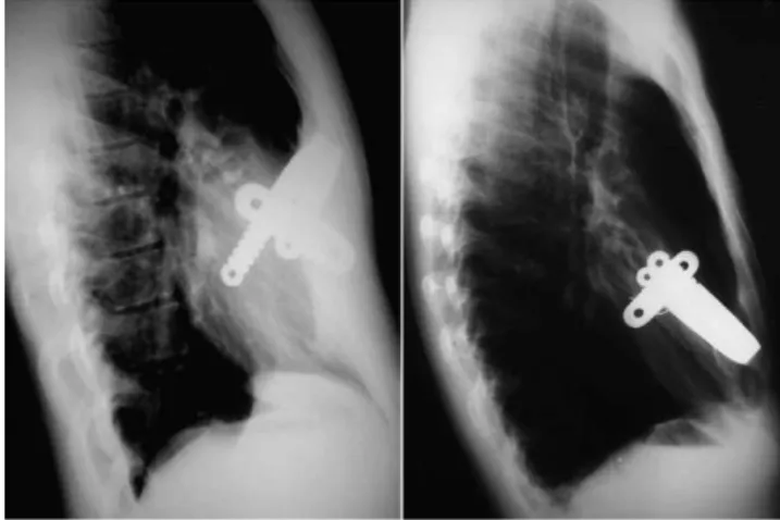
Bar displacement is a serious complication that can occur anytime, but occurs most frequently in the 30 days following the minimally invasive repair of PE.4 Following the initial acceptance and widespread use of the Nuss procedure, the rate of bar displacement was high. However, since the initial development of the technique, several modifications have been proposed in an effort to make the technique both safer and more effective. These modifications include the intro-duction and development of new positioning, fixation techniques, and stabilizers to replace pericostal sutures or other methods used to prevent bar displacement (Figure 1).5 The objective of this paper is to present our experience and compare bar displacement rates before and after the introduction of new methods, including using a shorter bar, a new model and a more medial positioning of the stabilizers throughout the execution of the Nuss procedure.
Copyrightß2011CLINICS– This is an Open Access article distributed under
the terms of the Creative Commons Attribution Non-Commercial License (http:// creativecommons.org/licenses/by-nc/3.0/) which permits unrestricted non-commercial use, distribution, and reproduction in any medium, provided the original work is properly cited.
No potential conflict of interest was reported.
CLINICS 2011;66(10):1743-1746 DOI:10.1590/S1807-59322011001000012
PATIENTS AND METHODS
After Ethics Committee approval, we retrospectively reviewed the patient data of 122 patients (109 males and 13 females) with a mean age of 17¡3 (range 5 to 37) years that had undergone pectus excavatum repair utilizing the Nuss procedure between May 2003 and June 2010. All data related to the hospital stay and follow-up results from 1 to 83 months post-operation were reviewed. During the study period, we used the basic Nuss technique with two technical modifica-tions. For this purposes of this study, the patients who were treated with the modified techniques were divided into two additional series, for an overall total of three patient series.
In the first series, which included the first 24 patients, we fixed the bar and stabilizers in a manner similar to the original Nuss procedure.6
In the second series, which included patients 25–71, we added the technical modification proposed by Hebra et al.3 in an effort to reduce the bar displacement rate. This modification, referred to as ‘‘third point fixation,’’ involves placing a suture around the bar and the underlying ribs.
Finally, our third series, which is a group of 51 patients (post-2008), underwent the Nuss procedure with additional technical modifications, including a smaller bar size and the use of a newly designed stabilizer model. This new model, with central grooves on the posterior surface, allows improved sliding of the stabilizer over the bar, regardless of its curvature. This ensures a more medial positioning of the stabilizer (Figure 2).5
Three patients were referred to us 7, 8, and 11 years post unsuccessful treatment with a modified Ravitch technique. Two of these patients were included in the second series, and one patient was included in the third.
Chest X-rays were taken postoperatively in all patients to both document the results and enable comparison of the position of the bar throughout the follow-up period. As no movement of the bar is expected, any position different from the initial location visualized in the X-rays was classified as bar displacement.
Postoperative pain management was achieved using epidural catheters and non-steroidal anti-inflammatory drugs. The follow-up protocol included outpatient visits at three weeks, three months, six months and annually for three years. Light physical activity was allowed three weeks after surgery, all kinds of sports except contact sports were allowed after three months, and all activity was allowed
after six months. The bar was removed after the three-year follow-up period.
Complications were classified as ‘‘major’’ if an organ injury occurred or if a secondary intervention became necessary or as ‘‘minor’’ if there was any need for clinical treatment and/or if evacuation of fluid or air from the thorax by drainage became necessary.
The data is presented as the frequency and percentage. To compare the incidence of complications in each of the series we used the Likelihood Ratio test. Ap-value less than 0.05 was considered significant. All statistical analyses were performed using SPSS Version 13 software (SPSS, Chicago, IL, USA).
RESULTS
There were no deaths in any of the series. The observed complications are listed in Table 1.
Minor complications occurred in six (4.9%) patients. There was pneumothorax with spontaneous resolution in two (1.6%) patients, with one in the first series and one in the second series. In addition, two (1.6%) cases in the first series suffered suture site infection. There were two (1.6%) cases of bar displacement that did not require surgery to remove the bar.
Major complications were observed in eight (6.5%) patients. Skin erosion/seroma occurred in one (0.8%) patient in the first series. In the second-series patients, we observed pleural effusion requiring drainage in one (0.8%) patient, a reaction to the bar (ABA foreign body reaction) in one (0.8%) patient, pneumonia and septic shock in one (0.8%) patient and cardiac perforation in one (0.8%) patient. Bar displacement that required bar removal occurred in three (2.5%) patients, two who were in the first series and one who was in the second series.
When the incidence of minor complications was com-pared among the three series, the only complication that
Figure 1 -A and B: Examples of bar displacement.
Figure 2 -New positioning of the stabilizer.
Table 1 -Minor and major complications observed in the three series.
Complication
First series (n= 24)
Second series (n= 47)
Third series (n= 51)
Pneumothorax 1 (4.1%) 1 (2.1%) 0 Suture infection 2 (8.3%) 0 0 Pleural effusion 0 1 (2.1%) 0 Reaction to the bar 0 1 (2.1%) 0 Skin erosion/seroma 1 (4.1%) 0 0
Pneumonia 0 1 (2.1%) 0
Cardiac perforation 0 1 (2.1%) 0 Bar displacement 4 (16.6%) 1 (2.1%) 0
Technique inPectus ExcavatumTreatment
Tedde ML et al. CLINICS 2011;66(10):1743-1746
presented a significant difference was suture infection (p= 0.036).
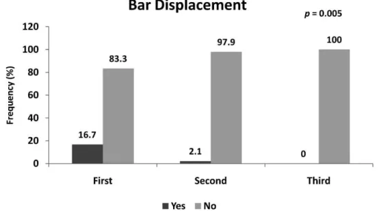
The incidence of bar displacement was the only major complication that was significantly different among the three series (Figure 3).
We were able to remove the bar after the three-year follow-up period in 37 (30.3%) of the 122 patients. In two (1.6%) patients the bar was removed within 2.5 years of placement. In one case, the removal was performed because of a persistent wound infection following a thoracic trauma with skin erosion. The other early removal was performed at the patient’s request, as it was his desire to join the Navy. In all of these patients, the contour of the chest wall obtained after surgical correction was maintained.
DISCUSSION
The rate of bar displacement following the original Nuss procedure was 15%; however, the introduction of stabilizers
reduced this rate to 5%. According to some authors, the rate can be lowered even further with the addition of pericostal sutures placed around the bar.1,7,8
Hebra et al.3were the first to advocate the placement of a suture around the bar and the underlying ribs, which they called ‘‘third point fixation.’’ In our second series of patients (patients 25–71), we used this technique. The main complaints reported by our patients in the first post-operative month were thoracic pain and discomfort close to the fixation points, perhaps due to compression of the intercostal bundle. With this technical modification to our procedure, the incidence of bar displacement dropped from four (16.6%) in the first series of patients to one (2.1%) in the second series.
Park et al.7described a variety of mechanisms governing bar displacement that we also observed in our cases, such as flipping, lateral sliding, and a backward shift. Flipping is most common at the hinge-point, with the bar sliding laterally towards the more depressed side and/or shifting
Figure 3 -The incidence of bar displacement in the three series.
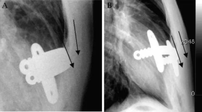
Figure 4 -A and B: Examples of the new technique.
CLINICS 2011;66(10):1743-1746 Technique inPectus ExcavatumTreatment
Tedde ML et al.
backward, and is often accompanied by a breakdown in the intercostal muscles. While they advocated a fifth wire stitched on the right side at the hinge-point or several pericostal wires to attain the desired firmness,8the rupture of the wire sutures used to secure the stabilizer and/or bar on the underlying rib is a common complication reported in 27.8% of the cases.1 Furthermore, the broken wire can hamper removal of the bar by requiring the surgeon to find and extract minute wire pieces. As a result, residual wire fragments remain embedded under the ribs of some patients. Therefore, dispensing with wire stitching is a welcome development in the Nuss technique.
Recently, the use of two bars has been reported4to increase stability and enhance the aesthetic results. Vergunta et al.2 advocates routinely placing two bars, with a stabilizing plate for each, on opposite sides of the chest. They justify this by noting that ‘‘a single bar can be inherently unstable because the deepest point of the sternum is balanced on only the center of one bar. Two bars placed above and below the midpoint of the deformity provides for increased stability’’. However, there are no data comparing the morbidity associated with one or two or more bars.
Instead of using two bars to avoid displacement, we chose to use one stabilizer at each side of the bar, distributing the forces to at least two ribs. The two stabilizers provide a stable basis for the correction we observed in all patients in our third series (Figure 4).
In our third series of patients, we followed the recom-mendations of Pilegaard et al.,9who, in 2008, concluded that their procedure may have reduced the incidence of bar displacement in 383 patients. These authors modified the original Nuss technique by using a shorter pectus bar and placing the stabilizer on the left side of the bar as close as possible to the entry of the thoracic cavity.
Several factors work against the lateral position of the stabilizer. According to Watanabe et al.,10due to its size, the use of a stabilizer in a lateral position increases the incidence of wound complications, such as seroma and dermatitis due
to pressure damage. In one patient in our first series, the stabilizer had to be removed to control seroma, dermatitis, and skin erosion due to pressure damage. When the stabilizer was placed more medially, it was at least partially covered with thepectoralis majormuscle.
To summarize, we would like to emphasize some con-cepts already published and that we have been following:10
1) small bars are probably more stable than large bars; 2) the placement of the bar should be at the deepest point
of the excavatum deformity (Figure 5);
3) older, asymmetric, or severe deformities may require placement of an additional bar or may require another method of correction;
4) bar should be secured with two stabilizers positioned medially, as close as possible to the hinge-point.
In conclusion, the use of these measures can prevent bar instability as demonstrated in our comparison of the incidence of bar displacement in our three series of patients (p,0.05).
ACKNOWLEDGMENTS
The authors would like to thank Aristides Tadeu Correa for technical assistance with the statistical analyses.
REFERENCES
1. Castellani C, Schalamon J, Saxena AK, Hoellwarth ME. Early complica-tions of the Nuss procedure for pectus excavatum: a prospective study. Pediatr Surg Int. 2008;24:659-66, doi: 10.1007/s00383-008-2106-z. 2. Vegunta RK, Pacheco PE, Wallace LJ, Pearl RH. Complications
associated with the Nuss procedure: continued evolution of the learning curve. Am J Surg. 2008;195:313-7, doi: 10.1016/j.amjsurg.2007.12.015. 3. Hebra A, Gauderer MW, Tagge EP, Adamson WT, Othersen HB Jr. A
simple technique for preventing bar displacement with the Nuss repair of pectus excavatum. J Pediatr Surg. 2001;36:1266-8, doi: 10.1053/jpsu. 2001.25791.
4. Nuss D. Minimally invasive surgical repair of pectus excavatum. Semin Pediatr Surg. 2008;17:209-17, doi: 10.1053/j.sempedsurg.2008.03.003. 5. de Campos JR, Das-Neves-Pereira JC, Lopes KM, Jatene FB. Technical
modifications in stabilizers and in bar removal in the Nuss procedure. Eur J Cardiothorac Surg. 2009;36:410-2, doi: 10.1016/j.ejcts.2009.03.061 6. de Campos JR, Fonseca MH, Werebe Ede C, Velhote MC Jatene FB.
Technical modifications of the Nuss operation for the correction of pectus excavatum. Clinics. 2006;61:185-6, doi: 10.1590/S1807-59322006000200018.
7. Park HJ, Lee SY, Lee CS, Youm W, Lee KR. The Nuss procedure for pectus excavatum: evolution of techniques and early results on 322 patients. Ann Thorac Surg. 2004;77:289-95, doi: 10.1016/S0003-4975(03)01330-4.
8. Uemura S, Choda Y. Nuss procedure for pectus excavatum and the operative results. Jpn J Pediatr Surg. 2003;35:665-71.
9. Pilegaard HK, Licht PB. Early results following the Nuss operation for pectus excavatum- a single-institution experience of 383 patients. Interact Cardiovasc Thorac Surg. 2008;7:54–7, doi: 10.1510/icvts.2007.160937. 10. Watanabe A, Watanabe T, Obama T, Ohsawa H, Mawatari T, Ichimiya Y,
et al. The use of a lateral stabilizer increases the incidence of wound trouble following the Nuss procedure. Ann Thorac Surg. 2004;77:296-300, doi: 10.1016/S0003-4975(03)01335-3.
11. Park HJ, Lee IS, Kim KT. Extreme eccentric canal type pectus excavatum: morphological study and repair techniques. Eur J Cardiothorac Surg. 2008;34:150-4, doi: 10.1016/j.ejcts.2008.03.044.
Figure 5 - A) One stabilizer, with two different, non-parallel planes between the bar and internal surface of the sternum. B) Two stabilizers, with two different, parallel planes.
Technique inPectus ExcavatumTreatment
Tedde ML et al. CLINICS 2011;66(10):1743-1746