Fisioter. Mov., Curitiba, v. 30, n. 3, p. 433-441, Jul./Sep. 2017 Licenciado sob uma Licença Creative Commons DOI: http://dx.doi.org/10.1590/1980-5918.030.003.AO01
Analysis of pain, functional capacity, muscular strength and
balance in young women with Patellofemoral Pain Syndrome
Análise da dor, funcionalidade, comportamento muscular e equilíbrio
em mulheres jovens com Síndrome da Dor Femoropatelar
Camile Ludovico Zamboti[a], Rubens Alexandre da Silva Junior[b], Cynthia Gobbi[a], Leonardo Shigaki[a], Christiane de Souza Guerino Macedo[a]*
[a] Universidade Estadual de Londrina (UEL), Londrina, PR, Brazil [b] Universidade Norte do Paraná (UNOPAR), Londrina, PR, Brazil
[R]
Abstract
Introduction: Patellofemoral Pain Syndrome (PFPS) is associated with anterior knee pain, changes in func-tional capacity, balance and muscle strength disorders. Objective: To quantify pain, functional capacity, strength in quadriceps (Q), gluteus medium (GM), hip external rotator (ER) muscles and balance in sedentary women with PFPS. Methods: Twenty sedentary women, aged 18 to 25 years, were divided into two groups: PFPS (N=10) and control group (N=10). All the volunteers answered the items of the Visual Analogue Scale (VAS), the Lysholm Knee Score Scale, the Anterior Knee Pain Scale (AKPS), and the Lower Extremity Functional Scale (LEFS). The participants performed the following tests: maximal voluntary isometric contraction mea-sured by dynamometry; postural balance using the Star Excursion Balance Test (SEBT) and a force platform. Statistical analyses were performed using the Shapiro Wilk test, the Mann Whitney U test and Spearman’s
correlation coefficient. Data were submitted to SPSS 20 software. Results: The PFPS group presented greater pain, balance impairment and higher average velocity of oscillation; however, no differences were observed in Q, GM and RE muscle strength and in balance analyzed by SEBT. Conclusion: Women with PFPS exhibited
*CLZ: undergrad, e-mail:[email protected]
RASJ: PhD, e-mail: [email protected]
CG: MS, e-mail: [email protected] LS: MS, e-mail: [email protected]
434
greater pain, worse functional capacity and body balance. Moderate correlation between both balance tests suggests the use of SEBT when the force platform is not available, which could facilitate and highlight the im-portance of clinical diagnosis with regard to postural balance.
Keywords: Patellofemoral Pain Syndrome. Knee. Physical Therapy Specialty.
Resumo
Introdução: A Síndrome da Dor Femoropatelar (SDFP) está relacionada à dor anterior do joelho, alteração de funcionalidade, déficits de equilíbrio e força muscular. Objetivo: Quantificar a dor, funcionalidade, força mus-cular do quadríceps (Q), glúteo médio (GM), rotadores externos de quadril (RE) e o equilíbrio em mulheres com SDFP. Métodos: Avaliou-se 20 voluntárias, sedentárias, entre 18 e 25 anos; divididas em grupo SDFP (N=10) e controle (N=10). Todas responderam a Escala Visual Analógica de Dor, Lysholm Knee Score Scale, Anterior Knee Pain Scale e Lower Extremity Functional Scale, realizaram a contração isométrica voluntária máxima dos músculos Q, GM e RE por meio de dinamometria e análise do equilíbrio postural pelo Star Excursion Balance Test (SEBT) e plataforma de força. Para análise estatística utilizou-se os testes Shapiro Wilk, Mann Whitney e Correlação de Spearmann, pelo SPSS® 20. Resultados: O grupo SDFP apresentou maior dor, incapacidade e velocidade média de oscilação do centro de pressão, entretanto não foram observadas diferenças para a força muscular de Q, GM e RE e equilíbrio analisado pelo SEBT. Estabeleceu-se correlação moderada entre SEBT e Centro de Oscilação de Pressão (COP) unipodal. Conclusão: Mulheres com SDFP apresentam maior dor, pior funcionalidade e alterações no equilíbrio corporal. A correlação moderada entre SEBT e o COP-unipodal aponta a possibilidade de uso deste teste funcional quando a plataforma de força não é acessível, o que facilita e destaca a importância do diagnóstico clínico do equilíbrio postural.
Palavras-chave: Síndrome da Dor Patelofemoral. Joelho. Fisioterapia.
Introduction
Patellofemoral pain syndrome (PFPS) is described as retropatellar or anterior knee aching pain and characterized by crepitation in the patellofemoral joint during and after activities, such as squatting, walking up or down stairs and sitting for prolonged periods (1). Sedentary women are more likely to de-velop PFPS (2). It affects 1 in 4 individuals, with 20 to 40% of the population with ages between 15 and 35 years (3). Some anatomical variations, namely pelvic width, femoral antevesion, quadriceps angle, tibial torsion, quadriceps strength and knee joint lax-ity, are seen more commonly in women (4). Factors believed to contribute to production of patellar pain include poor alignment between the joint surfaces, muscle shortening, weak vastus medialis oblique (VMO), different recruitment patterns between VMO and vastus lateralis (VL), weak quadriceps muscles, vigorous training and incongruity between patella and femoral trochlear groove (5 - 8).
The quadriceps muscle is responsible for activat-ing the forces exerted on the patella, whose function is to control the position of the patella in relation to the trochlea (9). Improper positioning of the patella, differences in quadriceps muscle recruitment and pain can affect balance and motor control in PFPS (10). The VMO opposes the lateral vector force of the VL. The imbalance between these muscles may contribute to the emergence of symptoms as it chang-es the biomechanics of the knee and increaschang-es the patellofemoral compression forces. Another factor related to PFPS is the weakness of the GM and the ER muscles (11, 12). Fukuda et al. (13) determined that the strengthening of these muscles may reduce pain and improve functional capacity.
435 contracture of the iliotibial band, retraction iliotibial tract, excessive subtalar pronation, patellar hypo — or hypermobility, patellar malalignment and lateral tibial torsion; report of three or more functional conditions including squatting, going up and down the stairs, kneeling, running, sitting for a long period of time; pain intensity of 3 or more in the VAS. The volunteers in the control group should not present any pain intensity in the knee joint or anywhere else in the last six months, but the presence of up to two signs of postural changes was accepted. The exclusion criteria for both groups were: recent history of knee surgery; cardiovascular, neurological or rheumato-logical diseases; diabetes or alteration of plantar sen-sitivity; use of medication; previous physical therapy; and inability to perform the functional tests proposed in this study or to attend the scheduled assessments.
- Procedures
The flowchart of the experimental procedures is presented in Figure 1. On the first day of data collec
-tion, all the participants filled out a form with name,
age, height and weight and marked the items on the VAS-pain (17, 18), the Anterior Knee Pain Scale — AKPS) (19), Lysholm Knee Score Scale (20) and the Lower Extremity Functional Scale (LEFS) (21, 22). The volunteers were informed about the objectives of the study and agreed to participate by providing their free and informed consent.
Prior to performing all measurements, the sub-jects were familiarized with the maximum voluntary isometric contraction (CIVM), followed by the mea-surement of Q, GM and ER muscle strength using a dynamometer (EMG System do Brasil® Ltda, São José dos Campos – SP, Brazil).
Considering the high prevalence of women with PFPS that seek for physical therapy, the biomechan-ical changes previously reported in the literature, the disagreement between results established by research on PFPS, and the possibility of using SEBT as a functional test for the analysis of balance in this population, it is believed that studies are still needed to establish and determine the biomechanical stan-dards for assessment and rehabilitation.
Therefore, the main purpose of this study was to quantify pain, functional capacity, and quadriceps (Q), gluteus medium (GM), hip external rotator (ER) muscle strength and balance in sedentary women with PFPS.
Methods
- Ethical aspects
This was a cross-sectional study approved by the Research Ethics Committee of the institution (Case N. 251637/2013).
- Sample
The sample size calculation was performed us-ing the Power and Sample Size software (Vanderbilt School of Medicine Biostatistics, Nashville, TN), with
95% confidence interval, 5% level of significance and
80% power, considering the mean and standard de-viation of the parameter data obtained before SEBT (16). The volunteers were 20 sedentary, university female students divided into two groups: the PFPS group (n = 10) and the control group (n = 10).
The inclusion criteria for the PFPS group, accord-ing to Santos et al. (5), were: presence of at least three clinical signs, such as patellar crepitus, high patella,
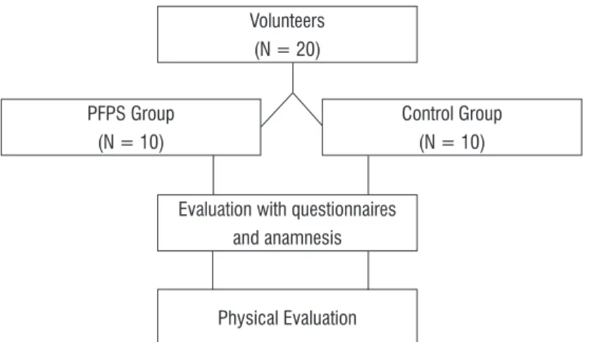
Figure 1 - Flow chart for sample selection and data collection.
Volunteers (N = 20)
PFPS Group (N = 10)
Control Group (N = 10)
Evaluation with questionnaires and anamnesis
436
distance, LL the length of the lower limb and the re-sult is the percentage of the three directions.
(Anterior + Posteromedial + Posterolateral)
3 X Limb Length X 100
Finally, after a week of the initial assessment, bal-ance was tested on a force platform (BIOMEC400-EMG System do Brasil® Ltda, São José dos Campos – SP, Brazil). The participants stood with eyes open, under unipodal condition performed with the limb with pain (in PFPS group) and leg preferred for 30s. There was a rest period of approximately 30s between each trial. The procedure was repeated three times and the mean was retained for analysis (26). The parameters associated with movements of the center of pressure (COP) were: the ellipse area (95%) of the COP (A-COP in cm2) and mean velocity (VEL in cm/s) of the COP sway in the mediolateral (M/L) direction.
- Statistical Analysis
Normal data distribution was verified using the
Shapiro-Wilk test. Student's t-test was used for between-group comparisons for each variable. The
Spearman’s correlation coefficient was used to exam
-ine the relationship between the balance data. The statistical analysis was performed using SPSS version 20 for Windows (SPSS Inc.; Chicago, IL, USA), with a
5% level of significance (p < 0 .05).
Results
The groups were homogeneous regarding height, weight and body mass index (BMI). Both groups pre-sented mean values and standard deviation, respec-tively: age 21.1m (1.10) and 22.0 (1.59) (p = 0.019); height 1.65 (0.06) and 1.64 (0.03) (p = 0.379); weight 60.2 (8.07) and 63.0 (7.40) (p = 0.739); and BMI 21.8 (1.79) and 23.25 (3.20) (p = 0.721). Mean and stan-dard deviation of pain in the PFPS group were 3.75 years and 1.24 years, respectively. In the CG, it was zero. The results obtained through the questionnaires and functional scales showed that the PFPS group
had increased pain and disability, with significant
differences compared to the control group (Table 1).
No significant difference was observed between
the groups regarding the quadriceps, hip external rotator and gluteus medium muscle strength and balance analyzed using SEBT (Table 2). The analysis For the measurement of quadriceps strength, the
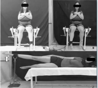
volunteer sat on a chair with the back supported and
fixed, knee in open kinetic chain in 60° flexion for
greater muscle activation of VL and VMO muscles (23). An ankle strap connected to a force transducer was fastened to the volunteer, who was verbally en-couraged to strengthen while extending the knee and maintain it during 10 seconds of MVIC (Figure 2A).
For the MVIC of the hip lateral rotators, the chair was positioned laterally to the force transducer (Figure 2B). For the GM muscle, the volunteer re-mained in decubitus contralaterally to the member
to be tested, while performing MVIC abduction at 30°
(Figure 2C). All tests were carried out following a sequence protocol for data collection: quadriceps, ex-ternal rotators and gluteus medium. The mean of the three values found was considered for the analysis.
Figure 2A - Analysis of quadriceps muscle strength. B -
Analysis of hip external rotator muscle strength. C - Analysis of gluteus medium muscle strength.
On the first collection day, the SEBT was used to
437 in the CG (Table 2). The correlation was considered moderate for COP-unipodal and SEBT (p = 0.046, r = 0.451) (Table 3).
of postural balance measures obtained using a force
platform showed significant differences between the
groups for the mean velocity of the COP sway in the mediolateral direction, with better balance control
Table 1 - Results of questionnaires and functional scales for the PFPS and Control groups using the Mann Whitney U test
Instruments
PFPS N=10 Median (min/max)
CONTROL N=10
Median (min/max) p
VAS 4 (3-7) 0 0.014
AKPS 80 (48-98) 100 (94 - 100) 0.000
Lysholm 89 (47 - 100) 100 (95 - 100) 0.000
LEFS 75.5 (53 - 98) 79.5 (78 - 80) 0.004
Note: PFPS- Patellofemoral Pain Syndrome; VAS- Visual Analogue Scale; AKPS- Anterior Knee Pain Syndrome; LEFS- Lower Extremity Func-tional Scale.
Table 2 - Analysis of muscle strength and balance between women with PFPS and control group using the Mann Whitney U test
Muscles
PFPS N = 10 Median (min/max)
CONTROL N = 10 Median (min/max)
P
Gluteus medium (kgf) 4.74 (3 - 7) 5.48 (2 - 7) 0.241
Quadriceps (kgf) 24.63 (14 - 34) 24.34 (20 - 38) 0.427
External rotators (kgf) 8.7 (5 - 11) 8.65 (6 - 10) 0.449
SEBT-Anterior 82.5 (71 - 86) 81.25 (70 - 87) 1.0
SEBT-Posteromedial 71 (48 - 79) 72.5 (62 - 83) 0.353
SEBT- Posterolateral 77.7 (56 - 87) 81.5 (68 - 87) 0.218
SEBT- General 89.53 (68 - 95) 92.74 (80 - 98) 0.393
COP Unipodal (cm2) 8 (2 - 13) 6.17(4 - 8) 0.143
MV mediolateral Unipodal (cm/s) 2.85 (1 - 4) 0.64 (1 - 3) 0.000 Note: PFPS- Patellofemoral Pain Syndrome; SEBT-Star Excursion Balance Test; kgf – Kilogram-force; COP- Center of Oscillation of Pressure; MV- Mean Velocity.
Table 3 - Spearman’s correlation coefficients between SEBT, mean unipodal velocity and COP unipodal for the PFPS group
Instruments SEBT MV UNIPODAL COP UNIPODAL
SEBT 1.000 (p = 0.000) 0.383 (p = 0.093) 0.451 (p = 0.046)
MV UNIPODAL 0.383 (p = 0.093) 1.000 (p = 0.000) 0.614 (p =0.004)
COP UNIPODAL 0.451 (p = 0.046) 0.614 (p = 0.004) 1.000 (p = 0.000)
438
Discussion
The present study showed that sedentary women with patellofemoral pain syndrome exhibited greater pain, functional impairment and velocity of the COP sway in the mediolateral direction. Patellofemoral
pain is multifactorial and is therefore difficult to ex -amine and treat.
This study proved that pain and balance impair-ment are important aspects to be considered in the rehabilitation of this dysfunction. Pain is the main symptom of PFPS and can be subjectively evaluated using VAS which also appears to be a valid and reli-able instrument (17). The results of the present study
showed that women with PFPS revealed a signifi
-cantly greater improvement in the intensity of pain. This result agrees with Souza et al. (27) that have also observed an increase in the intensity of pain in individuals with PFPS compared to controls. Piazza et al. (4) added that there was exacerbation of pain during activities, such as squatting, going up or down
stairs or ramp, and walking on flat surface.
With pain increase in daily life activities, the func-tional capacity should be considered. The use of ques-tionnaires and the AKPS, Lysholm and LEFS scales
has established significantly lower values for the
PFPS group. The AKPS was validated and translated into Brazilian Portuguese for measuring PFPS (19). It can also predict pain and functional limitations of the individuals. Bolgla et al. (12) and Fukuda et al. (28) showed that subjects with PFPS yielded a score between 64.5 and 80 in AKPS, which corroborate the score of 80 points obtained in the current study for this group. The LEFS can be used to evaluate the func-tional impairment of a patient with a disorder of one or both lower extremities. The results showed lower values, indicating worse functional capacity in the lower limb of women with PFPS.
With regard to functional capacity, the results ob-tained using the Lysholm Knee Scale questionnaire also showed lower values for the PFPS group, as well
as the results obtained by Dickschas et al. (29), who
found similar scores to those found for individuals with chronic patellar instability.
The study has also assessed the strength of the hip abductor and external rotator muscles in women
with PFPS. This assessment can be justified by the
association of the weakness of these muscles with that of the knee valgus, as well as the association be-tween the dynamic valgus and the unipodal support
during functional activities and sports. It is believed that the knee valgus position gives support to the patellar lateral region with the external condyle of femur leading to chondral lesion and causing pain (28). Bolgla et al. (12) hypothesized that women with
PFPS would demonstrate significantly less hip abduc
-tor and hip external rota-tor strength when compared to controls. A similar result was found by Meira et al. (30) with regard to hip strength muscles.
Other authors claimed that there is less hip strength as well as greater impairment in
quadri-ceps muscle strength (31). However, no significant
differences were found in quadriceps muscle strength in the study carried out by Souza et al. (26) and in our study. Barton et al. (8) reported that the
recruit-ment of the GM muscle occurs significantly late dur
-ing functional activities, therefore, the dysfunction could appear in muscle activation and not in muscle
strength. These results may have been influenced
by the sample that included young volunteers with normal daily activities, with complaints of pain, and also by a sample that obtained a maximum score of 80 (AKPS), as established by Fukuda et al. (28).
The present study investigated static postural control using the SEBT (16). It is a lower extremity functional test that can be easily applied in the clini-cal and research settings by physiclini-cal therapists when a force platform, the gold standard for the analysis of body balance, is not available. The SEBT has been shown to be a reliable measure and has validity as a dynamic test to predict risk of lower extremity injury,
to identify dynamic balance deficits in patients with
a variety of lower extremity conditions (32). Chen at al. (16) observed that individuals with chronic
ankle instability had significantly smaller reaching
distance in the posterior-lateral and posterior-medial
during SEBT. This finding differed from that of our study, which did not show any significant differences,
even in the normalization of all directions. The results obtained have established a moderate correlation between the SEBT and COP-unipodal provided by the force platform; this means that the SEBT can be considered useful for the analysis of balance, mainly in clinical practice.
While using the force platform, Citaker et al. (15) observed that the static unipodal standing balance in
the PFPS group was significantly worse, similar to our
439 the difference for the ellipse area of the COP was not
significant.
Our findings are in agreement with data reported
by Lee et al. (33), who compared postural stability between women with PFP and pain-free controls in unipodal standing on a force platform and showed worse performance and higher mean velocity of oscil-lation in the PFPS group. The literature demonstrates that in great lower limb impairment or imbalance
caused by neuromuscular deficits, the main muscle
groups used in mediallateral performance are the hip adductors and abductors returning to symmetrical stance in centre of gravity (34). In addition, Gribble and Hertel (35) found worse performance in the PFPS
group, with significant difference regarding the el
-lipse area of the COP after testing sessions of iso-kinetic fatigue of the hip abductors. In contrast, our
study did not observe significant differences in COP
and in GM muscle strength.
The present study focused on the results for the characterization of functional capacity and pain in individuals with PFPS, as well as of balance impair-ment compared to the control group, which should be considered in further investigations. The use of the SEBT in our study is worth mentioning, consid-ering that it is a functional and inexpensive postural control measurement tool that does not require high technology, which determines the usefulness
of SEBT in the management of PFPS. As a significant
contribution to clinical practice, it is suggested that evaluation and rehabilitation techniques that create balanced lower body strength in women with PFPS should be conducted. In addition, clinical tests for PFPS assessment such as AKPS, Lysholm, LEFS and SEBT should be used to reestablish the functional capacity of these patients.
However, it must be taken into consideration that the results from the present study are not consistent with those reported in recent literature probably be-cause of the small sample size and the high degree of functional capacity observed in the PFPS group, which comprised young women with a mean score of 4 on the VAS for pain.
Conclusion
The results obtained showed no significant
differences in the SEBT and in the ellipse area of the COP for the gluteus medium, quadriceps and
hip external rotator muscle strength and balance. However, women with PFPS exhibited greater pain, worse functional capacity and body balance provided by the AKPS, Lysholm and LEFS, also greater balance impairment, higher mean velocity of oscillation in single-leg stance. Moreover, the SEBT proved to be a useful clinical parameter as it established a moderate correlation with the COP-unipodal to evaluate the dy-namic balance of women with PFPS. However, further investigations should be carried out with inclusion criteria to establish higher scores for pain and lower for functional capacity.
References
1. Bevilaqua-Grossi D, Felício LR, Silvério GWP. Início da atividade elétrica dos músculos estabilizadores
da patela em indivíduos com SDPF. Acta Ortop Bras.
2009;17(5):297-9.
2. Bevilaqua-Grossi D, Felício LR, Simões R, Coqueiro KRR, Monteiro-Pedro V. Avaliação
eletromiográ-fica dos músculos da patela durante exercício iso
-métrico de agachamento em indivíduos com sín -drome da dor femoropatelar. Rev Bras Med Esporte. 2005;11(3):159-63.
3. Levinger P, Gilleard W. The heel strike transient dur-ing walkdur-ing in subjects with patellofemoral pain syn-drome. Phys Ther Sport. 2005;6(2):83-8.
4. Piazza L, Lisboa ACA, Costa V, Brinhosa GCS, Vidmar
MF, Oliveira LFB, et al. Sintomas e limitações funcio
-nais de pacientes com síndrome da dor patelofemoral. Rev Dor. 2012;13(1):50-4.
5. Santos GM, Say KG, Pulzato F, Oliveira AS,
Bevillaqua-Grossi D, Pedro VM. Relação eletromiográfica inte
-grada dos músculos vasto medial oblíquo e vasto lateral longo na marcha em sujeitos com e sem sín -drome de dor femoropatelar. Rev Bras Med Esporte. 2007;13(1):17-21.
6. Ribeiro ACS, Bevilaqua-Grossi D, Foerster B, Can
-dolo C, Pedro VM. Avaliação eletromiográfica e res
-sonância magnética do joelho de indivíduos com síndrome da dor femoropatelar. Rev Bras Fisioter.
440
7. Garcia FR, Azevedo FA, Alves N, Carvalho AC, Pado-vani CR, Negrão Filho RF. Efeitos da eletroestimulação
do músculo vasto medial oblíquo em portadores de síndrome da dor patelofemoral: uma análise eletro
-miográfica. Rev Bras Fisioter. 2010;14(6):477-82.
8. Barton CA, Lack S, Maliaras P, Morrisey D. Gluteal mus -cle activity and patellofemoral pain syndrome: a sys-tematic review. Br J Sports Med. 2013;47(4):207-14.
9. Nobre TL. Comparação dos exercícios em cadeia cinética aberta e cadeia cinética fechada na reabili-tação da disfunção femoropatelar. Fisioter Mov. 2011;24(1):167-72.
10. Saad MC, Felício LR, Masullo CL, Liporaci RF, Bev
-ilaqua-Grossi D. Analysis of the center of pressure
displacement, ground reaction force and muscular activity during step exercises. J Electromyogr Kinesiol. 2011;21(5):712-8.
11. Souza RB, Powers CM. Differences in hip kinematics, muscle strength, and muscle activation between sub-jects with and without patellofemoral pain. J Orthop Sports Phys Ther. 2009;39(1):12-9.
12. Bolgla LA, Malone TR, Umberger BR, Uhl TL. Hip strength and hip and knee kinematics during stair descent in females with and without patellofemo-ral pain syndrome. J Orthop Sports Phys Ther. 2008;38(1):12-8.
13. Fukuda TY, Rosseto FM, Silva RM, Apolinário A. For-talecimento dos músculos do quadril no tratamento da dor anterior do joelho. Ciência&Saúde. 2009; especial:96.
14. Duarte M, Freitas SMSF. Revisão sobre posturografia baseada em plataforma de força para avaliação do
equilíbrio. Rev Bras Fisioter. 2010;14(3):183-92.
15. Citaker S, Kaya D, Yuksel I, Yosmaoglu B, Nyland J, Atay OA, et al. Static balance in patients with patellofemoral pain syndrome. Sports Health. 2011;3(6):524-7.
16. Chen CY, Hsu AT, Guo LY, Lin CF, Chen YA. Postural control and lower extremity contribution during star excursion balance test in athletes with chronic ankle instability. Formosan Journal of Physical Therapy. 2011;36(4):263-73.
17. Thommé R, Augustsson J, Karlsson J. Patellofemoral Pain Syndrome: A review of current issues. Sports Med. 1999;28(4):245-62.
18. Price DD, McGrath PA, Rafii A. The validation of visual analogue scales as ratio scale measures for chronic and experimental pain. Pain. 1983;17(1):45-56.
19. Aquino VS, Falcon SFM, Neves LMT, Rodrigues CR,
Sendin FA. Tradução e adaptação cultural para a lín -gua portuguesa do questionário scoring of patello-femoral disorders: estudo preliminar. Acta Ortop Bras. 2011;19(5):273-9.
20. Peccin MS, Ciconelli R, Cohen, M. Questionário
espe-cífico para sintomas do joelho “Lysholm knee scoring scale” – tradução e validação para a língua portuguesa.
Acta Ortop Bras. 2006;14(5):268-72.
21. Binkley JM, Stratford PW, Riddle DL. The lower ex -tremity functional scale (LEFS): scale development, measurement properties, and clinical application. North American Orthopaedic Rehabilitation Research Network. Phys Ther. 1999;79(4):371-83.
22. Pereira LM, Dias M, Mazuquin BF, Castanhas LG, Menacho MO, Cardoso JR. Translation, cross-cultural adaptation and analysis of the psycometric proper-ties of the lower extremity functional scale (LEFS): LEFS- BRAZIL. Bras J Phys Ther. 2013;17(3):272-80.
23. Javadpour SM, Finegan PL, O’ Brien M. The anatomy of the extensor mechanism and its clinical relevance. Clin J Sport Med. 1991;1(4):229-35.
24. Filipa A, Byrnes R, Paterno MV, Myer GD, Hewett TE. Neuromuscular training improves performance on the Star Excursion Balance Test in young female ath-letes. J Orthop Sports Phys Ther. 2010;40(9):551-8.
25. Hertel J, Braham RA, Hale SA, Olmsted-Kramer LC. Simplifying the Star Excursion Balance Test: Analyses of Subjects with and without Chronic Ankle Instabil-ity. J Orthop Sports Phys Ther. 2006;36(3):131-7.
26. Rabello LM, Macedo CSG, Oliveira MR, Fregueto JH,
Ca-margo MZ, Lopes LD, et al. Relação entre testes funcio
-nais e plataforma de força nas medidas de equilíbrio
em atletas. Rev Bras Med Esporte. 2014;20(3):219-22.
27. Souza AC. Síndrome da dor femoropatelar - Análise
eletromiográfica, isocinética, ressonância magnética,
441
28. Fukuda TY, Magalhaes E, Sacramento SR, Forgas A, Cohen M, Abdalla RJ. A comparison of hip strength between sedentary females with or without patel-lofemoral pain syndrome. J Orthop Sports Phys Ther. 2010;40(10):641-7.
29. Dickschas J, Harrer J, Pefefferkorn R, Strecker W. Operative treatment of patellofemoral maltracking with torsional osteotomy. Arch Orthop Trauma Surg. 2012;132(3):289-98.
30. Meira EP, Brumitt J. Influence of the Hip on patients with patellofemoral pain syndrome: a systematic re-view. Sports Health. 2011;3(5):455-65.
31. Waryasz GR, McDermott AY. Patellofemoral pain syn -drome: a systematic review of anatomy and potential
risk factors. Dyn Med. 2008;7:9.
32. Gribble PA, Hertel J. Using the Star Excursion Balance
Test to assess dynamic postural-control deficits and
outcomes in lower extremity injury: a literature and systematic review. J Athl Train. 2012;47(3):339-57.
33. Lee SP, Souza RB, Powers CM. The influence of hip abductor muscle performance on dynamic postural stability in females with patellofemoral pain. Gait Posture. 2012;36(3):425-9.
34. Shigaki L, Rabello LM, Camargo MZ, Santos VBC, Gil
AWO, Oliveira MR, et al. Análise comparativa do equilí
-brio bipodal em atletas de ginástica rítmica. Rev Bras
Med Esporte. 2013;19(2):104-7.
35. Gribble PA, Hertel J. Effect of hip and ankle muscle fatigue on unipedal postural control. J Electromyogr Kinesiol. 2004;14(6)641-6.
Received in 09/11/2015
Recebido em 11/09/2015
Approved in 08/11/2016