Research Article
Protective Effect of
Baccharis trimera
Extract on Acute Hepatic
Injury in a Model of Inflammation Induced by Acetaminophen
Bruno da Cruz Pádua,
1,2Joamyr Victor Rossoni Júnior,
1Cíntia Lopes de Brito Magalhães,
1,3Míriam Martins Chaves,
4Marcelo Eustáquio Silva,
1,5Maria Lucia Pedrosa,
1,3Gustavo Henrique Bianco de Souza,
6Geraldo Célio Brandão,
6Ivanildes Vasconcelos Rodrigues,
7Wanderson Geraldo Lima,
1,3and Daniela Caldeira Costa
1,31Programa de P´os-graduac¸˜ao em Ciˆencias Biol´ogicas do N´ucleo de Pesquisas em Ciˆencias Biol´ogicas (NUPEB),
Universidade Federal de Ouro Preto (UFOP), 35.400-000 Ouro Preto, MG, Brazil
2Centro Federal de Educac¸˜ao Tecnol´ogica de Minas Gerais (CEFET/MG), 35.790-970 Curvelo, MG, Brazil
3Departamento de Ciˆencias Biol´ogicas (DECBI), Instituto de Ciˆencias Exatas e Biol´ogicas, Universidade Federal de Ouro Preto (UFOP),
35.400-000 Ouro Preto, MG, Brazil
4Departamento de Bioqu´ımica e Imunologia, Instituto de Ciˆencias Biol´ogicas, Universidade Federal de Minas Gerais (UFMG),
Cx. Postal 486, 30.161-970 Belo Horizonte, MG, Brazil
5Departamento de Alimentos, Escola de Nutric¸˜ao, Universidade Federal de Ouro Preto (UFOP), 35.400-000 Ouro Preto, MG, Brazil
6Programa de P´os-graduac¸˜ao em Ciˆencias Farmacˆeuticas (CIPHARMA), Escola de Farm´acia,
Universidade Federal de Ouro Preto (UFOP), 35.400-000 Ouro Preto, MG, Brazil
7N´ucleo de Pesquisas em Produtos Naturais e Sint´eticos, Departamento de F´ısica e Qu´ımica,
Faculdade de Ciˆencias Farmacˆeuticas de Ribeirao Preto, Universidade de S˜ao Paulo (USP), 14040-903 S˜ao Paulo, SP, Brazil
Correspondence should be addressed to Bruno da Cruz P´adua; brunobioufop@yahoo.com.br
Received 7 July 2014; Revised 13 September 2014; Accepted 8 October 2014; Published 12 November 2014
Academic Editor: Muzamil Ahmad
Copyright © 2014 Bruno da Cruz P´adua et al. This is an open access article distributed under the Creative Commons Attribution License, which permits unrestricted use, distribution, and reproduction in any medium, provided the original work is properly cited.
Background. Acetaminophen (APAP) is a commonly used analgesic and antipyretic. When administered in high doses, APAP
is a clinical problem in the US and Europe, often resulting in severe liver injury and potentially acute liver failure. Studies have demonstrated that antioxidants and anti-inflammatory agents effectively protect against the acute hepatotoxicity induced by APAP overdose.Methods. The present study attempted to investigate the protective effect ofB. trimeraagainst APAP-induced hepatic damage in rats. The liver-function markers ALT and AST, biomarkers of oxidative stress, antioxidant parameters, and histopathological changes were examined.Results. The pretreatment withB. trimeraattenuated serum activities of ALT and AST that were enhanced by administration of APAP. Furthermore, pretreatment with the extract decreases the activity of the enzyme SOD and increases the activity of catalase and the concentration of total glutathione. Histopathological analysis confirmed the alleviation of liver damage and reduced lesions caused by APAP.Conclusions. The hepatoprotective action ofB. trimeraextract may rely on its effect on reducing the oxidative stress caused by APAP-induced hepatic damage in a rat model.General Significance. These results make the extract ofB. trimeraa potential candidate drug capable of protecting the liver against damage caused by APAP overdose.
1. Introduction
The liver is the major site of detoxification and the primary
target of drug exposure in the body [1]; therefore,
drug-induced liver injury is a significant public health problem,
accounting for over half of all cases of acute liver failure
[2].
Acetaminophen (APAP) is a commonly used analgesic and antipyretic agent. At therapeutic doses, it is usually safe and well tolerated. However, acute acetaminophen overdose Volume 2014, Article ID 196598, 14 pages
causes severe and fatal hepatotoxicity [3]. Some individuals experience APAP toxicity even at therapeutic doses of less than 4 g/day, and, in pediatric populations, the majority of
APAP overdoses are unintentional [4]. Moling et al. [5]
reported a case of severe hepatotoxicity after therapeutic doses of APAP in an adult man. In fact, APAP overdose is the most common cause of drug-induced acute liver failure
in the United Kingdom and the United States [6,7].
The toxicity of APAP is related to its bioactivation by cytochrome P450 to the electrophilic metabolite N-acetyl benzoquinone imine (NAPQI). At therapeutic doses, NAPQI is efficiently detoxified by glutathione (GSH) and eliminated through urine or bile; however, at supratherapeutic doses, both the glucuronidation and sulfation pathways become saturated, and extensive bioactivation of APAP depletes the
hepatic GSH pool and causes oxidative stress [8]. This
oxida-tive stress may trigger signaling pathways to act through
mi-tochondrial toxicity, ultimately causing cell death [1]. A
sig-nificant amount of evidence has pointed to the potential
in-volvement of oxidative stress in acetaminophen toxicity [9,
10].
Several studies have shown that antioxidants and anti-inflammatory agents effectively protect against the acute
hepatotoxicity induced by acetaminophen overdose [11–13].
Several herbal medicines, their active constituents and formulations, are used in the treatment of a wide variety of clinical diseases and provide benefit to societies. Their protec-tive action is through antioxidant enzymes (e.g., SOD, CAT, GST, and GR), which modify many pathways and proteins,
including the DNA damage/repair processes [14], Nrf-2 [15],
and xenobiotic response elements [16], thus maintaining the
prooxidant/antioxidant balance in the body [1].
In this context, we highlightBaccharis trimera, popularly
known as “carqueja,” a member of the Asteraceae family and a native shrub from South Brazil, Paraguay, Uruguay, and Argentina. Medicinal teas prepared from the aerial parts of this shrub are used in folk medicine to treat not only gastro-intestinal and liver diseases but also inflammatory processes
[17]. Because of these biological effects, research on the
chem-ical composition ofB. trimera was conducted and
demon-strated that this plant has many bioactive compounds, such
as flavonoids, diterpenes, and triterpenes [18]. Triterpenes
have been reported to be primarily responsible for its
anti-inflammatory activity [19, 20], while the flavonoids, due to
their antioxidant activity, have been linked to protecting the
body against reactive oxygen species (ROS) [21].
This study was conducted to investigate the effect ofB.
trimerain the modulation of oxidative stress and to evaluate
the preventive effect ofB. trimerain acetaminophen-induced
liver damage.
2. Materials and Methods
2.1. Reagents. The chemical reagents, including DTNB [5,5 -dithio-bis (2-nitrobenzoic acid)], 2,4-dinitrophenylhydra-zine (DNP), and thiobarbituric acid (TBA), were purchased from Sigma-Aldrich (St. Louis, MO, USA). Acetaminophen (APAP) (200 mg/mL) was obtained from Janssen-Cilag
Pharmaceuticals, Brazil. The kit for measuring serum ala-nine aminotransferase (ALT) and aspartate aminotransferase (AST) was from Diagnostic Labtest, Brazil.
2.2. Collection of Plant Material. The aerial parts ofB. trimera
were collected during August 2011 in the city of Ouro Preto, Minas Gerais, Brazil. The specimen, voucher number OUPR 22.127, was identified by Professor Viviane R. Scanlon and deposited in the Herbarium Jos´e Badini, UFOP.
2.3. Preparation of Extract. The aerial parts of the plant were dried in a ventilated oven, sprayed in a mechanical mill, and stored in plastic bottles. To obtain the hydroethanolic extract, approximately 100 g of the plant was extracted with distilled water and 70% alcohol at a ratio of 1 : 1 for 24 h. Vacuum filtration and evaporation of the solvent in a rotovap were then performed. The crude extract that formed, with a percentage yield of 8-9%, was then diluted with phosphate-buffered saline (PBS, pH 7.4); a concentration of 600 mg/kg
body weight was used in vivo. The methodology for the
extract preparation was based on the work of Grance et al.
[22] with some modifications.
2.4. LC-DAD-ESI-MS Analyses. Analyses were performed using an UPLC Acquity (Waters) ion trap mass spectrometer equipped with an atmospheric pressure chemical ioniza-tion (APCI) interface operated in the following condiioniza-tions: positive and negative ion mode; capillary voltage, 3500 V;
capillary temperature, 320∘C; source voltage, 5 kV; vaporizer
temperature, 320∘C; corona needle current, 5 mA; and sheath
gas, nitrogen, 27 psi. Analyses were run in the full scan mode (100–2000 u). The ESI-MS/MS analyses were additionally performed in an UPLC Acquity (Waters) with helium as the collision gas, and the collision energy was set at 30 eV. Chromatographic separation was done on ACQUITY UPLC
BEH (1.7𝜇m, 50×2 mm i.d.) (Waters). The mobile phase
consisted of water 0.1% formic acid (solvent A) and ace-tonitrile 0.1% formic acid (solvent B). The elution protocol was 0–11 min, linear gradient from 5% to 95% B. The flow
rate was 0.3 mL min−1, and the sample injection volume was
4.0𝜇L. The UV spectra were registered from 190 to 450 nm.
Mass spectrometry analysis was performed on quadrupole instrument fitted with an electrospray source in the negative
mode (Figure 1). Ion spray voltage:−4 kV; orifice voltage:−60
V.
2.5. In Vitro Test: Cell Culture. The cell strains were acquired at the Cell Bank, Federal University of Rio de Janeiro (UFRJ).
Liver cells were cultured in 75 cm2growth vials (SARDEST)
containing MEM culture medium. HEPES, 10% (v/v) bovine fetal serum, and 1% (v/v) of a mix of penicillin (200 U/mL)
and streptomycin (200𝜇g/mL) were added to the medium.
The vials were stored in oven at 37∘C and humidified
with 5% carbon dioxide (CO2). This medium was replaced
1 2 3 4 5 6
Compound 5 Compound 6
Compound 4 Compound 3 Compound 2 Compound 1 135.0 120.0 105.0 90.0 75.0 60.0 45.0 30.0 15.0 0.0 −7.5 (A U) (A U) (A U) (A U) (A U) (A U) (A U)
2.0 4.0 6.0 8.0 10.0 12.0
(min)
(min)
7.0e − 1 6.0e − 1 5.0e − 1 4.0e − 1 3.0e − 1 2.0e − 1 1.0e − 1 0.0
220 240 260 280 300 320 340 360 380 400
(min)
220 240 260 280 300 320 340 360 380 400
(min)
220 240 260 280 300 320 340 360 380 400
(min)
220 240 260 280 300 320 340 360 380 400
(min)
220 240 260 280 300 320 340 360 380 400
(min)
220 240 260 280 300 320 340 360 380 400
RT= 2.36
1.1 1.0 9.0e − 1 8.0e − 1 7.0e − 1 6.0e − 1 5.0e − 1 4.0e − 1 3.0e − 1 2.0e − 1 1.0e − 1 0.0
RT= 3.83
6.5e − 1 6.0e − 1 5.5e − 1 5.0e − 1 4.5e − 1 4.0e − 1 3.5e − 1 3.0e − 1 2.5e − 1 2.0e − 1 1.5e − 1 1.0e − 1 5.0e − 2 0.0
RT= 5.14 1.4 1.3 1.2 1.1 1.0 9.0e − 1 8.0e − 1 7.0e − 1 6.0e − 1 5.0e − 1 4.0e − 1 3.0e − 1 2.0e − 1 1.0e − 1 0.0
RT= 5.34
3.0e − 1 2.8e − 1 2.6e − 1 2.4e − 1 2.2e − 1 2.0e − 1 1.8e − 1 1.6e − 1 1.4e − 1 1.2e − 1 1.0e − 1 8.0e − 2 6.0e − 2 4.0e − 2 2.0e − 2 0.0
RT= 6.07 2.8 2.6 2.4 2.2 2.0 1.8 1.6 1.4 1.2 1.0 8.0e − 1 6.0e − 1 4.0e − 1 2.0e − 1 0.0
RT= 7.08 245.13 271.13 234.13 210.13 255.13 371.13 216.13 275.13 300.13 215.13 274.13 341.13 210.13 275.13 340.13 294.19 274.19
Figure 1: RP-UPLC-DAD profiles of hydroethanolic extract ofB. trimera. Conditions: CHS130 100 RP-18 column (1.7𝜇m, 50×3 mm i.d.). Elution was carried out with a linear gradient of water 0.1% formic acid (A) and acetonitrile 0.1% formic acid (B) (from 5% to 95% of B in 11 min) and the UPLC fingerprints were registered on a ACQUITY Waters apparatus with a UV-DAD detector (Waters 2996). Operating parameters of the mass spectrometer were capillary temperature 320∘C; spray needle voltage set at 3.50 kV; ES capillary voltage +3 and−47 V for positive and negative polarity, respectively; and tube lens offset 0 and−25 V for positive and negative polarity, respectively. Nitrogen was used as a sheath gas with a flow of 50 arbitrary units. Mass analysis was carried out in full-scan mode from 100 to 1.500 amu, in both positive and negative mode. UV spectra (190–450 nm) from the main peaks are shown inside on the chromatogram.
with phosphate-buffered saline solution without calcium and without magnesium (PBS). Later, to detach the monolayers, a solution of 0.20% trypsin and 0.02% EDTA was used. Next, the cell count was carried out with Trypan blue 0.3% in a Neubauer chamber.
2.6. Toxicity Assay with MTT. The cell line was acquired from the Cell Bank, Federal University of Rio de Janeiro
(UFRJ). We added 5.0 × 10³ HEPG2 in culture medium
MEM (10% v/v bovine fetal serum and 1% v/v mixture of penicillin/streptomycin). The plates were incubated in a
humidified chamber with 5% CO2 at 37∘C for 1–72 hours.
After this time, the supernatant was removed, and 20𝜇L MTT
was added and incubated for 30 minutes. The absorbance was read at 570 nm in a microplate reader (Thermo Plate). HEPG2
was incubated in the presence of 1, 2, and 3% (50𝜇L) of the
B. trimeraextract for 1 and 24 hours to assess cell viability. The calculation used to assess the percentage of cell viability was
(absorbance of treated cells/absorbance control)×100. The
control was assigned 100% viability.
2.7. In Vivo Test: Animals. The Laboratory of Experimental Nutrition of the Federal University of Ouro Preto (UFOP) provided the male albino Fischer rats used in the experiment; the animals were approximately 12 weeks old and weighed approximately 180 g. All animals were kept in individual cages placed in an environment with controlled temperature, light, and humidity, and the animals received both commercial
rat chow and water ad libitum. This work was conducted
approved by the Ethics Committee on Animal Use (CEUA) of UFOP (OF 166/2011 protocol 2011/82).
For the experiments, thirty-two rats were divided into four groups according to their treatment. The control group
(C) received 1.0 mL PBS, theB. trimera(Bt) group received
600 mg/kg of the B. trimera extract, the acetaminophen
(APAP) group received a single dose of 835 mg/kg
acetamino-phen, and theB. trimera+ acetaminophen (Bt + APAP) group
received 600 mg/kg of theB. trimeraextract and a single dose
of 835 mg/kg acetaminophen an hour later. All treatments were administered by gavage and the interval between the
B. trimerapre-treatments and APAP dose were based on the
work of Meotti et al. [23] and Ajith et al. [24]. The animals
were anesthetized and euthanized twenty-four hours after the APAP dose. The dose of APAP used and the experimental
lining were based on the work of Yen et al. [25]. The animals
that received 835 mg/kg APAP orally demonstrated, by the kit LabTest, a higher activity of ALT and AST. This increase in activity reached a peak 24 hours after the administration of APAP.
2.8. Preparation of Liver Tissue. The liver tissue was collected immediately after euthanasia of animals. To determine car-bonyl protein concentrations, 200 mg of tissue was homoge-nized in 50 mM phosphate buffer (pH 6.7) and 1 mM EDTA. To determine the concentration of thiobarbituric acid reac-tive substances (TBARS), catalase and superoxide dismutase activity, 100 mg of liver tissue was homogenized in phosphate buffer (pH 7.4). Similarly, to determine total glutathione concentration, 100 mg of liver tissue was homogenized in 5% sulfosalicylic buffer. After homogenization, the samples were
centrifuged at 10.000 g for 10 minutes at 4∘C. The supernatant
was collected and used as the biological sample.
2.9. Determination of Oxidative Stress Markers
2.9.1. Thiobarbituric Acid Reactive Substances (TBARS). The TBARS concentration was determined based on thiobarbi-turic acid (TBA) binding to oxidized lipids. This
measure-ment was performed according to Buege and Aust [26].
2.9.2. Carbonylated Protein. Protein oxidation by ROS leads to the formation of carbonyl derivatives, which can be measured by sensitive methods. Methods that use 2,4-dinitrophenylhydrazine (DNPH), which reacts with carbonyl groups to generate the corresponding hydrazone and can then be analyzed spectrophotometrically, are especially use-ful. Measurements of carbonylated protein were performed
according to Levine et al. [27].
2.10. Determination of Antioxidant Defenses
2.10.1. Superoxide Dismutase Activity (SOD). The activity of total superoxide dismutase (SOD) was measured using a kit (Cayman Chemical Company, MI, USA). Briefly, hepatic tissue was homogenized in cold 20 mM HEPES (pH 7.2) con-taining 1 mM EGTA, 210 mM mannitol, and 70 mM sucrose. Ten microliters of the supernatant was used in the test. The
reaction was initiated by adding xanthine oxidase. The plate was incubated on a shaker for 20 min at room temperature, and the absorbance was measured at 450 nm using a plate reader (Biotek ELx808).
2.10.2. Catalase (CAT). Catalase activity was determined
based on its ability to convert hydrogen peroxide (H2O2) into
water and molecular oxygen. The assays were performed as
described by Aebi [28].
2.10.3. Total Glutathione Concentration. Glutathione is present in cells mainly in its reduced form (GSH), which represents approximately 90% of the total glutathione in the cell. The remaining amount is in the form of oxidized glutathione (GSSG). To determine the levels of total glutathione (GSG + GSSG) in our biological samples, we used a Sigma kit that employs a kinetic method based on
the reduction of DTNB [5,5-dithiobis(2-nitrobenzoic acid)]
to TNB, which can be spectrophotometrically measured at 412 nm. A solution of reduced glutathione (G4251-Sigma) was used to determine the standard curve. Total glutathione is expressed in nmoles per mL of sample.
2.10.4. Glutathione Peroxidase Activity (GPx). The Glutathi-one Peroxidase Cellular Activity Assay kit (Sigma-Aldrich, St. Louis, MO, USA) was used to measure glutathione peroxidase activity in tissue extracts. The decrease in NADPH absorbance measured at 340 nm during the oxidation of NADPH to NADP is indicative of glutathione peroxidase activity because the enzyme is the rate-limiting factor of the coupled reactions. The enzyme activity is expressed as units/mL.
2.10.5. Glutathione Reductase Activity (GR). Glutathione reductase (GR) activity was measured using a kit (Sigma-Aldrich, St. Louis, MO, USA). Glutathione reductase is essential for the glutathione redox cycle to maintain adequate levels of reduced cellular GSH, which serves as an antioxidant that reacts with free radicals and organic peroxides. The activity of glutathione reductase was measured by following the increase in absorption caused by the reduction of DTNB at 412 nm. The enzyme activity is expressed as units/mL.
2.11. Real-Time Quantitative RT-PCR Assay. The total RNA was extracted from 50 mg tissue using Trizol reagent (Invitro-gen Life Technologies, CA, USA) according to the
manufac-turer’s protocol and resuspended in 30𝜇L RNase-free water.
The concentration and purity of RNA were estimated spec-trophotometrically from the A260/A280 ratio (NanoVue,
GE Healthcare, UK). A total of 1𝜇g RNA was converted to
cDNA using oligo (dT) and a High-Capacity cDNA Reverse Transcription Kit (Applied Biosystems, Foster City, CA, USA) according to the manufacturer’s recommendations.
Quantitative real-time PCR (qPCR) was performed using thePower SYBR Green PCR Master Mix reagent (Applied Biosystems, Foster City, CA, USA) in a final reaction volume
of 12𝜇L. The reaction included 0.1𝜇g cDNA and 0.5𝜇L
Table 1: Flavonoids identified in the hydroethanolic extract ofB. trimeraby LC-DAD-ESI-MS.
Peak Compound RT (min) UV (nm) LC-MS [M−H]−
(m/z)
LC-MS [M + H]+
(m/z)
1 5,3-dihydroxy-4-methoxy-7-O-pyranosyl-furanosyl flavone 2.36 271.13;
332.13 563.49 565.38
2 Quercetin 3.83 255.13;371.13 301.23 303.26
3 3,5-Dihydroxy-4,7-dimethoxyflavone 5.14 275.13;
333.13 313.32 315.28
4 3,5-Dihydroxy-4,6,7-trimethoxyflavone 5.34 274.13;
341.13 343.24 345.40
5 5-hydroxy-6,7,3,4-tetramethoxyflavone 6.07 275.13;
340.13 357.49 359.31
6 Unidentified 7.08 274.13 — 301.36
(—) protonated species not detected.
and reverse primer sequences for Zn-SOD, Mn-SOD, CAT, glutathione peroxidase (GPx), and gamma-glutamylcysteine
(𝛿-GCS) were obtained from published nucleotide sequences
[29]. The reactions were performed using the ABI Prism 7300
Sequence Detector (Applied Biosystems) under the following
conditions: 50∘C for 2 min, 95∘C for 10 min and 40 cycles of
95∘C for 15 s, and 60∘C for 1 min.
The specificity of the products obtained was confirmed by analysis of the dissociation curves of the amplified product. As an internal control, the expression of the housekeeping gene 18S was used. The data obtained were analyzed using
the comparative CTmethod. All analyses were performed in
triplicate.
2.12. Histological Evaluation. Liver fragments not exceeding 4 mm in diameter were fixed in 10% formaldehyde solution and then dehydrated, diaphanized, and embedded in paraffin.
Paraffin sections of approximately 4𝜇m were obtained by
sectioning embedded fragments on a rotary microtome. The sections were mounted on cleaned and degreased glass slides. The slides were stained with hematoxylin and eosin for visualization of histological damage.
2.13. Statistical Analysis. The data are expressed as the mean
± standard deviation (SD). All data were subjected to a
normality test. After determining that the data were normally
distributed, we chose to use Student’s𝑡test. A𝑃value<0.05
was considered significant. The tests were performed using GraphPad Prism version 4.00 for Windows (San Diego, CA, USA).
3. Results
3.1. LC-DAD-ESI-MS Analysis of Hydroethanolic Extract.
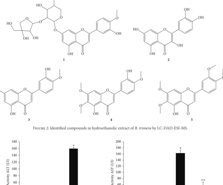
In hydroethanolic extract of B. trimera we identify five
flavonoids by LC-DAD-ESI-MS obtained of chromatogram
in LC-DAD, as shown inTable 1.
3.2. In Vitro Assays: Toxicity of the Hydroethanolic Extract of B. trimera in Liver Cells (HEPG2). After incubation for
Table 2: The toxicity of the hydroethanolic extract ofB. trimera
on hepatic cells (HEP G2) and viability of cells treated with the extract. The data are expressed as a percentage. HepG2 cells were incubated in the absence ofB. trimeraextract, and Hep G2 + Cq cells correspond to the hepatic cell culture incubated withB. trimera
extract.
Strain 1 hour 24 hours
Viability Toxicity Viability Toxicity
HEP G2 100% 0% 100% 0%
HEP G2 + Cq 89.0% 11.0% 98.6% 1.4%
1 and 24 hours (Table 2), it was observed that cells treated
with the extract maintained high viability of 89.0% and 98.6%, respectively, without exhibiting significant toxicity.
3.3. In Vivo Assays: Effect of B. trimera on Biomarkers of
Hepatocellular Damage. Figure 3shows the activity of serum
transaminases in the different treatment groups. The ALT and
AST transaminases were significantly enhanced (𝑃 < 0.05) by
9.8- and 6.3-fold, respectively, in APAP-intoxicated animals.
Treatment withB. trimerasignificantly inhibited the elevation
of the activity of serum transaminases ALT and AST, which was found to be 5.0- and 3.6-fold lower than that in the APAP-intoxicated animals, respectively.
3.4. Effect of B. trimera on APAP-Induced Oxidative Damage.
We evaluated the oxidative damage to proteins and lipids by measuring oxidative stress markers in hepatic tissue. Specifically, we analyzed carbonylated protein and TBARS in
this tissue. The results shown inFigure 4indicate a significant
increase in the concentration of carbonylated protein in the livers of animals intoxicated with APAP compared to the nonintoxicated control group. However, treatment of
APAP-intoxicated rats withB. trimeraresulted in the reduction of
hepatic carbonylated protein concentration compared to the
untreatedB. trimeragroup. Moreover, we observed increased
lipid peroxidation in rats intoxicated with APAP compared
to nonintoxicated animals, andB. trimeratreatment was also
the untreated group. Thus, these results demonstrate that
treatment withB. trimerais capable of minimizing oxidative
damage.
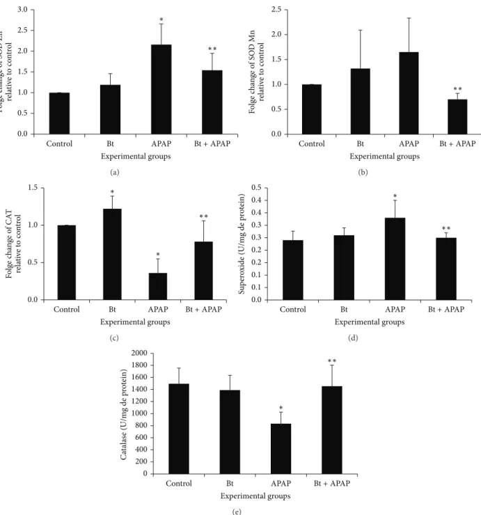
3.5. Effect of B. trimera on Antioxidant Status. To investigate the involvement of antioxidant enzymes in mediating the
radical-scavenging activity ofB. trimera, the mRNA levels
and activities of intracellular antioxidant enzymes were mea-sured in the different groups. The mRNA levels and activities
of SOD and CAT are shown inFigure 5.
SOD and CAT function coordinately to remove superox-ide radicals from the cellular system. The APAP-intoxicated
group showed an increase in Zn-SOD (Figure 5(a)) and
Mn-SOD (Figure 5(b)) expression compared to control animals.
This increase in the expression of different isoforms of SOD was accompanied by an increase in the total activity of this
enzyme in the group intoxicated with APAP (Figure 5(d))
(0.33 ± 0.07units/mg protein) compared to the control (0.24 ± 0.036units/mg protein). In the APAP-intoxicated
group treated withB. trimera, the mRNA level and activity
were significantly lower than their respective control (APAP group). In this study, the mRNA levels and activity of catalase
decreased significantly (𝑃 < 0.05) in the APAP-treated rats,
and this decrease in expression and activity was prevented in
the group intoxicated with APAP and treated withB. trimera.
Figure 6shows that there was a significant increase in𝛿
-GCS (Figure 6(a)) gene expression in the livers of animals
intoxicated with APAP. Treatment withB. trimerawas able
to reverse this profile, decreasing the expression of𝛿-GCS.
Despite the increased expression of 𝛿-GCS, there was a
decrease in the concentration of total glutathione in the livers of animals intoxicated with APAP. However, treatment with
B. trimerawas able to reverse this decrease (Figure 6(b)).
The results inFigure 7show a decrease in the mRNA and
enzyme activity of GPx (Figures 7(a)and 7(b)) and in the
activity of the enzyme GR (Figure 7(c)) in the livers of
APAP-intoxicated rats compared to control rats.B. trimerawas able
to increase the mRNA levels of GPx and GR activities. No change was observed in the activity of GPx and GR in the livers of rats that received only the extract.
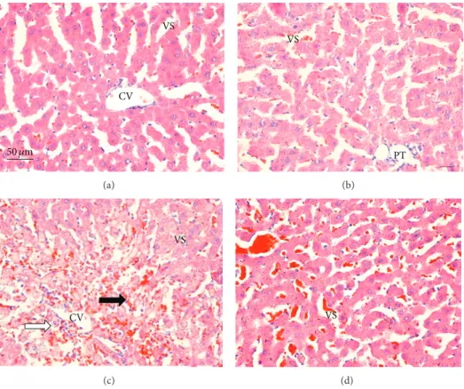
3.6. Histopathology. Microscopic observations revealed nor-mal histology with regular morphology of the liver tissue in
the control (Figure 8(a)) andB. trimeragroups (Figure 8(b)).
In the APAP-intoxicated group (Figure 8(c)), cellular damage
was visible in the form of hydropic degeneration,
inflamma-tion, and hemorrhage. TheB. trimeratreatment (Figure 8(d))
considerably improved the liver morphology in comparison to the APAP-intoxicated rats.
4. Discussion
Research aiming to propose new strategies of therapeutic intervention increasingly includes the use of plant extracts and other natural products. Studies carried out in our lab have demonstrated the beneficial potential of the hydroalcoholic
extract ofB. trimeraon the toxicity induced by APAP [30]. In
the peripheral neutrophils of rats intoxicated with this drug,
the extract was able to regulate the production of reactive oxygen species (ROS) and reactive nitrogen species (RNS). Williams et al. (2014) studied the activation of neutrophils during APAP-induced hepatotoxicity and demonstrated that ROS generation mediated by NADPH oxidase is not a critical event during liver injury caused by this drug. However, they also found that neutrophils present in peripheral blood are activated after administration of APAP. Although the contri-bution of inflammatory cells to the hepatic damage induced
by APAP is still controversial [31], the innate immune
response and the production of reactive species from different sources are correlated with many diseases that affect the liver. For example, neutrophils are associated with hepatic injury during ischemia, endotoxemia, and obstructive cholestasis in
animal models [9,32].
The pharmacological effect of the hydroalcoholic extract ofB. trimerawas investigated in our lab. A LC-DAD-ESI-MS analysis of the hydroalcoholic extract revealed presence of flavonoids, flavone, and glucosides of flavone compounds (Figure 2).
Many studies have found that the hepatotoxicity induced by APAP is the result of oxidative stress, which causes alterations in mitochondrial proteins due to a depletion of glutathione, leading to an inhibition of cellular respiration
with consequent cell death [33]. Because of the biological
effects described for flavonoids found in the extract, this study
was conducted to investigate the effect ofB. trimerain the
modulation of oxidative stress and to evaluate the preventive
effect ofB. trimerain acetaminophen-induced liver damage.
Despite the lack of studies related to the toxicity of B.
trimera, histopathological alterations have recently been found in the livers of pregnant rats treated with a
hydroeth-anolic extract of the plant [22]; moreover, Rodrigues et al.
[21] showed thatB. trimera produced some genotoxic and
mutagenic effects after consumption of high doses of the extract.
In this context, to analyze its toxicity, hepatic HEP G2
cells were incubated in the presence or absence ofB. trimera
extract to obtain the cellular viability/toxicity relationship. After the assays, it was observed that the hydroalcoholic extract did not present significant toxicity, and cells main-tained viability of 89.0% and 98.6% after incubation periods of 1 h and 24 h, respectively. These values show no statistical difference.
Based on these results, we wondered whether the B.
trimeraextract would be able to reverse the hepatotoxicity induced by APAP. To do this, initially, we analyzed the activity of the hepatic enzymes AST and ALT. Both AST and ALT are intracellular enzymes present in large amounts in the cyto-plasm of hepatocytes. Lesions or destruction of hepatic cells
releases these aminotransferases into the circulation [34].
Our study showed that APAP caused a significant increase in the activity of ALT and AST. However, pretreatment with the plant extract restored this activity to values similar to those of the control. High doses of APAP have been associated
with higher activity of ALT and AST [35, 36]. The ability
2
3 4 5
O
O
O
O
O O
OH O
O
O
O O
OH
OH O
O
O
O
OH
OH
O
O
OH
OH
OH
OH
HO O
O
O O
O O
OH OH
OH
OH OH
HO
HO
O
1
Figure 2: Identified compounds in hydroethanolic extract ofB. trimeraby LC-DAD-ESI-MS.
0 20 40 60 80 100 120 140 160 180 200
Control Bt APAP
A
cti
vi
ty AS
T (UI)
Experimental groups 0
20 40 60 80 100 120 140 160 180
A
cti
vi
ty AL
T (UI)
Bt+APAP
Control Bt APAP
Experimental groups
Bt+APAP
∗
∗∗ ∗
∗∗
Figure 3: Effect ofB. trimerahydroethanolic extract on ALT and AST activity in serum of rats 24 h after treatment with APAP. The rats were treated with 600 mg/kgB. trimera1 h before administration of 835 mg/kg APAP. The data are expressed as the mean±SD (𝑛 = 8).∗𝑃 < 0.05 compared to the control,∗∗𝑃 < 0.05compared to APAP.
In addition to analyzing the activities of these hepatic enzymes, the levels of products of oxidative stress have also been described to demonstrate the occurrence of oxidative
damage [37]. Among these products are substances reactive
to thiobarbituric acid (TBAR) and carbonylated proteins. The levels of these substances are used as markers of redox balance in terms of lipid peroxidation and protein oxidation,
respectively, in hepatic cells [38].
In our results, animals intoxicated with APAP presented high levels of carbonylated proteins and TBAR. However,
pretreatment withB. trimeraextract was able to reduce the
levels of these compounds to values similar to those of their respective controls. It is known that plants, because they present antioxidant constituents, are efficient in reducing the lipid peroxidation and protein oxidation induced by APAP
[39,40]. One of the explanations for this phenomenon is that
these phytochemicals are able to minimize the oxidative stress in the livers of animals intoxicated with high doses of this
drug [41].
BecauseB. trimerahas shown such a significant effect on
0.0 0.5 1.0 1.5 2.0 2.5 3.0 3.5 4.0 4.5 5.0
TB
AR (U/mL/m
g p
ro
t.)
0 5 10 15 20 25 30
P
ro
tein ca
rb
o
n
yl
at
ed
(nmo
les/m
g o
f
p
ro
tein)
Control Bt APAP
Experimental groups
Bt+APAP
Control Bt APAP
Experimental groups
Bt+APAP
∗
∗∗ ∗
∗∗
Figure 4: Effect ofB. trimerahydroethanolic extract on the level of carbonylated protein and TBARS in the livers of rats 24 h after treatment with APAP. The rats were treated with 600 mg/kgB. trimera1 h before administration of 835 mg/kg APAP. The data are expressed as the mean±SD (𝑛 = 8).∗𝑃 < 0.05compared to the control,∗∗𝑃 < 0.05compared to APAP.
The protective action of several herbal medicines and their active constituents occurs through antioxidant enzymes (e.g., SOD, CAT, GPx, and GR), which maintain the prooxi-dant/antioxidant balance in the body. To eliminate ROS from the cellular system, SOD and CAT function coordinately to
remove superoxide radicals [1]. Our results showed that the
livers of animals intoxicated with APAP presented higher expression of the SOD-Zn and SOD-Mn isoforms. However,
treatment withB. trimerawas able to regulate this expression
to values similar to those of the livers of the control animals. This increase in gene expression was followed by a higher SOD enzymatic activity. Although SOD is an antioxidant enzyme, some studies have suggested that its overexpression
is in fact harmful to cells [42]. The toxic effect of ROS
that has been observed in many cells overexpressing SOD
has been linked to elevated levels of H2O2 and oxidative
damage accompanying hydroxyl radical formation [43]. The
implication is that SOD upregulation results in high H2O2
turnover.
Our results showed that the liver of animals intoxicated with APAP presented lower expression of CAT. However,
B. trimeratreatment was able to regulate this expression to values similar to those of the livers of control animals. This decrease in gene expression was followed by a lower CAT enzymatic activity. CAT activity was found to be significantly
decreased after a toxic APAP dose [44]. Our results were
similar in that CAT activity was significantly diminished fol-lowing toxic APAP insult. This would allow for the accumu-lation of ROS and hydrogen peroxide, which can exacerbate the hepatocellular damage initiated by NAPQI. Treatment
with the B. trimera extract abrogated the effect of APAP
and induced an increase in CAT activity. This suggests the involvement of their antioxidant constituents in facilitating the rapid and efficient consumption of reactive oxygen
spe-cies generated by APAP-mediated P450 bioactivation [36].
Glutathione (GSH/GSSG) is regarded as the main redox buffer in cells. Glutathione plays an important role in the removal of ROS and protects the thiols in biomacromolecules
[45]. Under normal conditions, glutathione is mainly found
in its reduced form (GSH) and in much smaller amounts
in its oxidized form (GSSG) [46]. Depletion of glutathione
has been associated with enhanced toxicity to chemicals,
including APAP [47]. The results of our present study
showed that the livers of animals intoxicated with APAP,
even presenting high𝛿-GCS activity, presented levels of total
glutathione less than those of the livers of control animals.
However, pretreatment with B. trimera increased the level
of total glutathione in APAP-treated animals. These results
suggested thatB. trimeracould exert its hepatoprotective and
radical-scavenging activities by preventing the formation of free radicals originating from APAP metabolism as well as peroxidation products and enhance the antioxidant defense system. This hypothesis is supported by recent findings that demonstrate that the antioxidant and hepatoprotective activities of extract might be mediated through augmentation of antioxidant defenses and increase in free radical inhibition
due to the presence of important antioxidative factors [48].
The antioxidant effects of glutathione are directly related to GPx and GR, which are key enzymes in the maintenance of redox homeostasis via protecting cells from free
radical-generated toxicity [1]. Our results show a decrease in the
mRNA and enzyme activity of GPx and in the activity of the enzyme GR in the livers of rats 24 h after treatment with APAP compared to control rats. GR is an enzyme that plays a critical role in oxidative stress by APAP; a decrease in its activity will lead to interruption of the cycling between GSSG and GSH and, thus, to a shortage of GSH. Although the impairment of GR activity by APAP is not well understood, at least two hypotheses have been put forth to explain this occurrence, one invoking direct action of ROS or toxic aldehydes and another ascribing the effect to the NAPQI-GSH conjugate that forms in the presence of glutathione
S-transferase [49].
The low activity of GPx is one of the early consequences of a disturbance of the prooxidant/antioxidant balance in
0.0 0.5 1.0 1.5 2.0 2.5 3.0 F o lg e c h an ge o f SO D Z n re la ti ve t o c o n tro l
Control Bt APAP
Experimental groups
Bt+APAP
∗ ∗∗ (a) 0.0 0.5 1.0 1.5 2.0 2.5 F o lg e c h an ge o f SO D M n re la ti ve t o c o n tro l
Control Bt APAP
Experimental groups
Bt+APAP
∗∗ (b) 0.0 0.5 1.0 1.5 F o lg e c h an ge o f CA T re la ti ve t o co n tr o l
Control Bt APAP
Experimental groups
Bt+APAP
∗ ∗ ∗∗ (c) 0.0 0.1 0.1 0.2 0.2 0.3 0.3 0.4 0.4 0.5 Su p er o xide (U/m
g de p
ro
tein)
Control Bt APAP
Experimental groups
Bt+APAP
∗ ∗∗ (d) 0 200 400 600 800 1000 1200 1400 1600 1800 2000 C at alas e (U/m
g de p
ro
tein)
Control Bt APAP
Experimental groups
Bt+APAP
∗
∗∗
(e)
Figure 5: Effect ofB. trimerahydroethanolic extract on the mRNA expression of the enzymes Zn-SOD (a), Mn-SOD (b), and CAT (c) and on the SOD (d) and CAT (e) activity in the livers of rats 24 h after treatment with APAP. The rats were treated with 600 mg/kgB. trimera1 h before administration of 835 mg/kg APAP. The data are expressed as the mean±SD (𝑛 = 8).∗𝑃 < 0.05compared to the control,∗∗𝑃 < 0.05 compared to APAP.
susceptibility of hepatocytes to paracetamol toxicity, indicat-ing that a component of paracetamol’s toxic effect involves the formation of species that are detoxified by GPx enzymes
[51]. Flavonoids have been demonstrated to protect against
paracetamol toxicity by inhibiting lipid peroxidation and
increasing glutathione concentration [52]. Hence, the ability
of plant extracts to restore the loss of GPx activity is most likely due to the presence of flavonoids. The striking increase
in the GPx activity of theB. trimera-treated group compared
with the group that received only APAP may be a result of the presence of quercetin and flavones in addition to other antioxidants in the plant.
Due to the inhibition of GPx, this study concluded that hepatic cell injury was the result of an increase in the
steady-state level of H2O2 and hydroperoxides [53]. While this
0.00 0.50 1.00 1.50 2.00 2.50 3.00
F
o
lg
e c
h
an
ge
o
f
𝛿
-GCS
re
la
ti
ve
t
o
c
o
n
tro
l
∗
∗∗
Control Bt APAP
Experimental groups
Bt+APAP
(a)
0 50 100 150 200 250 300 350 400 450
Gl
u
ta
thio
ne t
o
ta
l (nmo
l/mL)
∗
∗∗
Control Bt APAP
Experimental groups
Bt+APAP
(b)
Figure 6: Effect ofB. trimerahydroethanolic extract on mRNA expression of𝛿-GCS (a) and on total glutathione in rat livers 24 h after treatment with APAP (b). The rats were treated with 600 mg/kgB. trimera1 h before administration of 835 mg/kg APAP. The data are expressed as the mean±SD (𝑛 = 8).∗𝑃 < 0.05compared to the control,∗∗𝑃 < 0.05compared to APAP.
0.0 0.2 0.4 0.6 0.8 1.0 1.2 1.4 1.6
F
o
lg
e c
h
an
ge o
f GPx
re
la
ti
ve
t
o
co
n
tr
o
l
Control Bt APAP
Experimental groups
Bt+APAP
∗
∗∗
(a)
0.0 0.1 0.2 0.3 0.4 0.5 0.6
Gl
u
ta
thio
ne p
er
o
xidas
e (U/mL)
Control Bt APAP
Experimental groups
Bt+APAP
∗
(b)
0.0 0.2 0.4 0.6 0.8 1.0 1.2 1.4 1.6
Gl
u
ta
thio
ne r
ed
u
ct
as
e (U/mL)
Control Bt APAP
Experimental groups
Bt+APAP
∗
∗∗
(c)
Figure 7: Effect ofB. trimerahydroethanolic extract on mRNA expression of GPx (a) and on the activity of glutathione peroxidase (GPx) (b) and activity of glutathione reductase (c) in the livers of rats 24 h after treatment with APAP. The rats were treated with 600 mg/kgB. trimera
1 h before administration of 835 mg/kg APAP. The data are expressed as the mean±SD (𝑛 = 8).∗𝑃 < 0.05compared to the control,∗∗𝑃 < 0.05 compared to APAP.
the depletion of GSH [54]. In other words, the levels of
antioxidant enzymes, if analyzed concomitantly, allow us to infer that the animals intoxicated with APAP presented high
concentration of hepatic H2O2, given that high SOD activity
leads to a high production of ERO, and low CAT and GPx
activities prevent the H2O2generated from being neutralized.
To confirm the hepatoprotective effect ofB. trimera
50 𝜇m
VS
CV
(a)
VS
PT
(b)
VS
CV
(c)
VS
(d)
Figure 8: Sections of the livers of rats 24 hours after administration of PBS (a), extract ofB. trimera(b), APAP (c), andB. trimeraextract and APAP (d), showing (a) normal rats liver with no significant hepatic abnormalities; (b) normal rat liver with no significant hepatic abnormalities; (c) hepatic lesions, hydropic degeneration, inflammation (white arrow) and hemorrhage (black arrow); (d) well-formed polygonal hepatocytes and relatively reduced hydropic degeneration. CV: central vein; VS: venous sinuses; PT: portal triads; liver sections were stained with H & E (400x).
NAPQI
GSSG
Hepatic injury
GSSG
NAPQI APAP
H2O+O2 H2O+O2
O2∙ O2∙
B. trimera+APAP ↑ALT
↑AST
↑TBARS
↑Protein carbonylated
↑H2O2
↓GPx
↓CAT
↑SOD
↓GR
↓GSH
↓ALT
↓AST
↓TBARS
↓Protein carbonylated
↓H2O2
GPx
↑CAT
↓SOD
↑GR
↑GSH
H2O H2O
analyses were performed. APAP-intoxicated animals treated
with the B. trimera extract had improved histopathology
compared to the APAP-intoxicated group without treatment. In conclusion, the present study demonstrated that the
hydroalcoholic extract ofB. trimera has a hepatoprotective
effect against APAP-induced hepatotoxicity in rats. The enhanced levels of antioxidant enzymes and reduced amount of peroxidation products are suggested to be the major
mechanisms by which theB. trimerahydroalcoholic extract
prevents the development of liver damage induced by APAP (Figure 9). Furthermore, a recent study published by our
research group shows that the extract of B. trimerais also
capable of modulating the activity of NADPH oxidase in
peripheral neutrophils of rats intoxicated with APAP [55].
Conflict of Interests
The authors declare no conflict of interests.
Acknowledgments
This research was supported by the Fundac¸˜ao de Amparo `a Pesquisa do Estado de Minas Gerais (FAPEMIG), Con-selho Nacional de Desenvolvimento Cient´ıfico e Tecnol´ogico (CNPq), Coordenac¸˜ao de Aperfeic¸oamento de Pessoal de N´ıvel superior (CAPES-PNPD), Centro Federal de Educac¸˜ao Tecnol´ogica de Minas Gerais (CEFET-MG), and Universi-dade Federal de Ouro Preto (UFOP), Brazil. The authors are grateful to Dr. Maria Terezinha Bahia of the Laboratory of Doenc¸a de Chagas, Ouro Preto, MG, Brazil, for the use of the ABI Prism 7300 Sequence Detector (Applied Biosystems).
References
[1] A. Kumari and P. Kakkar, “Lupeol prevents acetaminophen-induced in vivo hepatotoxicity by altering the Bax/Bcl-2 and oxidative stress-mediated mitochondrial signaling cascade,”
Life Sciences, vol. 90, no. 15-16, pp. 561–570, 2012.
[2] W. M. Lee, “Drug-induced hepatotoxicity,” The New England
Journal of Medicine, vol. 349, no. 5, pp. 474–485, 2003.
[3] W. C. Maddrey, “Drug induced hepatotoxicity,”Journal of
Clin-ical Gastroenterology, vol. 39, pp. 883–889, 2005.
[4] P. J. Amar and E. R. Schiff, “Acetaminophen safety and hepato-toxicity—where do we go from here?”Expert Opinion on Drug
Safety, vol. 6, no. 4, pp. 341–355, 2007.
[5] O. Moling, E. Cairon, G. Rimenti, F. Rizza, R. Prister´a, and P. Mian, “Severe hepatotoxicity after therapeutic doses of acetaminophen,”Clinical Therapeutics, vol. 28, no. 5, pp. 755– 760, 2006.
[6] W. M. Lee, “Acetaminophen and the U.S. acute liver failure study group: lowering the risks of hepatic failure,”Hepatology, vol. 40, no. 1, pp. 6–9, 2004.
[7] O. W. Morgan, C. Griffiths, and A. Majeed, “Interrupted time-series analysis of regulations to reduce paracetamol (acetamino-phen) poisoning,”PLoS Medicine, vol. 4, no. 4, article e105, 2007. [8] G. Dai, L. He, N. Chou, and J.-J. Y. Wan, “Acetaminophen metabolism does not contribute to gender difference in its
hepatotoxicity in mouse,”Toxicological Sciences, vol. 92, no. 1, pp. 33–41, 2006.
[9] L. P. James, P. R. Mayeux, and J. A. Hinson, “Acetaminophen-induced hepatotoxicity,”Drug Metabolism and Disposition, vol. 31, no. 12, pp. 1499–1506, 2003.
[10] H. Jaeschke, “Molecular mechanisms of hepatic ischemia-reperfusion injury and preconditioning,”The American Journal
of Physiology—Gastrointestinal and Liver Physiology, vol. 284,
no. 1, pp. G15–G26, 2003.
[11] H. S. Oz, C. J. McClain, H. T. Nagasawa, M. B. Ray, W. J. S. de Villiers, and T. S. Chen, “Diverse antioxidants protect against acetaminophen hepatotoxicity,”Journal of Biochemical
and Molecular Toxicology, vol. 18, no. 6, pp. 361–368, 2004.
[12] H. S. Oz and T. S. Chen, “Green-tea polyphenols downregulate cyclooxygenase and Bcl-2 activity in acetaminophen-induced hepatotoxicity,”Digestive Diseases and Sciences, vol. 53, no. 11, pp. 2980–2988, 2008.
[13] C. Girish, B. C. Koner, S. Jayanthi, K. Ramachandra Rao, B. Rajesh, and S. C. Pradhan, “Hepatoprotective activity of picro-liv, curcumin and ellagic acid compared to silymarin on paracetamol induced liver toxicity in mice,”Fundamental and
Clinical Pharmacology, vol. 23, no. 6, pp. 735–745, 2009.
[14] S. D. Ray, D. Zinkovsky, and E. Bulku, “Prevention of drug induced programmed andunprogrammed cell death by citrus flavonoids,” in Proceedings of the 228th ACS-Agriculture and
Food “Botanical Health Effects of Citrus”, ACS Symposium
Series, chapter 11, American Chemical Society, 2006.
[15] Y.-J. Surh and H.-K. Na, “NF-𝜅B and Nrf2 as prime molecular targets for chemoprevention and cytoprotection with anti-in-flammatory and antioxidant phytochemicals,”Genes and Nutri-tion, vol. 2, no. 4, pp. 313–317, 2008.
[16] C. Mitchell, M. A. Park, G. Zhang et al., “Extrinsic pathway- and cathepsin-dependent induction of mitochondrial dysfunction are essential for synergistic flavopiridol and vorinostat lethality in breast cancer cells,”Molecular Cancer Therapeutics, vol. 6, no. 12, pp. 3101–3112, 2007.
[17] M. J. Abad and M. Bermejo, “Baccharis(Compositae): a review update,”Arkivoc, vol. 7, pp. 76–96, 2007.
[18] L. G. Verdi, I. M. C. Brighente, and M. G. Pizzolatti, “Gˆenero
Baccharis(Asteraceae): aspectos qu´ımicos, econˆomicos e
bio-l´ogicos,”Quimica Nova, vol. 28, no. 1, pp. 85–94, 2005. [19] R. Della-Loggia, A. Tubaro, S. Sosa, H. Becker, S. Saar, and O.
Isaac, “The role of triterpenoids in the topical anti-inflammato-ry activity ofCalendula officinalisflowers,”Planta Medica, vol. 60, no. 6, pp. 516–520, 1994.
[20] T. Akihisa, K. Yasukawa, H. Oinuma et al., “Triterpene alcohols from the flowers of compositae and their anti- inflammatory effects,”Phytochemistry, vol. 43, no. 6, pp. 1255–1260, 1996. [21] C. R. F. Rodrigues, J. H. Dias, R. N. de Mello, M. F. Richter, J.
N. Picada, and A. B. F. Ferraz, “Genotoxic and antigenotoxic properties ofBaccharis trimerain mice,”Journal of
Ethnophar-macology, vol. 125, no. 1, pp. 97–101, 2009.
[22] S. R. M. Grance, M. A. Teixeira, R. S. Leite et al., “Baccharis trimera: effect on hematological and biochemical parame-ters and hepatorenal evaluation in pregnant rats,”Journal of
Ethnopharmacology, vol. 117, no. 1, pp. 28–33, 2008.
[23] F. C. Meotti, J. M. Rosa, P. S. Brocardo et al., “Protective effect of crude extract from Wedelia paludosa(Asteraceae) on the hepatotoxicity induced by paracetamol in mice,” Journal of
[24] T. A. Ajith, U. Hema, and M. S. Aswathy, “Zingiber officinale
Roscoe prevents acetaminophen-induced acute hepatotoxicity by enhancing hepatic antioxidant status,”Food and Chemical
Toxicology, vol. 45, no. 11, pp. 2267–2272, 2007.
[25] F. L. Yen, T. H. Wu, L. T. Lin, T. M. Cham, and C. C. Lin, “Nanoparticles formulation of Cuscuta chinensis prevents acetaminophen-induced hepatotoxicity in rats,” Food and
Chemical Toxicology, vol. 46, no. 5, pp. 1771–1777, 2008.
[26] J. A. Buege and S. D. Aust, “Microsomal lipid peroxidation,”
Methods in Enzymology, vol. 52, pp. 302–310, 1978.
[27] R. L. Levine, J. A. Williams, E. R. Stadtman, and E. Shacter, “Carbonyl assays for determination of oxidatively modified proteins,”Methods in Enzymology, vol. 233, pp. 346–357, 1994. [28] H. Aebi, “[13] Catalasein vitro,”Methods in Enzymology, vol.
105, pp. 121–126, 1984.
[29] Q. Xiong, P. Xie, H. Li et al., “Acute effects of microcystins exposure on the transcription of antioxidant enzyme genes in three organs (liver, kidney, and testis) of male Wistar rats,”
Journal of Biochemical and Molecular Toxicology, vol. 24, no. 6,
pp. 361–367, 2010.
[30] B. D. C. P´adua, L. D. Silva, J. V. Rossoni et al., “Antioxidant properties ofBaccharis trimerain the neutrophils of Fisher rats,”
Journal of Ethnopharmacology, vol. 129, no. 3, pp. 381–386, 2010.
[31] H. Jaeschke, “Innate immunity and acetaminophen-induced liver injury: why so many controversies?”Hepatology, vol. 48, no. 3, pp. 699–701, 2008.
[32] H. Jaeschke and T. Hasegawa, “Role of neutrophils in acute inflammatory liver injury,”Liver International, vol. 26, no. 8, pp. 912–919, 2006.
[33] H. Jaeschke, C. D. Williams, A. Ramachandran, and M. L. Bajt, “Acetaminophen hepatotoxicity and repair: the role of sterile inflammation and innate immunity,”Liver International, vol. 32, no. 1, pp. 8–20, 2012.
[34] V. T. Motta,Bioqu´ımica Cl´ınica Para o Laborat´orio: Princ´ıpios
e Interpretac¸˜oes, Editora M´edica Missau, Caxias do Sul, Brazil,
2003.
[35] E. Kozer, S. Evans, J. Barr et al., “Glutathione, glutathione-dependent enzymes and antioxidant status in erythrocytes from children treated with high-dose paracetamol,”British Journal of
Clinical Pharmacology, vol. 55, no. 3, pp. 234–240, 2003.
[36] M. T. Olaleye and B. T. J. Rocha, “Acetaminophen-induced liver damage in mice: effects of some medicinal plants on the oxidative defense system,” Experimental and Toxicologic
Pathology, vol. 59, no. 5, pp. 319–327, 2008.
[37] G. Perry, A. K. Raina, A. Nunomura, T. Wataya, L. M. Sayre, and M. A. Smith, “How important is oxidative damage? Lessons from Alzheimer’s disease,”Free Radical Biology and Medicine, vol. 28, no. 5, pp. 831–834, 2000.
[38] M. F. Beal, “Oxidatively modified proteins in aging and disease,”
Free Radical Biology & Medicine, vol. 32, no. 9, pp. 797–803,
2002.
[39] T. H. Tseng, E. S. Kao, C. Y. Chu, F. P. Chou, H. W. Lin Wu, and C. J. Wang, “Protective effects of dried flower extracts ofHibiscus
sabdariffaL. against oxidative stress in rat primary hepatocytes,”
Food and Chemical Toxicology, vol. 35, no. 12, pp. 1159–1164, 1997.
[40] P. Picerno, G. Autore, S. Marzocco, M. Meloni, R. Sanogo, and R. P. Aquino, “Anti-inflammatory activity of verminoside from
Kigelia africanaand evaluation of cutaneous irritation in cell
cultures and reconstituted human epidermis,”Journal of Natural
Products, vol. 68, no. 11, pp. 1610–1614, 2005.
[41] S. D. Ray, N. Patel, N. Shah, A. Nagori, A. Naqvi, and S. J. Stohs, “Pre-exposure to a novel nutritional mixture containing a series of phytochemicals prevents acetaminophen-induced programmed and unprogrammed cell deaths by enhancing BCL-XL expression and minimizing oxidative stress in the liver,”Molecular and Cellular Biochemistry, vol. 293, no. 1-2, pp. 119–136, 2006.
[42] R. Gardner, A. Salvador, and P. Moradas-Ferreira, “Why does SOD overexpression sometimes enhance, sometimes decrease, hydrogen peroxide production? A minimalist explanation,”Free
Radical Biology & Medicine, vol. 32, no. 12, pp. 1351–1357, 2002.
[43] J. B. de Haan, F. Cristiano, R. Iannello, C. Bladier, M. J. Kelner, and I. Kola, “Elevation in the ratio of Cu/Zn-superoxide dismutase to glutathione peroxidase activity induces features of cellular senescence and this effect is mediated by hydrogen peroxide,”Human Molecular Genetics, vol. 5, no. 2, pp. 283–292, 1996.
[44] R. Bhattacharjee and C. S. Parames, “The protein fraction of
Phyllanthus niruriplays a protective role against acetaminophen
induced hepatic disorder via its antioxidant properties,”
Phy-totherapy Research, vol. 20, no. 7, pp. 595–601, 2006.
[45] A. Pastore, G. Federici, E. Bertini, and F. Piemonte, “Analysis of glutathione: implication in redox and detoxification,”Clinica
Chimica Acta, vol. 333, no. 1-2, pp. 19–39, 2003.
[46] C. Cereser, J. Guichard, J. Drai et al., “Quantitation of reduced and total glutathione at the femtomole level by high-per-formance liquid chromatography with fluorescence detection: application to red blood cells and cultured fibroblasts,”Journal
of Chromatography B: Biomedical Sciences and Applications, vol.
752, no. 1, pp. 123–132, 2001.
[47] R. P. Hewawasam, K. A. P. W. Jayatilaka, C. Pathirana, and L. K. B. Mudduwa, “Protective effect ofAsteracantha longifolia
extract in mouse liver injury by carbon tetrachloride and parac-etamol,”Journal of Pharmacy and Pharmacology, vol. 55, no. 10, pp. 1413–1418, 2003.
[48] M. T. Olaleye, A. C. Akinmoladun, A. A. Ogunboye, and A. A. Akindahunsi, “Antioxidant activity and hepatoprotective property of leaf extracts of Boerhaavia diffusa Linn against acetaminophen-induced liver damage in rats,”Food and
Chem-ical Toxicology, vol. 48, no. 8-9, pp. 2200–2205, 2010.
[49] T. Rouˇsar, P. Paˇr´ık, O. Kuˇcera, M. Bartoˇs, and Z. Cervinkov´a, “Glutathione reductase is inhibited by acetaminophen-glutathi-one conjugate in vitro,”Physiological Research, vol. 58, pp. 239– 246, 2009.
[50] H. Benabdeslam, H. Abidi, I. Garcia, G. Bellon, R. Gilly, and A. Revol, “Lipid peroxidation and antioxidant defenses in cystic fibrosis patients,”Clinical Chemistry and Laboratory Medicine, vol. 37, no. 5, pp. 511–516, 1999.
[51] G. M. Adamson and A. W. Harman, “A role for the glutathione peroxidase/reductase enzyme system in the protection from paracetamol toxicity in isolated mouse hepatocytes,”
Biochemi-cal Pharmacology, vol. 38, no. 19, pp. 3323–3330, 1989.
[52] M. Lahouel, S. Boulkour, N. Segueni, and J. P. Fillastre, “The pro-tective effect of flavonoids against vinblastine cyclophamide and paracetamol toxicity by inhibition of lipid-peroxidation and increasing liver glutathione concentration,”Pathologie Biologie, vol. 52, pp. 314–322, 2006.
[53] S. L. Arnaiz, S. Llesuy, J. C. Cutrin, and A. Boveris, “Oxidative stress by acute acetaminophen administration in mouse liver,”
Free Radical Biology and Medicine, vol. 19, no. 3, pp. 303–310,
[54] M. L. Bajt, T. R. Knight, J. J. Lemasters, and H. Jaeschke, “Acetaminophen-induced oxidant stress and cell injury in cultured mouse hepatocytes: protection by N-acetyl cycteine,”
Toxicological Sciences, vol. 80, no. 2, pp. 343–349, 2004.
[55] B. D. C. P´adua, J. V. Rossoni Jr., C. L. D. B. Magalh˜aes et al.,
“Baccharis trimeraimproves the antioxidant defense system and
Submit your manuscripts at
http://www.hindawi.com
Stem Cells
International
Hindawi Publishing Corporation
http://www.hindawi.com Volume 2014
Hindawi Publishing Corporation
http://www.hindawi.com Volume 2014
INFLAMMATION
Hindawi Publishing Corporation
http://www.hindawi.com Volume 2014
Behavioural
Neurology
Endocrinology
International Journal ofHindawi Publishing Corporation
http://www.hindawi.com Volume 2014
Hindawi Publishing Corporation
http://www.hindawi.com Volume 2014
Disease Markers
Hindawi Publishing Corporation
http://www.hindawi.com Volume 2014
BioMed
Research International
Oncology
Journal of Hindawi Publishing Corporationhttp://www.hindawi.com Volume 2014
Hindawi Publishing Corporation
http://www.hindawi.com Volume 2014
Oxidative Medicine and Cellular Longevity
Hindawi Publishing Corporation
http://www.hindawi.com Volume 2014
PPAR Research
The Scientific
World Journal
Hindawi Publishing Corporationhttp://www.hindawi.com Volume 2014
Immunology Research
Hindawi Publishing Corporation
http://www.hindawi.com Volume 2014 Journal of
Obesity
Journal ofHindawi Publishing Corporation
http://www.hindawi.com Volume 2014
Hindawi Publishing Corporation
http://www.hindawi.com Volume 2014 Computational and Mathematical Methods in Medicine
Ophthalmology
Journal ofHindawi Publishing Corporation
http://www.hindawi.com Volume 2014
Diabetes Research
Journal ofHindawi Publishing Corporation
http://www.hindawi.com Volume 2014
Hindawi Publishing Corporation
http://www.hindawi.com Volume 2014
Research and Treatment
AIDS
Hindawi Publishing Corporation
http://www.hindawi.com Volume 2014
Gastroenterology Research and Practice
Hindawi Publishing Corporation
http://www.hindawi.com Volume 2014
Parkinson’s
Disease
Evidence-Based Complementary and Alternative Medicine
Volume 2014