Facial morphology and obstructive sleep apnea
Anderson Capistrano1, Aldir Cordeiro2, Leopoldino Capelozza Filho3, Veridiana Correia Almeida4, Priscila Izabela de Castro e Silva5, Sandra Martinez6, Renata Rodrigues de Almeida-Pedrin3
1 Professor of Occlusion and Orthodontics, Faculdade de Odontologia de Recife
(FOR-PE), Recife, Pernambuco, Brazil.
2 MSc in Orthodontics, Universidade Sagrado Coração (USC), Bauru, São
Paulo, Brazil.
3 Professor, Universidade Sagrado Coração (USC), Undergraduate and Graduate
programs, Bauru, São Paulo, Brazil.
4 Postgraduate student, Cooperativa dos Odontologistas de Pernambuco
(COOPE), Recife, Pernambuco, Brazil.
5 Student, Sindicato dos Odontologistas no Estado de Pernambuco
(SOEPE), Postgraduate program in Orthodontics, Recife, Pernambuco, Brazil.
6 Specialist in Sleep Medicine, Universidade de São Paulo (USP), São Paulo, São
Paulo, Brazil, and Stanford University, Stanford, California, USA.
Submitted: December 21, 2014 - Revised and accepted: June 30, 2015
How to cite this article: Capistrano A, Cordeiro A, Capelozza Filho L, Almei-da VC, Castro e Silva PI, Martinez S, Almeida-Pedrin RR. Facial morphology and obstructive sleep apnea. Dental Press J Orthod. 2015 Nov-Dec;20(6):60-7. DOI: http://dx.doi.org/10.1590/2177-6709.20.6.060-067.oar
» The authors report no commercial, proprietary or financial interest in the products or companies described in this article.
» Patients displayed in this article previously approved the use of their facial and in-traoral photographs.
Contact address: Anderson Capistrano E-mail: [email protected]
Objective: This study aimed at assessing the relationship between facial morphological patterns (I, II, III, Long Face and Short Face) as well as facial types (brachyfacial, mesofacial and dolichofacial) and obstructive sleep apnea (OSA) in patients attending a center specialized in sleep disorders. Methods: Frontal, lateral and smile photographs of 252 patients (157 men and 95 women), randomly selected from a polysomnography clinic, with mean age of 40.62 years, were evalu-ated. In order to obtain diagnosis of facial morphology, the sample was sent to three professors of Orthodontics trained to classify patients’ face according to five patterns, as follows: 1) Pattern I; 2) Pattern II; 3) Pattern III; 4) Long facial pattern; 5) Short facial pattern. Intraexaminer agreement was assessed by means of Kappa index. The professors ranked patients’ facial type based on a facial index that considers the proportion between facial width and height. Results: The multiple linear regression model evinced that, when compared to Pattern I, Pattern II had the apnea and hypopnea index (AHI) worsened in 6.98 episodes. However, when Pattern II was compared to Pattern III patients, the index for the latter was 11.45 episodes lower. As for the facial type, brachyfacial patients had a mean AHI of 22.34, while dolichofacial patients had a significantly statistical lower index of 10.52. Conclusion: Patients’ facial morphology influences OSA. Pattern II and brachyfacial patients had greater AHI, while Pattern III patients showed a lower index.
Keywords:Diagnosis. Face. Obstructive sleep apnea. DOI: http://dx.doi.org/10.1590/2177-6709.20.6.060-067.oar
Objetivo: o objetivo deste estudo foi avaliar a associação entre os padrões morfológicos faciais e tipos faciais (braquifacial,
mesofacial e dolicofacial) com a Apneia Obstrutiva do Sono (AOS) em pacientes de um centro especializado em dis-túrbios do sono. Métodos: foram utilizadas fotografias faciais de frente, perfil e sorriso de 252 indivíduos, selecionados
aleatoriamente entre pacientes que procuraram uma clínica especializada em polissonografia. Para o diagnóstico morfo-lógico facial, a amostra foi enviada a três professores de Ortodontia treinados na classificação do padrão facial, e cada um recebeu a orientação para classificar o padrão facial da seguinte forma: 1 = Padrão I, 2 = Padrão II, 3 = Padrão III, 4 = Padrão Face Longa e 5 = Padrão Face Curta. A concordância interexaminadores foi avaliada por meio do Índice Kappa. O diagnóstico do tipo facial foi estabelecido por meio de um índice facial que leva em consideração a proporção entre a largura e altura da face. Resultados: no modelo de regressão linear múltipla, ficou evidenciado que o Padrão II teve a
capacidade de agravar o índice de apneia e hipopneia (IAH) em 6,98, enquanto os pacientes do Padrão III tinham esse índice atenuado em 11,45. Para o tipo facial, os pacientes braquifaciais apresentaram um IAH médio de 22,34, enquanto o grupo classificado como dolicofacial mostrou um índice menor, de 10,52, com significância estatística. Conclusão: o
desenho morfológico facial se mostrou um considerável fator de agravamento ou proteção da SAOS, onde os indivíduos Padrão II e braquifaciais tiveram IAH maiores, enquanto nos pacientes Padrão III esse índice foi reduzido.
INTRODUCTION
In the last 20 years, Dentistry has discussed cases of snoring and obstructive sleep apnea (OSA). Within an interdisciplinary approach, neurologists, otolaryngolo-gists, physiotherapists, speech therapists and clinicians have all recognized the importance of assessing these patients from the point of view of Dentistry, not only in terms of therapeutic control, but also in prevent-ing it by means of treatprevent-ing potential malocclusions that could increase the risk of airway disorders, particularly when they are associated with predisposing factors
such as obesity, hypertension and aging.1
Obstructive sleep apnea (OSA) is a common disor-der associated with snoring, upper airway collapse at sleep, oxygen desaturation and fragmented sleep. It is also associated with cardiovascular morbidity, risk of
car accidents and general mortality.1 OSA diagnosis is
not simple, since polysomnography requires patient’s monitoring by a specialist at a sleep laboratory, which renders the procedure relatively expensive and diffi-cult. In order to simplify diagnosis, a study aimed at assessing differences in craniofacial phenotype among caucasian patients with OSA. The study found that detailed anatomic data, such as facial width, distance between eyes, as well as mandibular length and chin-neck angle, were useful in predicting OSA with a
sensitivity index of 86%.2
Anatomical airway narrowing is among the etio-logical factors of snoring and OSA. It consists in soft tissues excess, macroglossia and retrognathism. This condition causes great resistance that hinders air flow and engenders negative intraluminal pressure while inspiring, thereby favoring breathing collapse. The risk of developing this disorder significantly in-creases with weight gain, aging, increased neck cir-cumference and alcohol consumption. The following systemic conditions also appear as predisposing fac-tors: systemic hypertension, untreated
hypothyroid-ism, acromegaly and nasal obstruction.3
OSA recognition in the overall population
re-mains low and most patients are not diagnosed.1
Thus, there is a critical, clinical need to develop bet-ter methods that allow OSA recognition and diag-nosis. The present study was conducted to assess the relationship between facial morphological pattern, within a contemporary context of genetic determin-ism, and OSA. This relationship might stand for an
important clinical evidence of quick, inexpensive and simple diagnosis of a disease that significantly impairs patients’ quality of life.
The literature2-7 has demonstrated a relationship
between craniofacial dimensions and upper airway structures in patients with OSA. These results give support to the potential role facial measurements play in the anatomical phenotype of OSA.
Craniofacial morphology seems to be among the predisposing factors for the development of OSA. Mandibular deficiency and increased anterior-inferior
facial height highlight such possibility.7 The majority
of publications7-12 used cephalometric measurements
to define craniofacial morphology, which may cause considerable doubt and contradiction. In an attempt to avoid it, the present study aims at assessing a diag-nosis system that considers the genetic determinism of craniofacial morphology, with specific methods centered around a diagnosis concept based on facial
patterns,13,14 in addition to studying its relationship
with obstructive sleep apnea in adult patients.
MATERIAL AND METHODS Sample calculation
For sample size calculation, alpha = 5% and the power of the test was of 80%. Calculation was based on multiple linear regression analysis, with AHI as the dependent variable; and sex, age, BMI, facial type (three categories) and facial pattern (five categories) as independent variables, thereby totaling 11 predic-tor variables. Effect size was set at 0.10 and minimal sample size was of 178 cases. Calculation was carried
out using the software developed by Soper,15 in 2014.
Sample selection
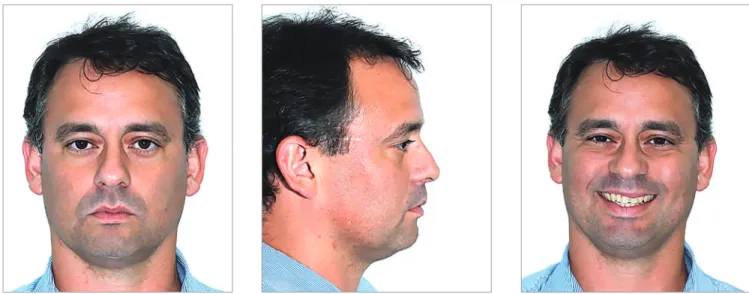
Figure 1 - Example of photograph set-up for diagnosis.
Ater interviewing an average of 900 patients, some of them were excluded based on the following criteria:
» BMI over 40.
» Impaired posterior occlusal support.
» Patients with a beard that hindered facial
analy-sis.
» Patients using continuous positive airway
pres-sure (CPAP) or intraoral appliance and who were be-ing subjected to the examination, so as to assess the therapeutic effectiveness of the devices.
» Craniofacial syndromic patients.
» Patients with chronic obstructive pulmonary
diseases (CPOC) or neurological or mental disorders who were affected by upper airway infection.
» Patients with history of orthognathic surgery or
any other type of airway surgery.
» Patients under 18 and above 62 years old.
» Patients who, for any reason, did not agree in
taking part in the research.
» After facial pattern analysis, other patients were
also excluded for presenting three different diagnoses. The 252 individuals comprising the sample were divided into two groups according to polysomnog-raphy results. The group without OSA (Group I, 77 patients) and the group with OSA (Group II, 175 patients) which presented an AHI value greater than five episodes of apnea per hour of sleep, enough to characterize the individuals as having the disease.
Frontal, lateral and smile standardized photographs were used to assess patients’ facial morphological
pat-tern (Fig 1). The photographs were inserted in a
Pow-erPointTM slide presentation and sent via
WeTrans-ferTM to three experienced orthodontic professors.
Each examiner was advised to classify patients’ facial pattern according to the following: 1) Pattern I; 2) Pat-tern II; 3) Pat2) Pat-tern III; 4) Long Facial Pat2) Pat-tern; 5) Short Facial Pattern. Examiners did not have access to pa-tients’ reports and, for this reason, were unaware of those with and without OSA.
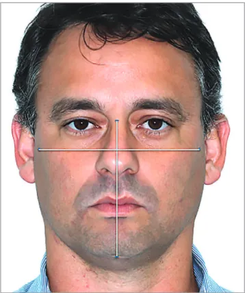
Examiners reached facial type diagnosis through a
facial index (n-gn/zy-zy)16 that considers the
propor-tion between facial width and height, with a mean value of 88.5 for men and 86.2 for women (Table 1). Measurements were obtained with the aid of
Photo-shopTM CS4 software (Fig 2).
Patients were classified as mesofacial, brachyfacial and dolichofacial, as shown in Table 1.
Error of the method
To assess the error of classifying both facial pattern
and type, Kappa index17 was used (Table 2). For facial
pattern, the diagnoses of three examiners were crossed for each patient. Patients with three diferent diagno-ses were excluded from this study. Four patients were excluded by means of this criterion. As for facial type, 30% of the sample was randomly selected, so as to re-assess patients’ facial proportions ater 30 days.
All assessments reached an agreement value that ranged between strong and nearly perfect. Landis &
Table 1 - Facial index.
Table 2 - Agreement percentage and Kappa’s values for intraexaminer agree-ment assessagree-ment.
Figure 2 - One of the patients comprising the sample. Image used to illus-trate how facial type was obtained.
To assess the combined effect of sex, age, BMI, facial type and facial pattern on the AHI value, step-wise backward multiple linear regression analysis
was used. Significance level was set at 5% (p < 0.05)
for all tests. All statistical procedures were carried out by means of Statistica version 12 (StatSoft Inc., Tulsa, USA) software.
RESULTS
Of the 289 patients, 29 were excluded based on the exclusion criteria. A total of 260 patients were analyzed, four of which were excluded for present-ing three different morphological diagnoses, one for not having polysomnography concluded and three for not having polysomnography results sent for analysis within a reasonable time. Thus, 252 patients were in-cluded in the statistical analysis.
In terms of facial morphological pattern and OSA prevalence, patients were classified according to data presented in Table 3. Data analysis revealed statistical difference between Pattern II and long face. Despite no relevant statistically significant difference, the per-centage of short face individuals with OSA is great-er than expected, as it totaled 77.8% against 22.2% for individuals without the disorder. Both percent-ages are significantly near those found for Pattern II, 80.3% and 19.7%, respectively. When facial patterns were assessed by Kruskal-Wallis test, according to the absolute AHI value, there was statistically significant difference between Pattern II and long face (Table 4). Importantly, it is worth noting that the mean AHI value for the Pattern III group (11.4) was nearly half that of the Pattern II group (22.51).
Table 5 shows no statistically significant differences for distribution of facial types for groups I and II. Nevertheless, when groups were classified according to the AHI value (Table 6), there was statistically sig-nificant difference between brachyfacial patients with a mean AHI of 22.34, and dolichofacial patients with a mean AHI value of 10.52.
Results reveal that facial pattern influenced the apnea and hypopnea index (AHI) severity when mul-tiple linear regression analysis was used. This is be-cause apnea is a multifactorial disorder in which the interaction among variables, such as BMI, sex and age, also influence AHI values. Importantly, multiple linear regression analysis is considered adequate for
Men Women
Mesofacial 83.4 – 93.6 81.6 – 90.8 Brachyfacial < 83.4 < 81.6 Dolichofacial > 93.6 > 90.8
Measurement % of agreement Kappa
Examiner 1 versus Examiner 2 77.8 0.64 Examiner 1 versus Examiner 3 77.4 0.62 Examiner 2 versus Examiner 3 81.4 0.68 Facial type 1 versus Facial type 2 90.4 0.90
Statistical analysis
Data were arranged in tables and charts using ab-solute (n) and relative (%) frequencies. Quantitative variables were described by mean and standard devia-tion parameters.
Chi-square (p = 0.009* – Pattern II ≠ Long Face).
Table 3 - Relationship between facial pattern and OSA.
Facial pattern
Group I Group II Total
n % n %
Pattern I 46 32.6 95 67.4 141 Pattern II 15 19.7 61 80.3 76 Pattern III 7 43.8 9 56.3 16
Short face 2 22.2 7 77.8 9
Long face 7 70.0 3 30.0 10
Table 4 - Comparison among the five facial patterns with regard to AHI mea-surements based on Kruskal-Wallis test.
Facial
pattern
n Mean SD p-value
Pattern I 141 17.97 21.4
Pattern II 76 22.51 23.05 0.027 (P II ≠ L.F.) Pattern III 16 11.4 13.31
Short face 9 16.03 16.31 Long face 10 6.22 9.94
Table 5 - Relationship between facial type and OSA.
Chi-square (p = 0.505 ns).
Facial type Without OSA With OSA Total
n % n %
Mesofacial 44 32.6 91 67.4 135 Brachyfacial 26 26.5 72 73.5 98 Dolichofacial 7 36.8 12 63.2 19
Table 6 - Comparison among the three facial types with regard to AHI mea-surements based on Kruskal-Wallis test.
Facial type n Mean SD p-value
Mesofacial 135 16.63 19.51
Brachyfacial 98 22.34 23.87 0.044* B ≠ D Dolichofacial 19 10.52 14.65
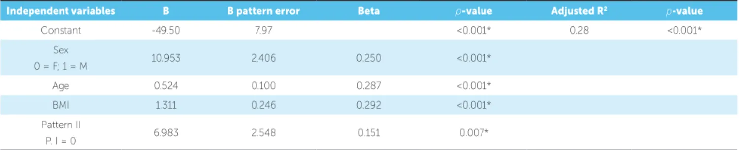
Table 7 - Stepwise backward multiple linear regression analysis with AHI as dependent variable; and sex, age, BMI, facial type and facial pattern as independent variables.
* statistically significant (p < 0.05).
Independent variables B B pattern error Beta p-value Adjusted R2 p-value
Constant -49.50 7.97 <0.001* 0.28 <0.001*
Sex
0 = F; 1 = M 10.953 2.406 0.250 <0.001*
Age 0.524 0.100 0.287 <0.001*
BMI 1.311 0.246 0.292 <0.001*
Pattern II
P. I = 0 6.983 2.548 0.151 0.007*
data analysis, since it allows assessment of interaction between two or among more than two variables.
Table 7 shows that when using Pattern I as a pattern of comparison, Pattern II had AHI sever-ity worsened in 6.98 episodes. Conversely, Table 8 shows that, when Pattern II was used as reference,
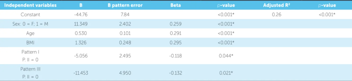
Pattern I individuals had the AHI value reduced in 5.06 while Pattern III patients had the AHI re-duced in 11.45.
Table 8 - Stepwise backward multiple linear regression analysis with AHI as dependent variable; and sex, age, BMI, facial type and facial pattern as independent variables.
* - statistically significant (p < 0.05).
Independent variables B B pattern error Beta p-value Adjusted R2 p-value
Constant -44.76 7.84 <0.001* 0.26 <0.001*
Sex: 0 = F; 1 = M 11.349 2.402 0.259 <0.001*
Age 0.530 0.101 0.291 <0.001*
BMI 1.326 0.248 0.295 <0.001*
Pattern I
P. II = 0 -5.056 2.495 -0.118 0.044*
Pattern III
P. II = 0 -11.453 4.950 -0.132 0.021*
DISCUSSION
Craniofacial morphology plays an important role
in OSA2,4-7 in adult patients. Increased gonial angle,
changes in anterior and posterior facial height, de-creased anterior cranial base and mandibular deiciency
seem to contribute to pharyngeal airway narrowing.7
Cephalometry is, without a doubt, the method most used in current literature to analyze facial morphology
and its relationship with OSA.7-12 Nevertheless, with a
view to rendering diagnosis easier, or at least suspect-ing the existence of the disease, front-view and proile photographs have been used to assess anatomical data, such as facial width, distance between the eyes,
chin-neck angle and mandibular length.2,4
Morphological expression used for diagnosis in Orthodontics might be better assessed and under-stood by means of facial soft tissues analysis. In this sense, cephalometry plays a secondary and supple-mentary role and could not be used as a primary diag-nostic tool to determine facial morphology. The re-producibility and reliability of the method still allow assessment of the influence of growth and therapeutic
actions on facial morphology.3,4
Analysis of facial morphology was based on the clinical experience of three professors of Orthodon-tics. Intraexaminer agreement yielded good results (78.9%) with a Kappa index of 0.65 (strong agree-ment). The methods employed in the present study
were based on Reis et al19 who found an
intraexam-iner agreement of 72%, with Kappa index of 0.65 (strong agreement). Both values were close to those found for the present study. With a view to render-ing the relationship between facial morphology, as-sessed within a subjective approach, and OSA, four
individuals were excluded from the sample due to having three different diagnoses.
A total of 30% of the sample was randomly select-ed and measurselect-ed again after 30 days, so as to reassess patients’ facial proportions. Agreement was of 90.4% with a Kappa index of 0.90 (nearly perfect), thereby resulting in high reliability of research results.
Due to lack of studies focusing on establishing a relationship between facial morphology and obstruc-tive sleep apnea, based on the subjecobstruc-tive determina-tion of facial morphological pattern and facial type as well as analysis of proportion between facial width and height, comparison of results is usually made with facial anthropometric measurements and, most of times, by means of cephalometry.
One of the most common findings in the literature addressing patients’ facial morphology is the relation-ship established between OSA and convex facial
pro-file,12,15,20,21 even though Katyal et al’s systematic
re-view8 minimizes such association, at least in children.
Although no statistically significant difference was found, Pattern II patients present greater OSA preva-lence (80.3%) in comparison to patients without the disorder (19.7%). In terms of AHI severity, Pattern II individuals present the greatest incidence, with 22.51 episodes of apnea per hour of sleep in comparison to 11.40 episodes for Pattern III individuals who are
morphologically opposite to Pattern II.13 These
re-sults reveal a tendency of OSA worsening in Pattern II patients, even though statistically significant differ-ence was only observed when the Pattern II group was compared to Long Face patients.
are signiicantly diferent from dolichofacial patients who yielded a mean AHI value of 10.52. This ind-ing disagrees with the results commonly found in the literature which does not establish a strong association
between dolichofacial patients and OSA.6,7,9,10,22
How-ever, this fact is not unanimous;8,23 it is supported by
Grauer et al20 who used cone-beam computed
tomog-raphy and did not ind any diferences in airspace vol-ume for dolichofacial patients. Moreover, the study by
Haskell et al23 assessed 50 patients by means of
cone-beam computed tomography and found greater air-space in vertical patients.
With a view to highlighting the importance of the factors assessed herein (facial pattern and type), mul-tiple linear regression analysis was employed to assess the relevance of the present variables in comparison to other OSA predicting factors, such as obesity
mathe-matically measured by BMI.4,10,11,21 In the present
anal-ysis, Pattern II patients had the AHI value increased in 6.98 episodes of apnea per hour of sleep when com-pared to Pattern I individuals. Conversely, Pattern III patients had the AHI value reduced in 11.45 when compared with Pattern II patients. Data analysis sug-gests that while Pattern II might render OSA more se-vere, Pattern III seems to protect patients against the sleeping disorder. This statistical analysis did not reveal any relationship between facial type and OSA.
The low prevalence of long face (4.0%) and short face (3.6%) in the sample studied did not allow us to make further inference with regard to the occurrence of OSA in either one of these morphological groups. Likewise, low prevalence also seems to be found in the overall population. In 2007, the study conducted by
Siécola24 comprised 151 children aged between 7 and
13 years old and who were enrolled in two diferent schools in the city of Bauru. The study found a preva-lence of 5.96% of long-face individuals and 1.98% of short-face individuals. Nevertheless, should the mor-phological diagnosis of long face be strongly associated
with OSA, as suggested by a few studies,6,7,9,10,22 this
group would be expected to appear in a higher number in a sample selected at a clinics specialized in sleeping disorders where 69.5% of individuals had OSA.
Due to being a multifactorial disease, the eti-ology of OSA is too diverse and complex to be ex-plained by a simple relationship established between facial morphology and the development of the disease.
However, therapeutic orthodontic actions should re-spect the tendency towards these results and include functional aspects related to OSA in their therapeutic practice. The importance of considering patients’ cra-niofacial morphology as a factor that protects or worsens OSA is conirmed by asystematic literature review
car-ried out by Pirklbauer et al15 in 2011. The authors found
that the most efective surgical approach employed to treat OSA is advancement of the maxilla and mandible, i.e., exactly when a drastic facial morphological change occurs. Post-operative polysomnography results can be compared to those yielded by CPAP therapy.
Adult brachyfacial patients and Pattern II due to mandibular deiciency should be included in the ben-eits of decompensatory orthodontic treatment for surgeries of mandibular advancement: a functional breathing beneit that widens the upper airways while reducing the anatomical risk of OSA development. Meanwhile, if patients comprising this diagnostic group have incomplete growth, compensatory treat-ment with reduction in intraoral volume, such as upper premolars extraction, should be avoided, as it could re-sult in maintenance or exacerbation of anatomical dis-advantages likely to lead to the development of OSA in the long term, particularly when associated with other
predisposing factors such as obesity and aging.1,3
Adult Pattern III patients, however, should have mandible position preserved, in order to avoid poten-tial mandibular setback, unless supported by progna-thism severity and impaired esthetics. Thus, when-ever recommending advancement of the maxilla, clinicians should consider including the benefit of widening upper airways in patients’ orthodontic and surgical planning, thereby preserving the functional benefits these patients seem to have with regard to the development of OSA. For growing patients, the protocol of widening the maxilla is also supported by the gain in breathing volume, which minimizes the potential for developing OSA, thereby providing pa-tients with undeniable functional benefits.
1. Hiestand DM, Britz P, Goldman M, Phillips B. Prevalence of symptoms and risk of sleep apnea in the US population: results from the national sleep foundation sleep in America 2005 poll. Chest. 2006 Sep;130(3):780-6.
2. Lee RW, Petocz P, Prvan T, Chan AS, Grunstein RR, Cistulli PA. Prediction of obstructive sleep apnea with craniofacial photographic analysis. Sleep. 2009 Jan;32(1):46-52.
3. Caldas SGFR, Ribeiro AA, Santos-pinto L, Martins LP, Matoso RM. Efetividade dos aparelhos intrabucais de avanço mandibular no tratamento do ronco e da síndrome da apneia e hipopneia obstrutiva do sono (SAHOS): revisão sistemática. Rev Dental Press Ortod Ortop Facial. 2009 Jul-Ago;4(14):74–82.
4. Lee RW, Chan AS, Grunstein RR, Cistulli PA. Craniofacial phenotyping in obstructive sleep apnea—a novel quantitative photographic approach. Sleep. 2009 Jan;32(1):37-45.
5. Lee RW, Sutherland K, Chan AS, Zeng B, Grunstein RR, Darendeliler MA, et al. Relationship between surface facial dimensions and upper airway structures in obstructive sleep apnea. Sleep. 2010 Sep;33(9):1249-54.
6. Albajalan OB, Samsudin AR, Hassan R. Craniofacial morphology of Malay patients with obstructive sleep apnoea. Eur J Orthod. 2011 Oct;33(5):509-14.
7. Di Francesco R, Monteiro R, Paulo ML, Buranello F, Imamura R. Craniofacial morphology and sleep apnea in children with obstructed upper airways: diferences between genders. Sleep Med. 2012 Jun;13(6):616-20. 8. Katyal V, Pamula Y, Martin AJ, Daynes CN, Kennedy JD, Sampson WJ.
Craniofacial and upper airway morphology in pediatric sleep-disordered breathing: systematic review and meta-analysis. Am J Orthod Dentofacial Orthop. 2013 Jan;143(1):20-30.e3.
9. Kikuchi M, Higurashi N, Miyazaki S, Itasaka Y. Facial patterns of obstructive sleep apnea patients using Ricketts’ method. Psychiatry Clin Neurosci. 2000 Jun;54(3):336-7.
10. Kubota Y, Nakayama H, Takada T, Matsuyama N, Sakai K, Yoshizawa H, et al. Facial axis angle as a risk factor for obstructive sleep apnea. Intern Med. 2005 Aug;44(8):805-10.
11. Chang ET, Shiao GM. Craniofacial abnormalities in Chinese patients with obstructive and positional sleep apnea. Sleep Med. 2008 May;9(4):403-10. Epub 2007 Jul 19.
12. Banabilh SM, Asha’ari ZA, Hamid SS. Prevalence of snoring and craniofacial features in Malaysian children from hospital-based medical clinic population. Sleep Breath. 2008 Aug;12(3):269-74.
REFERENCES
13. Capelozza Filho L. Diagnóstico em Ortodontia. Maringá: Dental Press; 2004. 14. Reis SAB, Abrão J, Claro CAA, Capelozza Filho L. Evaluation of the determinants
of facial proile aesthetics. Dent Press J Orthod. 2011;16(1):57-67.
15. Soper DS. A-priori sample size calculator for multiple regression [Software]. 2014. Available from http://www.danielsoper.com/statcalc.
16. Farkas LG, Posnick JC, Hreczko TM. Growth patterns of the face: a morphometric study. Cleft Palate Craniofac J. 1992 Jul;29(4):308-15. 17. Fleiss JL. Statistical methods for rates and proportions. New York: John Wiley &
Sons; 1973.
18. Landis JR, Koch GG. The measurement of observer agreement for categorical data. Biometrics. 1977 Mar;33(1):159-74.
19. Reis SAB, Abrão J, Claro CAA, Fornazari RF, Capelozza Filho L. Agreement among orthodontists regarding facial pattern diagnosis. Dent Press J Orthod. 2011;16(4):60-72.
20. Grauer D, Cevidanes LS, Styner MA, Ackerman JL, Proit WR. Pharyngeal airway volume and shape from cone-beam computed tomography: relationship to facial morphology. Am J Orthod Dentofacial Orthop. 2009 Dec;136(6):805-14. 21. Banabilh SM, Samsudin AR, Suzina AH, Dinsuhaimi S. Facial proile shape,
malocclusion and palatal morphology in Malay obstructive sleep apnea patients. Angle Orthod. 2010 Jan;80(1):37-42.
22. Huynh NT, Morton PD, Rompré PH, Papadakis A, Remise C. Associations between sleep-disordered breathing symptoms and facial and dental morphometry, assessed with screening examinations. Am J Orthod Dentofacial Orthop. 2011 Dec;140(6):762-70.
23. Haskell JA, Haskell BS, Spoon ME, Feng C. The relationship of vertical skeletofacial morphology to oropharyngeal airway shape using cone beam computed tomography: possible implications for airway restriction. Angle Orthod. 2014 May;84(3):548-54.
24. Siecola GS. Prevalência de padrão facial e má oclusão em populações de duas escolas diferentes de Ensino Fundamental [dissertação]. Bauru (SP): Universidade de São Paulo; 2007.
25. Pirklbauer K, Russmueller G, Stiebellehner L, Nell C, Sinko K, Millesi G. Maxillomandibular advancement for treatment of obstructive sleep apnea syndrome: a systematic review. J Oral Maxillofac Surg. 2011 Jun;69(6):e165-76.
CONCLUSIONS
Based on the results of this study, it is reasonable to conclude that:
1) Pattern II seems to worsen OSA, whereas Pat-tern III seems to decrease its severity;
2) Brachyfacial type was more associated with se-vere apnea than the dolichofacial type;
3) The following aspects influence AHI in an as-cending order: facial morphological pattern, sex, age and BMI.
Acknowledgements