Evolution of short cognitive test performance
in stroke patients with vascular cognitive
impairment and vascular dementia
Baseline evaluation and follow-up
Nilton Custodio1,2, Rosa Montesinos1,3, David Lira1,2, Eder Herrera-Perez1,4,5, Yadira Bardales1,6, Lucia Valeriano-Lorenzo1,7
ABSTRACT. There is limited evidence about the progression of cognitive performance during the post-stroke stage. Objective:
To assess the evolution of cognitive performance in stroke patients without vascular cognitive impairment (VCI), patients with vascular mild cognitive impairment (MCI), and patients with vascular dementia (VD). Methods: A prospective cohort of stroke outpatients from two secondary medical centers in Lima, Peru was studied. We performed standardized evaluations at definitive diagnosis (baseline evaluation), and control follow-ups at 6 and 12 months, including a battery of short cognitive tests: Clinical Dementia Rating (CDR), Addenbrooke’s Cognitive Examination (ACE), and INECO Frontal Screening (IFS).
Results: 152 outpatients completed the follow-up, showing progressive increase in mean score on the CDR(0.34 to 0.46), contrary to the pattern observed on the ACE and IFS (78.18 to 76.48 and 23.63 to 22.24). The box plot for the CDR test showed that VCI patients had progressive worsening (0.79 to 0.16). Conversely, this trend was not observed in subjects without VCI. The box plot for the ACE and IFS showed that, for the majority of the differentiated stroke types, both non-VCI and VCI patients had progressive worsening. Conclusion: According to both ACE and IFS results during a 1-year follow-up, the cognitive performance of stroke patients worsened, a trend which was particularly consistent in infarction-type stroke patients.
Key words: vascular dementia, vascular cognitive impairment, Addenbrooke’s Cognitive Examination, INECO Frontal Screening, cohort.
EVOLUÇÃO DO DESEMPENHO EM TESTES COGNITIVOS BREVES DE PACIENTES COM ACIDENTE VASCULAR CEREBRAL COM COMPROMETIMENTO COGNITIVO VASCULAR E DEMÊNCIA VASCULAR: AVALIAÇÃO INICIAL E ACOMPANHAMENTO RESUMO. Há evidências limitadas sobre a progressão do desempenho cognitivo durante o estágio pós- acidente vascular cerebral (AVC). Objetivo: Avaliar a evolução do desempenho cognitivo em pacientes com AVC sem comprometimento cognitivo vascular (SCCV), pacientes com comprometimento cognitivo leve vascular (CCL-V) e pacientes com demência vascular (DV). Métodos: Coorte prospectivo de pacientes ambulatoriais com AVC de dois centros médicos secundários de Lima, Peru. Realizamos avaliações padronizadas no diagnóstico definitivo (avaliação inicial) e controles aos 6 e 12 meses depois, incluindo um conjunto de testes cognitivos breves: Clinical Dementia Rating (CDR), Addenbrooke’s Cognitive Examination (ACE), and INECO Frontal Screening (IFS). Resultados: Completaram o estudo 152 pacientes ambulatoriais, mostrando que as médias de pontuação da CDR apresentaram aumento progressivo (0,34 a 0,46), contrariamente ao observado com ACE e IFS (78,18 a 76,48 e 23,63 a 22,24). A soma das caixas do teste CDR mostrou que os pacientes com comprometimento cognitivo vascular (CCL-V e DV) apresentaram piora progressiva (0,79 a 0,16). Por outro lado, em indivíduos SCCV, essa tendência não foi observada. O gráfico box-plot para ACE e IFS mostrou que, para a maioria dos tipos de AVC observados, tanto os pacientes SCCV como aqueles com CCV apresentaram piora progressiva. Conclusão:
De acordo com os resultados de ACE e IFS durante o acompanhamento de 1 ano, o desempenho cognitivo em pacientes com AVC piora, o que é particularmente consistente em pacientes com AVC tipo infarto.
Palavras-chave: demencia vascular, comprometimento cognitivo vascular, Exame Cognitivo de Addenbrooke, INECO Frontal Screening, coorte.
This study was conducted at the Servicio de Neurología. Instituto Peruano de Neurociencias.
1Unidad de Diagnóstico de Deterioro Cognitivo y Prevención de Demencia. Instituto Peruano de Neurociencias. Lima, Peru. 2Servicio de Neurología. Instituto
Peruano de Neurociencias. Lima, Peru. 3Servicio de Medicina de Rehabilitación. Instituto Peruano de Neurociencias. Lima, Peru. 4Unidad de Diseño y Elaboración
de Proyectos de Investigación. Instituto Nacional de Salud del Niño. Lima, Peru; 5GESID. Lima, Peru. 6Unidad de Geriatría. Instituto Peruano de Neurociencias. Lima,
Peru. 7Unidad de Neuropsicología. Instituto Peruano de Neurociencias. Lima. Peru.
Nilton Custodio. Instituto Peruano de Neurociencias – Bartolomé Herrera 161, Lince – Lima – Peru. E-mail: [email protected] Disclosure: The authors report no conflicts of interest.
Received September 22, 2017. Accepted in final form November 21, 2017.
INTRODUCTION
C
erebrovascular diseases (CVD) (such as infarc-tion, hemorrhage, large artery disease, cardiac embolism, small-vessel disease, among others) cause cerebral vascular injury (CVI) that can lead to vascular cognitive impairment (VCI), a syndrome in which cog-nitive impairment can be attributed to vascular disor-ders.1 hus, there are two main clinical forms of VCI,including VCI caused by recent and symptomatic stroke (post-stroke VCI) and VCI caused by “covert” CVI (VCI without recent stroke), such as silent brain infarction, hemorrhage, and white matter lesions, detectable only on neuroimaging or at autopsy.2
According to its severity, VCI may be subdivided into either mild cognitive impairment (MCI) or dementia.3,4
MCI is deined based on the presence of acquired cogni-tive problems, representing a decline from a previous level of functioning, accompanied by objective evidence of impairment on cognitive testing but with essentially preserved activities of daily living. In dementia, impair-ments are suiciently severe to afect activities of daily living.
Some autopsy studies have shown that small clini-cally unrecognized infarctions are very common and associated with dementia,11-14 consequently, many cases
of both vascular or mixed dementia are misclassiied as Alzheimer’s disease (AD).2 Despite this, vascular
demen-tia (VD) is the second-most-common type of demendemen-tia,5
accounting for up to 17 million cases worldwide with annual costs of up to 200 billion U.S. dollars.6 hus,
dementia caused by vascular disease as well as AD, i.e. mixed dementia, is the most common cause of later life dementia.7
Despite its importance for public health, there is lim-ited literature about the progression of cognitive perfor-mance in patients during the post-stroke stage. hus, the aim of this study was to assess the evolution, during one year, of cognitive performance in stroke patients without VCI, patients with vascular MCI (VCI without dementia), and patients with VD.
METHODS
Study design. A prospective, 12-month cohort study was conducted between July 2011 and June 2017 involving stroke outpatients from two secondary medical centers in Lima, Peru (the “Clínica Internacional” and the “Instituto Peruano de Neurociencias”).
Selection of patients. Eligible patients were individ-uals over 55 years old, with Spanish as their native language, at least 4 years of education, and at least
30 days post-cerebral stroke. We excluded patients with any neurodegenerative disease (mild cognitive impairment, AD, Parkinson’s disease, fronto-temporal dementia (FTD), or Lewy body dementia), dementia secondary to medical causes, history of psychiatric disorders (depression or schizophrenia), aphasia or consciousness impairment that hindered communica-tion ability, and with structural and/or funccommunica-tional dei-cits (motor, visual or auditory) because these can afect performance on the neuropsychological tests. During the follow-up, we excluded patients who sufered addi-tional cerebral stroke events, did not complete the eval-uations/tests of the study, or dropped out of the study.
Because depression and apathy are potential con-founding variables, at baseline and during follow-up, we excluded probable cases, according to their results on screening tests: the Beck Depression Inventory–Second version (BDI-II).8-10 and Apathy scale (AS),11 respectively.
herefore, patients with a BDI-II score ≥ 14 and AS score ≥ 14 were excluded.
Definitions
Stroke. According to the criteria of the WHO MONICA
Project Investigators, we deined cerebral stroke as the existence of rapidly evolving clinical signs that are a consequence of focal/global disturbance of brain func-tion, with symptoms lasting more than one hour and consistent indings on brain CT scan or magnetic reso-nance imaging, in the context of absence of apparent non-vascular causes.12 hus, this study enrolled patients
with ischemic stroke, and patients with other CVD/CVI such as transient ischemic attack (atherothrombotic infarction, lacunar infarction, cardio embolic, and non-determined embolism) and hemorrhagic events (paren-chymal and subarachnoid hemorrhage).
Vascular Cognitive Impairment (VCI). Cognitive syndrome
caused by CVD/CVI, in which the manifestations of cognitive deicits exceed those of normal aging. Eventu-ally, VCI progresses to afect daily living activities and social/occupational functioning, i.e. VD.4,13
Procedures. his cohort was constructed by use of a dementia protocol providing a standardized approach to demented outpatients. Applying this protocol, the evolution of the disease was assessed at deinitive diag-nosis (baseline evaluation), and at 6 and 12 months later (irst and second control follow-ups).
Stroke diagnosis. Because in the setting of a stroke event
and sensory consequences,2 our team of neurologists
rigorously evaluated each case to determine the degree to which functional impairments were directly attrib-uted to cognitive impairment or were secondary to the consequences of stroke.
Based on neuroimaging and neuropathology stud-ies, the evidence shows that progressive accumulation of ‘covert’ CVD/CVI (i.e. clinically silent CVD/CVI) in the absence of evident stroke is suicient to cause clinically relevant cognitive impairment.14,15 hus, the absence
of a clinical history of stroke does not exclude VCI. For this reason, our sample comprised only stroke cases con-irmed by brain CT scan or magnetic resonance imaging, regardless of the history of apparent stroke.
Baseline evaluation. Following our study protocol, a
complete clinical evaluation was performed, including: 1) interview for sociodemographic information (age, sex, education) and history; 2) physical examination by neurologists to perform a complete neurological evalu-ation and measure weight, height, and waist circum-ference; 3) clinical examination by neurologists to apply the modiied Hachinski index, modiied Barthel index (MBI), National Institutes of Health Stroke Scale (NIHSS), and neuropsychological short tests; 4) neuro-psychological assessment by neuropsychologists; and 5) assessment of standard laboratory tests, including measurement of levels of hemoglobin, glucose, serum urea, serum creatinine, glutamic-oxalacetic and gluta-mic-pyruvic transaminases, albumin, globulin, vitamin B12, folic acid, thyroid proile (free T3 and T4, and ultra-sensitive TSH) and serum electrolytes (sodium, potassium and chloride), and serologic test for syphilis (VDRL) and HIV screening test (ELISA).
he cognitive battery for neuropsychological assess-ment encompassed the following tests: Rey Auditory Verbal Learning Test (Rey, 1941), Logical Memory – Subtest of the Wechsler Memory Scale-Revised (Wechsler, 1997), Trail Making Test A and B (Parting-ton et. al., 1949), Rey-Osterrieth Complex Figure Test (Rey, 1941), Boston Naming Test (Kaplan et. al., 1983), Wisconsin Card Sorting Test (Nelson, 1976), Letter-Number and Cubes Test – Subtests of the Wechsler Adult Intelligent Scale III (Wechsler, 1997), and copy of drawing (Strub and Black, 1988). he neuropsychiatric and functional assessments were performed using the Neuropsychiatric Inventory and the Alzheimer’s Disease Cooperative Study – Activities of Daily Living inventory (ADCS-ADL), respectively.
We applied the following neuropsychological short test: 1) Clinical Dementia Rating (CDR);16 2)
Adden-brooke’s Cognitive Examination (ACE);17,18 and 3)
INECO Frontal Screening (IFS).19,20
Diagnosis. Based on the diagnostic criteria of the
Amer-ican Heart Association/AmerAmer-ican Stroke Association,4
the team of neurologists and neuropsychologists reached a consensus for the following diagnostic catego-ries: cognitively normal patients without vascular cogni-tive impairment (non-VCI) and patients with VCI. he patients with VCI were diferentiated into: 1) vascular MCI, according to the criteria of the Canadian Study of Health and Aging;21 and 2) VD, according to the criteria of
the Stroke – Association Internationale pour la Recherche et l’Enseignement en Neurosciences (NINDS–AIREN).22
In contrast with older deinitions of dementia, newer deinitions of VD do not require evidence of memory impairment or impairments in more than one cognitive domain.3,4,23 his allows a diagnosis of VD to be
estab-lished in the not uncommon setting where a patient with VCI has severely impaired executive function but preserved memory and other cognitive domains.
Follow-up. We carried out control follow-ups at 6 and 12
months after diagnosis to re-evaluate neuropsycholog-ical status using the same tests.
Data analysis. Descriptive statistics were applied to all
variables, including measures of frequency distribution for qualitative variables, as well as central tendency (mean or median) and of statistical variability (stan-dard deviation or minimum and maximum values) for quantitative variables. Box plots were used to map the progression of performance on the cognitive short tests, diferentiated by etiology and by neuropsycho-logical diagnosis. For exploratory purposes, progres-sion of BDI-II was also assessed.
Analyses were performed using the statistical soft-ware package STATA, version 12.0 (StataCorp LP College Station, TX, USA). he information on patients was ano-nymized and de-identiied prior to analysis.
Ethics statement. Written informed consent was obtained
from all participants (or their relative in the case of dependent patients) whose clinical records were used for this study. he study protocol was approved by the ethics committee of the Universidad San Martín de Porres of Lima, Peru.
RESULTS
Table 1. Demographic characteristics, neuropsychological test scores and neuropsychological diagnosis in stroke patients from Lima-Peru, according to evaluation timepoint.
Evaluation timepoint
Baseline evaluation (n = 204) 1st Control (n = 181) 2nd Control (n = 152)
Sex: female 105 (51.47%) 91 (50.28%) 76 (50%)
Age, years* 69.12 (4.65) 68.78 (4.59) 68.82 (4.75)
Education, years* 11.57 (3.37) 11.69 (3.39) 11.69 (3.51) Barthel scale, score* 93.09 (4.10) 93.37 (4.02) 93.32 (4.21) NIH Stroke Scale/Score, score* 1.74 (1.41) 1.67 (1.38) 1.72 (1.39) Neuropsychological diagnosis Without VCI 124 (60.78%) 111 (61.30%) 77 (50.80%)
VCI without dementia 61 (29.90%) 48 (26.50%) 46 (30.20%) Vascular dementia 19 (9.31%) 22 (12.20%) 29 (19%)
CDR, score* 0.34 (0.51) 0.39 (0.58) 0.46 (0.63)
ACE, score* 78.18 (10.18) 77.24 (10.07) 76.48 (9.98)
IFS, score* 23.63 (4.23) 23.22 (4.24) 22.24 (4.67)
BDI-II, score* 5.22 (2.84) 5.23 (1.76) 5.58 (1.27)
Apathy scale, score* 8.81 (3.11) 8.05 (2.28) 7.05 (2.24)
VCI: Vascular cognitive impairment; CDR: Clinical Dementia Rating; ACE: Addenbrooke’s Cognitive Examination; IFS: INECO Frontal Screening; BDI-II: Beck Depression Inventory – Second version; *Data expressed as mean (standard deviation).
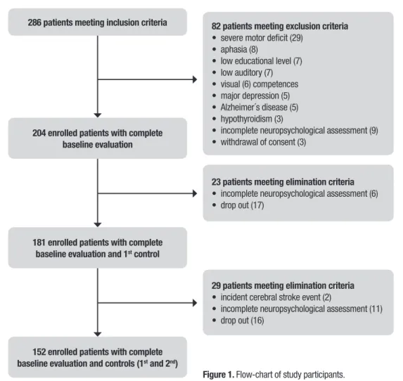
286 patients meeting inclusion criteria
204 enrolled patients with complete baseline evaluation
23 patients meeting elimination criteria
• incomplete neuropsychological assessment (6) • drop out (17)
181 enrolled patients with complete baseline evaluation and 1st control
29 patients meeting elimination criteria
• incident cerebral stroke event (2)
• incomplete neuropsychological assessment (11) • drop out (16)
152 enrolled patients with complete baseline evaluation and controls (1st and 2nd)
Figure 1. Flow-chart of study participants.
82 patients meeting exclusion criteria
• severe motor deficit (29) • aphasia (8)
• low educational level (7) • low auditory (7) • visual (6) competences • major depression (5) • Alzheimer´s disease (5) • hypothyroidism (3)
Supplementary table. Demographic characteristics and neuropsychological test scores at baseline evaluation in patients with CVI from Lima-Peru, according to neuropsychological diagnosis.
Neuropsychological diagnosis
Non-VCI (n = 124) VCI without dementia (n = 61) Vascular dementia (n = 19)
Sex: female 60 (48.39 %) 34 (55.74%) 11 (51.47%)
Age, years* 69.07 (4.53) 68.21 (4.57) 72.31 (4.46)
Education, years* 11.91 (3.47) 11.09 (3.20) 10.84 (3.13)
Barthel scale, score* 93.37 (4.33) 93.20 (3.26) 90.73 (4.42)
NIH Stroke Scale/Score, score* 1.77 (1.41) 1.51 (1.30) 2.32 (1.63)
CDR, score* 0.48 (0.15) 0.55 (0.15) 1.58 (0.61)
ACE, score* 84.79 (6.86) 69.32 (3.36) 63.52 (4.10)
IFS, score* 26.49 (1.52) 20.57 (1.82) 14.78 (2.18)
BDI-II, score* 4.37 (2.61) 5.97 (2.63) 8.32 (2.11)
Apathy scale, score* 9.18 (3.40) 8.26 (2.60) 8.16 (2.17)
VCI: Vascular cognitive impairment; CDR: Clinical Dementia Rating; ACE: Addenbrooke’s Cognitive Examination; IFS: INECO Frontal Screening; BDI-II: Beck Depression Inventory – Second version; *Data expressed as mean (standard deviation).
up was 25.5%. he cohort that completed follow-up comprised a sample that was 50% female with mean age of 68.82 years, 11.69 years of formal education, 93.32 on the Barthel scale, and 1.72 on the NIH Stroke Scale/Score. hese variables had similar values for all three evaluation timepoints (baseline, and irst and second control follow-ups) (Table 1).
During the follow-up, the proportion of VCI (without dementia) in the sample showed a progressive increase. hus, 30.20% and 19.00% of the patients evaluated at the 2nd control follow-up had VCI without dementia and
vascular dementia, respectively. Regarding the neuro-psychological short test, the data showed a progressive increase in average scores on the CDR and BDI-II, and a progressive decline in average scores on the ACE and IFS (Table 1).
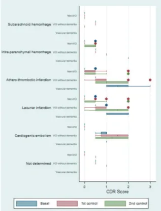
he box plot for the CDR test showed a diference in scores by etiology. he patients with atherothrombotic infarction had the highest values. For the majority of the diferentiated etiologies, the data showed that VCI patients had a progressive worsening relative to baseline status. Conversely, this trend was not observed in sub-jects without VCI (Figure 2).
he box plot for the ACE test showed similar progres-sion patterns among the diferent etiologies, particularly for atherothrombotic infarction and lacunar infarction. Patients with atherothrombotic infarction and lacunar infarction had the lowest values. For the majority of the diferentiated etiologies, the data showed that both
non-VCI and non-VCI patients had progressive worsening relative to baseline status. he only exception was subarachnoid hemorrhage (Figure 3). Similar results were observed for the IFS test (Figure 4).
he box plot for BDI-II showed similar progression among the diferent etiologies, particularly for athero-thrombotic infarction and lacunar infarction. Patients with atherothrombotic infarction and lacunar infarction had the highest values. For the majority of the difer-entiated etiologies, the data showed that both non-VCI and VCI patients had progressive worsening relative to baseline status. he only exception was subarachnoid hemorrhage (Figure 5).
Regarding expected progression patterns, some apparent inconsistences were observed in the group of patients with subarachnoid hemorrhage and VCI with-out dementia (Figures 2-5).
DISCUSSION
Figure 2. Box plot to graph of the evolution of the scores of Clinical Dementia Rating (CDR), differentiated by etiology and by neuropsycho-logical diagnosis.
Figure 3. Box plot to graph of the evolution of the scores of Adden-brooke’s Cognitive Examination (ACE), differentiated by etiology and by neuropsychological diagnosis.
Figure 4. Box plot to graph of the evolution of the scores of INECO Frontal Screening (IFS), differentiated by etiology and by neuropsycho-logical diagnosis.
he clinical and cognitive proile of VCI difers from other causes of cognitive impairment and dementia.3
In our study, we found that infarction-type stroke patients had similar cognitive proiles on both ACE and IFS tests. Because of the small size of these groups, we were unable to identify a clear cognitive pattern in the case of other etiologies. Future studies should focus on patients with subarachnoid hemorrhage, intra-paren-chymal hemorrhage, and cardiogenic embolism.
In general, there was an acceptable inter-individual variation on the cognitive performances across all the short tests. For infarction-type stroke patients, variabil-ity on the ACE test appeared to be less than on the IFS test. However, this variability progressively reduced only on the IFS test. his inding should be further evalu-ated by pooled longitudinal studies to establish both the reliability and consistency of these short cognitive tests over time.
Regarding mean scores at baseline evaluation, the ACE performance of stroke patients was higher than that previously reported in Peruvian patients with AD (67.00; SD = 5.26) and FTD (76.61 SD = 4.90). Simi-larly, IFS performance was higher than that previously reported in Peruvian patients with AD (20.02; SD = 2.10) and FTD (14.55; SD = 2.16).20 hese indings
sug-gest that the ACE could be a useful tool for discriminat-ing between VCI/VD and AD, and IFS for discriminatdiscriminat-ing between VCI/VD and FTD.
he MMSE is the most used short cognitive test but has shown shortcomings for detecting dysexecutive abnormalities, which are characteristic of VCI disor-ders. In this context, he ACE and IFS do not share these drawbacks and are potentially useful tests for both cog-nitively assessing and following up stroke patients.3,26
Our team has validated these short cognitive tests in Peruvian samples of AD and FTD patients.18,20 hus, we
recommended validating both the ACE and IFS tests in Latin American patients for discriminating VCI in stroke patients.
However, there is wide inter-individual variation in the manifestations of vascular brain injury along with overlap with other non-VCI causes of dementia, such that clinical signs and symptoms may suggest the diag-nosis of VCI but radiological or pathological conirma-tion of vascular brain injury is usually needed. In this review, VCI diagnostic criteria, epidemiology, and clini-cal manifestations were reviewed.
Limitations. Since comorbid pathologies such as AD are commonly identiied and pathological diagnosis is needed to rule out the existence of underlying neurode-generative disorder,27 it is unclear whether our sample
comprised only patients with pure VCI.
Because some VCI lesions are undetectable by the current technique of brain MR (such as microinfarcts, which are only visible under light microscopy), the fre-quency of VCI may have been underestimated.26 hus,
our study is biased toward VCI cases secondary to evi-dent CVD/CVI.
In conclusion, in the stroke patients, cognitive per-formance worsened during the 1-year follow-up, even in patients without VCI. his decline is consistent and pro-gressive, particularly in infarction-type stroke patients (atherothrombotic infarction and lacunar infarction), who showed similar cognitive proiles on both ACE and IFS tests. We believe that both the ACE and IFS can be good tests for discriminating VCI/VD from other neuro-degenerative disorders, such as AD and FTD. However, rigorous studies assessing this hypothesis should be conducted.
Author contributions. NC and DL conceived the study. All members of the research team designed the protocol, generated the data, and participated in the writing of the paper. EH interpreted the statistical data and partic-ipated in the writing of the paper. All members reviewed and approved the inal version of the manuscript.
REFERENCES
1. Moorhouse P, Rockwood K. Vascular cognitive impairment: current concepts and clinical developments. Lancet Neurol. 2008;7(3):246-55. 2. Smith EE. Clinical presentations and epidemiology of vascular dementia.
Clin Sci. 2017;131(11):1059-68.
3. Sachdev P, Kalaria R, O’Brien J, Skoog I, Alladi S, Black SE, et al. Diag-nostic criteria for vascular cognitive disorders: a VASCOG statement. Alzheimer Dis Assoc Disord. 2014;28(3):206-18.
4. Gorelick PB, Scuteri A, Black SE, Decarli C, Greenberg SM, Iadecola C, et al. Vascular contributions to cognitive impairment and dementia: a statement for healthcare professionals from the american heart associa-tion/american stroke association. Stroke. 2011;42(9):2672-713. 5. Plassman BL, Langa KM, Fisher GG, Heeringa SG, Weir DR, Ofstedal
MB, et al. Prevalence of dementia in the United States: the aging, demographics, and memory study. Neuroepidemiology. 2007;29(1-2): 125-32.
6. Wimo A, Jönsson L, Bond J, Prince M, Winblad B, Alzheimer Disease International. The worldwide economic impact of dementia 2010. Alzheimers Dement. 2013;9(1):1-11.e3.
7. Schneider JA, Arvanitakis Z, Bang W, Bennett DA. Mixed brain patholo-gies account for most dementia cases in community-dwelling older persons. Neurology. 2007;69(24):2197-204.
9. Penley JA, Wiebe JS, Nwosu A. Psychometric properties of the Spanish Beck Depression Inventory-II in a medical sample. Psychol Assess. 2003;15(4):569-77.
10. Wiebe JS, Penley JA. A psychometric comparison of the Beck Depres-sion Inventory-II in English and Spanish. Psychol Assess. 2005;17(4): 481-5.
11. Starkstein SE, Mayberg HS, Preziosi TJ, Andrezejewski P, Leiguarda R, Robinson RG. Reliability, validity, and clinical correlates of apathy in Parkinson’s disease. J Neuropsychiatry Clin Neurosci. 1992;4(2):134-9. 12. WHO MONICA Project Principal Investigators. The world health
orga-nization monica project (monitoring trends and determinants in cardio-vascular disease): A major international collaboration. Journal of Clinical Epidemiology. 1988;41(2):105-14.
13. Gorelick PB, Counts SE, Nyenhuis D. Vascular cognitive impairment and dementia. Biochim Biophys Acta. 2016;1862(5):860-8.
14. Peters N, Herzog J, Opherk C, Dichgans M. A two-year clinical follow-up study in 80 CADASIL subjects: progression patterns and implications for clinical trials. Stroke. 2004;35(7):1603-8.
15. Prins ND, van Dijk EJ, den Heijer T, Vermeer SE, Jolles J, Koudstaal PJ, et al. Cerebral small-vessel disease and decline in information processing speed, executive function and memory. Brain. 2005;128(Pt 9):2034-41. 16. Morris JC. The Clinical Dementia Rating (CDR): current version and
scoring rules. Neurology. 1993;43(11):2412-4.
17. Mathuranath PS, Nestor PJ, Berrios GE, Rakowicz W, Hodges JR. A brief cognitive test battery to differentiate Alzheimer’s disease and fron-totemporal dementia. 2000;1613-20.
18. Custodio N, Lira D, Montesinos R, Gleichgerrcht E, Manes F. [Usefulness of the Addenbrooke’s Cognitive Examination (Spanish version) in Peru-vian patients with Alzheimer’s disease and Frontotemporal Dementia]. Vertex. 2012;23(103):165-72.
19. Marenco V, Roca M, Gleichgerrcht E, Manes F, Torralva T. The Utility of the INECO Frontal Screening (IFS) in the Detection of Executive Dysfunc-tion in Patients with Traumatic Brain Injury (TBI) (P07.194). Neurology. 2012;78(Meeting Abstracts 1):P07.194-P07.194.
20. Custodio N, Herrera-Perez E, Lira D, Roca M, Manes F, B?ez S, et al. Evaluation of the INECO Frontal Screening and the Frontal Assess-ment Battery in Peruvian patients with Alzheimer’s disease and behav-ioral variant Frontotemporal dementia. eNeurologicalSci. 2016;5: 25-9.
21. Ingles JL, Wentzel C, Fisk JD, Rockwood K. Neuropsychological predic-tors of incident dementia in patients with vascular cognitive impairment, without dementia. Stroke. 2002;33(8):1999-2002.
22. Román GC, Tatemichi TK, Erkinjuntti T, Cummings JL, Masdeu JC, Garcia JH, et al. Vascular dementia: diagnostic criteria for research studies. Report of the NINDS-AIREN International Workshop. Neurology. 1993;43(2):250-60.
23. Association AP, others. Diagnostic and statistical manual of mental disor-ders (DSM-5®) [Internet]. American Psychiatric Pub; 2013 [cited 2017 Sep 17]. Available from: https://books.google.com/books?hl = es&lr = &id = -JivBAAAQBAJ&oi = fnd&pg = PT18&dq = %22Diagnostic+and+St atistical+Manual+of+Mental+Disorders%22+2013&ots = ceNP54IIx9&sig = LtZxG9-pQCF9TeRuXQkqCmlxR_Q
24. Tham W, Auchus AP, Thong M, Goh M-L, Chang H-M, Wong M-C, et al. Progression of cognitive impairment after stroke: one year results from a longitudinal study of Singaporean stroke patients. J Neurol Sci. 2002;203-204:49-52.
25. Chaudhari TS, Verma R, Garg RK, Singh MK, Malhotra HS, Sharma PK. Clinico-radiological predictors of vascular cognitive impairment (VCI) in patients with stroke: a prospective observational study. J Neurol Sci. 2014;340(1-2):150-8.
26. Hachinski V, Iadecola C, Petersen RC, Breteler MM, Nyenhuis DL, Black SE, et al. National Institute of Neurological Disorders and Stroke-Canadian Stroke Network vascular cognitive impairment harmonization standards. Stroke. 2006;37(9):2220-41.