*e-mail: [email protected]
Polyvinyl Pyrrolidone-Assisted Synthesis of Crystalline Manganese Vanadate Microtubes
Li-Zhai Pei*, Yin-Qiang Pei, Yi-Kang Xie, Chang-Zhou Yuan, Dian-Kai Li, Qian-Feng Zhang
Key Laboratory of Materials Science and Processing of Anhui Province, Institute of Molecular Engineering and Applied Chemistry,
School of Materials Science and Engineering, Anhui University of Technology, Ma’anshan, Anhui 243002, P. R. China
Received: June 6, 2012; Revised: September 2, 2012
Manganese vanadate microtubes have been synthesized by a facile polyvinyl pyrrolidone-assisted hydrothermal route. X-ray diffraction pattern confirms that the microtubes are composed of monoclinic MnV2O6, tetragonal V2O5 and orthorhombic MnO2 phases. The outer diameter and inner diameter of the microtubes are about 300 nm-3 µm and 200 nm-1 µm, respectively. The tube wall thickness of the microtubes is about 50 nm-1 µm. The possible formation process of the manganese vanadate microtubes has been proposed as a polyvinyl pyrrolidone-assisted growth mechanism.
Keywords: manganese vanadate microtubes, crystal growth, photoluminescence, electron microscopy
1. Introduction
Recently, great research interest has been devoted to functional materials with solid rod-shaped structures and hollow tubular structures owing to their distinctive physical, chemical properties and potential application in the nanoscale devices1,2. Efforts have also been made
to synthesize rod-shaped and tubular vanadate structures because of their potential applications in the fields of lithium batteries, sensors and photocatalysis3. Different rod-shaped
and tubular vanadate structures, such as LiV3O8 nanorods4,
silver vanadate nanorods5,6, cerium vanadate nanorod
arrays7, FeVO
4 nanorods8 and bismuth vanadate nanotubes9
have been synthesized by hydrothermal route, nanoporous anodic aluminum oxide template via sol-gel method and wet chemical process.
Manganese (Mn) vanadate, as a kind of important transition metal vanadate, has been researched extensively for lithium ion rechargeable batteries10,11. Mn vanadate
powders have been prepared by solid state reaction process12,
solution method13,14. Tubular Mn vanadate instead of bulk
Mn vanadate may show novel physical and chemical properties due to special tubular structure for efficient electron transport and confinement effect. Therefore, it is important to synthesize crystalline tubular Mn vanadate by a facile route for the research of novel physical and chemical properties.
Functional materials with special morphologies, such as alumina nanotubes15, AlOOH nanotubes16, ZnO
nanowires/nanorods17,18 and copper nanowires19 have been
synthesized by a facile hydrothermal route using different surfactants. Surfactants can be used as versatile soft templates for the formation of functional materials with different morphologies. Polyvinyl pyrrolidone (PVP) is a kind of important surfactant which can assist the growth
of functional materials with different morphologies20-22.
In the paper, crystalline Mn vanadate microtubes have been successfully synthesized via a facile PVP-assisted hydrothermal route using sodium orthovanadate (Na3VO4) and Mn acetate (Mn(CH3COO)2·4H2O) as the raw materials, PVP as the surfactant. The possible growth process of the Mn vanadate microtubes has been discussed.
2. Experimental
Na3VO4 (AR grade, purity: ≥99.9%) and PVP (AR grade) were purchased from Aladdin Reagent Co., Ltd. of China. Mn(CH3COO)2·4H2O (AR grade, purity: ≥99.0%) was purchased from Sinopharm Chemical Reagent Co., Ltd. of China. In a typical procedure, Na3VO4, Mn(CH3COO)2·4H2O and PVP were dissolved in 60 mL distilled water. Then, the mixture was placed into a 100 mL autoclave with a Teflon liner. The autoclave was maintained at 180 °C for 24 hours. Subsequently the autoclave was cooled naturally in air. The resulting black precipitates were filtered, washed with distilled water for several times and dried at 60 °C in air. Finally, the black Mn vanadate powders were gained.
(Perkin Elmer PE, WQF-410 spectrometer) was used at room temperature in the range of 400-4000 cm–1. PL
measurement was carried out at room temperature using 212 nm as the excitation wavelength with a luminescence spectrometer (Cary Eclipse) in the range of 350-600 nm.
3. Results and Discussion
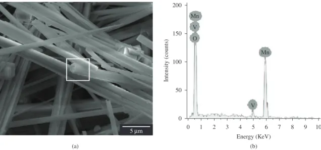
Figure 1a displays the SEM image of the Mn vanadate products grown from 180 °C for 24 hours using PVP as the surfactant. It is observed that the Mn vanadate products are composed of a large quantity of tubular structures with the length of about several dozens of micrometers. Some microtubes can be seen obviously which is designated by white arrows. No other structures are observed besides the tubular structures. The outer diameter and inner diameter of the Mn vanadate microtubes are about 300 nm to 3 µm and 200 nm to 1 µm, respectively. The tube wall thickness of the microtubes is about 50 nm to 1 µm. The cracked microtube also shows the curved structure (designated by black arrow). The morphology of the Mn vanadate microtubes is similar to that of ZnO microtubes23-25 and BiVO
4 microtubes26.The
results indicate that the PVP-assisted hydrothermal route is an effective method for preparing Mn vanadate microtubes. To analyze the role of the PVP on the formation of Mn vanadate microtubes, the experiment under same synthesis conditions without PVP was performed. The SEM image of the products is shown in Figure 1b. It is interesting that only irregular particles with the sub-microscale size are obtained. The irregular particles are very different from those obtained from the same hydrothermal conditions using PVP as the surfactant. Generally, rod-like Mn vanadate containing crystal water can easily obtained by hydrothermal method. For example, Inagaki et al.27 and Morishita et al.12 reported
the synthesis of rod-shaped MnV2O6 using Mn(CH3COO)2 and V2O5 with a metal ion concentration of 0.01-1.0 mol.L–1
at 135-200 °C under hydrothermal conditions. However, only irregular Mn vanadate particles are obtained without
PVP. In our experiment, Na3VO4 is used as the V raw material instead of V2O5 and PVP is used as the surfactant. PVP and Na3VO4 are considered to have the essential roles on the formation of the Mn vanadate microtubes under present hydrothermal conditions.
The composition of the microtubes has been analyzed using energy dispersive spectrometer (EDS) equipped in the FESEM. Figure 2b is the EDS spectrum of the microtubes corresponding to the white square in Figure 2a. It is clear that the Mn vanadate microtubes are composed of Mn, V and O. The phase of the Mn vanadate microtubes is identified by XRD which is shown in Figure 3a. Most diffraction peaks can be indexed to monoclinic MnV2O6 phase (JCPDS card, No. 40-0165). Some diffraction peaks of tetragonal V2O5 (JCPDS card, No. 45-1074) and orthorhombic MnO2 phase (JCPDS card, No. 39-0375) are also indexed besides monoclinic MnV2O6 phase. The V2O5 and MnO2 phases may originate from the residue decomposed from Na3VO4 and Mn(CH3COO)2. The XRD result shows that the Mn vanadate microtubes are composed of monoclinic MnV2O6, tetragonal V2O5 and orthorhombic MnO2 phases. The XRD pattern of the irregular particles obtained without PVP (Figure 3b) shows that the irregular particles are composed of orthorhombic MnV2O5 phase (JCPDS card, No. 51-0203). The phase is totally different from that obtained using PVP as the surfactant. The result shows that the PVP induces the phase transformation of the products from irregular particles to microtubes.
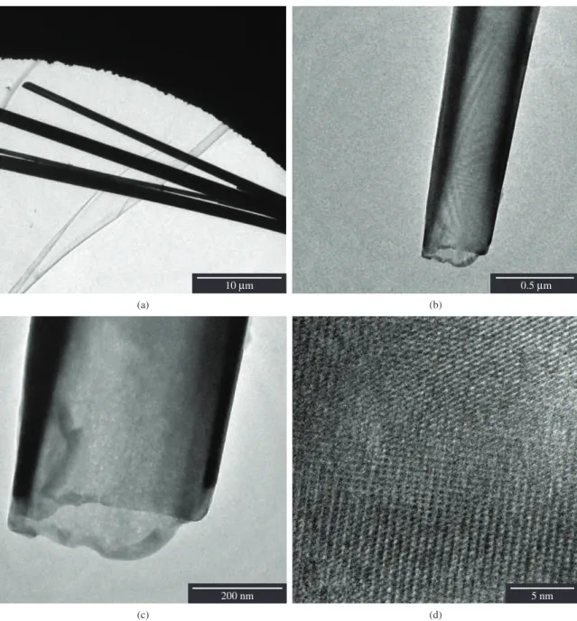
More structure formation of the Mn vanadate microtubes can be provided by TEM observations. Figure 4 is the TEM and HRTEM images of the Mn vanadate microtubes. From the general TEM image of the Mn vanadate microtubes (Figure 4a), the Mn vanadate microtubes with smooth surface exhibit the diameter of less than 3 µm and length of several dozens of micrometers. The morphology and size of the Mn vanadate microtubes are similar to those observed by SEM observation. Obviously, TEM image at the tip of the Mn vanadate microtube is shown in Figure 4b, c exhibiting
the tubular structure. The inner diameter and outer diameter are about 300 nm and 500 nm, respectively. The tube wall thickness of the microtube is about 50 nm. HRTEM analysis on the Mn vanadate microtubes may provide more structural information which may help to analyze the crystalline structure. However, the thickness of the Mn vanadate microtubes prevents the HRTEM observation. So only HRTEM image of the microtubes at the tip is measured
which is shown in Figure 4d. The HRTEM image obviously shows that the microtubes have good crystalline structure.
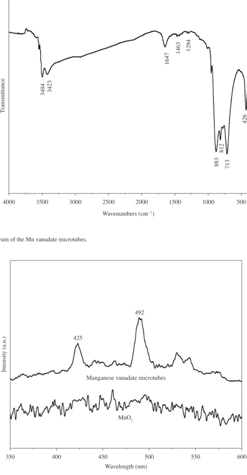
The IR spectrum at 400-4000 cm–1 of the Mn vanadate
microtubes obtained from 180 °C for 24 hours is shown in Figure 5. The broad absorption bands at 2800-3800 cm–1
with the absorption peaks of 3494 and 3423 cm–1 are the
characteristic stretching vibration of −OH originated from water. The absorption peaks at 1647, 1463 and 1294 cm–1
Figure 3. (a) XRD pattern of the Mn vanadate microtubes obtained from 180 °C for 24 hours using PVP as the surfactant, (b) XRD pattern of the samples obtained from 180 °C for 24 hours without PVP.
are assigned to the C=O stretching, CH2 bending and C-N stretching vibration, respectively in the PVP28. The
absorption peak at 713 cm–1 contributes to the Mn-O
vibration of the products29. Yamaguchi et al.30 reported
that YVO4 synthesized by the sol-gel procedure showed vibrations of V-O bonding at 870, 820 and 430 cm–1. IR
spectrum of LaVO4 also exhibited the vibration of V-O bonding at 432 cm–1 reported by Manca and Baran31. The
absorption peaks located at 883, 812 and 426 cm–1 are very
close to those reported by above literatures. Therefore, these absorption peaks at 883, 812 and 426 cm–1 are assigned to
the vibration of V-O bonding.
The room temperature PL spectrum of the Mn vanadate microtubes (Figure 6) shows violet and blue light emission
centered at 425 nm and 492 nm, respectively. A broad band emission from 400 to 700 nm was observed from PL spectrum of metavanadates AVO3 (A=K, Rb and Cs)32.
Broad band emission spectrum between 400 and 800 nm from the M2V2O7 (M=Ca, Sr and Ba) was also reported by Nakajima et al.33 The origin of the luminescence of
M2V2O7 phosphors may be the charge transfer transition from the oxygen 2p to vanadium 3d orbitals in the VO4 tetrahedra34. The violet and blue light emission centered
at 425 nm and 492 nm of the Mn vanadate microtubes are very similar to those of the above literatures. The Mn vanadate microtubes are mainly composed of monoclinic MnV2O6 and V2O5 phases besides MnO2 phase. Therefore, the PL spectrum of the Mn vanadate microtubes is
Figure 5. IR spectrum of the Mn vanadate microtubes.
synthesized by adding PVP under hydrothermal conditions. Therefore, PVP is considered to be a structure-directing agent for the growth of the Mn vanadate microtubes. PVP is a long chain polymer with each pyrrolidone unit chemically bonded to a polyethylene main chain47 forming PVP micelles.
The PVP micelles are filled with polyethylene chains and entrapped water which may have a good stability to solubilize nanoparticles. Under the hydrothermal conditions, MnV2O6 nanoparticles are generated and crystallize to form MnV2O6 nuclea. Hence MnV2O6 nanoparticles can exist either in water or in the PVP micelles. PVP which is used as a structure-directing agent plays a crucial role in controlling the distribution of MnV2O6 nanoparticles in the hydrothermal solution and leads to the formation of Mn vanadate microtubes following a polyol-assisted formation mechanism48,49. Only irregular particles are generated
without PVP. However, a plenty of micelles and Mn vanadate nanoparticles are filled in the PVP micelles. PVP micelles alter the surface energies of the Mn vanadate surfaces to promote the selective anisotropic growth of crystals leading to the formation of Mn vanadate microtubes.
4. Conclusions
In summary, novel crystalline Mn vanadate microtubes have been synthesized via a facile hydrothermal route in the presence of PVP. The Mn vanadate microtubes with the length of about several dozens of micrometers are composed of monoclinic MnV2O6, tetragonal V2O5 and orthorhombic MnO2 phases. The outer diameter and inner diameter of the Mn vanadate microtubes are about 300 nm-3 µm and 200 nm to 1 µm, respectively. The tube wall thickness of the microtubes is about 50 nm to 1 µm. PVP-assisted hydrothermal route is potentially extendable to other inorganic tubular materials.
Acknowledgements
This work was supported by the Natural Science Foundation of Anhui Province (1208085QE98), Innovative Research Foundation of Postgraduate of Anhui University of Technology (2011005) and National Natural Science Foundation of China (90922008).
considered to be originated from V-O bonding. The emission centered at 425 nm is also considered to be the overtones of the excitation wave, which is similar to that reported by McGinley et al.35 However, the PL spectrum is very
noisy. The samples present OH groups in the structure of material. This may be principal effect that luminescence is being suppressed. In addition, No PL peaks are observed from MnO2. So MnO2 in the Mn vanadate microtubes is also considered to contribute to the suppression of the luminescence.
The analysis on the formation mechanism of Mn vanadate microtubes is very important to understand the synthetic methods for the formation of tubular structure. Several models, such as curving followed by seaming of molecular layers36, helical nanobelt-twist-join-growth
process37 and rolling mechanism for the conversion from
nanosheets to nanotubes38 have been proposed which can
not explain the formation process of the Mn vanadate microtubes. Recently, Mo et al.39 reported the synthesis
of β-Mn2V2O7 microtubes with a length of 15-25 µm, 2.5-3.5 µm external diameter and 0.4 µm wall thickness, as well as β-Mn2V2O7 hollow microspheres in a suitable molar ratio of NH3VO3 and MnCO3 powders via a hydrothermal process without surfactants. They contribute to the tubular morphology caused by layered oxide structure. However, different from the raw materials of NH3VO3 and MnCO3 reported by Liuet al.37, Na
3VO4, Mn(CH3COO)2 and PVP
are used as the raw materials in our experiments. Therefore, the formation of the Mn vanadate microtubes may take a different formation process. In fact, crystalline MnV2O6 powders with irregular particles and rod-like particles have been synthesized via a hydrothermal process using Mn(CH3COO)2 and V2O5 as the raw materials without surfactants27,40. It is well known that surfactant-assisted
reaction to control the nucleation and growth is a simple and effective method. The surfactant molecules can modulate the kinetics of the crystal growth and determine the subsequent morphology of the product20,41-43.
In recent years, some research groups44-46 reported that
the surfactants could alter the surface energy of various crystallographic surfaces to promote selective anisotropic growth of crystals. Only Mn vanadate microtubes can be
References
1. Shao MW, Qian GX, Ban HZ, Li M, Hu H, Lu L et al. Synthesis and magnetic property of quasi one-dimensional Ni nanostructures via Si nanowire template. Scripta Materialia. 2006; 55:851-854. http://dx.doi.org/10.1016/j. scriptamat.2006.08.003
2. Qian JM, Wang JP, Hou GY, Qiao GJ and Jin ZH. Preparation and characterization of biomorphic SiC hollow fibers from wood by chemical vapor infiltration. Scripta Materialia. 2005; 53:1363-1368. http://dx.doi.org/10.1016/j. scriptamat.2005.08.029
3. Souza Filho AG, Ferreira OP, Santos EJG, Mendes Fiho J and Alves OL. Raman spectra in vanadate nanotubes revisited. Nano Letters. 2004; 4:2099-2104. http://dx.doi.org/10.1021/ nl0488477
4. Xu HY, Wang H, Song ZQ, Wang YW, Yan H and Yoshimura M. Novel chemical method for synthesis of LiV3O8 nanorods as cathode materials for lithium ion batteries. Electrochimica Acta. 2004; 49:349-353. http://dx.doi.org/10.1016/j. electacta.2003.08.017
5. Pan GT, Lai MH, Juang RC, Chung TW and Yang TCK. Preparation of visible-light-driven silver vanadates by a microwave-assisted hydrothermal method for the photodegradation of volatile organic vapors. Industrial and Engineering and Chemistry Research. 2011; 50:2807-2814. http://dx.doi.org/10.1021/ie1012932
well-controlled coaxial nanocables of silver/silica. Nano Letters. 2002; 2:427-430. http://dx.doi.org/10.1021/nl025508+ 22. Wang JW, Wang X, Peng Q and Li YD. Synthesis and
characterization of bismuth single-crystalline nanowires and nanospheres. Inorganic Chemistry. 2004; 43:7552-7556. http:// dx.doi.org/10.1021/ic049129q
23. Zhang W, Zhang D, Fan TX, Ding J, Guo QX and Ogawa H. Fabrication of ZnO microtubes with adjustable nanopores on the walls by the templating of butterfly wing scales. Nanotechnology. 2006; 17:840-844. http://dx.doi. org/10.1088/0957-4484/17/3/038
24. Jeong JS, Lee JY, Cho JH, Suh HJ and Lee CJ. Single-crystalline ZnO microtubes formed by coalescence of ZnO nanowires using a simple metal-vapor deposition method. Chemistry of Materials. 2005; 17:2752-2756. http://dx.doi.org/10.1021/ cm049387l
25. Vayssiers L, Kels K, Hagfeldt A and Lindquist SE. Three-dimensional array of highly oriented crystalline ZnO microtubes. Chemistry of Materials. 2001; 13:4395-4398. http://dx.doi.org/10.1021/cm011160s
26. Zhou L, Wang WZ, Zhang LS, Xu HL and Zhu W. Single-crystalline BiVO4 microtubes with square cross-sections: Microstructure, growth mechanism, and photocatalytic property. Journal of Physics and Chemistry C. 2007; 111:13659-13664. http://dx.doi.org/10.1021/ jp065155t
27. Inagaki M, Morishita T, Hirano M, Gupta V and Nakajima T. Synthesis of MnV2O6 under autogenous hydrothermal conditions and its anodic performance. Solid State Ionics. 2003; 156:275-282. http://dx.doi.org/10.1016/ S0167-2738(02)00679-3
28. Zhang TR, Lu R, Liu XL, Zhao YY, Li TJ and Yao JN. Photochromic polyoxotungstoeuropate K12[EuP5W30O110]/ polyvinylpyrrolidone nanocomposite films. Journal of Solid State Chemistry. 2003; 172:458-463. http://dx.doi.org/10.1016/ S0022-4596(03)00036-7
29. Wang HE, Lu ZG, Qian D, Li YJ and Zhang W. Single-crystal α-MnO2 nanorods: synthesis and electrochemical properties. Nanotechnology. 2007; 18:115616. http://dx.doi. org/10.1088/0957-4484/18/11/115616
30. Yamaguchi O, Mukaida Y, Shigeta H, Takemura H and Yamashita M. Preparation of alkoxy-derived YVO4. Materials Letters. 1988; 7:158-160. http://dx.doi.org/10.1016/0167-57 7X(88)90176-0
31. Manca SG and Baran EJ. Characterization of the monoclinic form of praseodymium chromate (V). Journal of Physics and Chemistry of Solids. 1981; 42:923-925. http://dx.doi. org/10.1016/0022-3697(81)90018-4
32. Nakajima T, Isobe M, Tsuchiya T, Ueda Y and Kumagai T. Direct fabrication of metavanadate phosphor films on organic substrates for white-light-emitting devices. Nature Materials. 2008; 7:735-740. http://dx.doi.org/10.1038/ nmat2244
33. Nakajima T, Isobe M, Tsuchiya T, Ueda Y and Manabe T. Photoluminescence property of vanadates M2V2O7 (M: Ba, Sr and Ca). Optical Materials. 2010; 32:1618-1621. http://dx.doi. org/10.1016/j.optmat.2010.05.021
34. Park KC and Mho SI. Photoluminescence properties of Ba3V2O8, Ba3(1-x)Eu2xV2O8 and Ba2Y2/3V2O8:Eu3+. Journal of Luminescence. 2007; 95:122-123.
35. M c G i n l ey E S a n d C r i m F F. H o m o g e n e o u s a n d inhomogeneous structure in the vibrational overtone spectrum of tetramethyldloxetane. Journal of Chemistry 7. Liu JF, Wang LL, Sun XM and Zhu XQ. Cerium vanadate nanorod
arrays from ionic chelator-mediated self-assembly. Angewandte Chemie International Edition. 2010; 49:3492-3495. http:// dx.doi.org/10.1002/anie.201000783
8. Ma H, Yang XJ, Tao ZL, Liang J and Chen J. Controllable synthesis and characterization of porous FeVO4 nanorods and nanoparticles. CrystEngComm. 2011; 13:897-901. http:// dx.doi.org/10.1039/c0ce00273a
9. Singh S, Kumari N, Varma KBR and Krupanidhi SB. Synthesis, structural characterization and formation mechanism of ferroelectric bismuth vanadate nanotubes. Journal of Nanoscience and Nanotechnology. 2009; 9:6549-6553. http:// dx.doi.org/10.1166/jnn.2009.1300
10. Andrukaitis E. Lithium intercalation into the copper, nickel or manganese vanadates Me(VO3)2·yH2O. Journal of Power Sources. 1997; 68:652-655. http://dx.doi.org/10.1016/ S0378-7753(96)02572-4
11. Andrukaitis E, Torlone GL and Hill IR. Study of Mex(VO3)2 vanadates, (Me=Co, Ni, Mn, 1<x<2) for lithium rechargeable cells. Journal of Power Sources. 1999; 81-82:651-655. http:// dx.doi.org/10.1016/S0378-7753(99)00094-4
12. Morishita T, Konno H, Izumi Y and Inagaki M. Oxidation state of vanadium in amorphous MnV2O6 formed during discharge–charge cycle and the improvement of its synthesis condition. Solid State Ionics. 2006; 177:1347-1353. http:// dx.doi.org/10.1016/j.ssi.2006.05.035
13. Leroux F, Piffard Y, Ourvard G, Mansot JL and Guyomard D. New amorphous mixed transition metal oxides and their Li derivatives: Synthesis, characterization, and electrochemical behavior. Chemistry of Materials. 1999; 11:2948-2959. http:// dx.doi.org/10.1021/cm991074g
14. Liao JH, Drezen T, Leroux F, Guyomard D and Piffard Y. Synthesis, structures and thermal analysis of MnV2O6×nH2O phases (n=1, 2 and 4). European Journal of Solid State Inorganic Chemistry. 1996; 33:411-427.
15. Qu LH, He CQ, Yang Y, He YL and Liu ZM. Hydrothermal synthesis of alumina nanotubes templated by anionic surfactant. Materials Letters. 2005; 59:4034-4037. http://dx.doi. org/10.1016/j.matlet.2005.07.059
16. Kuang DB, Fang YP, Liu HQ, Frommen C and Fenske D. Fabrication of boehumite AlOOH and γ–Al2O3 nanotubes via a soft solution route. Journal of Materials Chemistry. 2003; 13:660-662. http://dx.doi.org/10.1039/ b212885c
17. Sun GB, Cao MH, Wang YH, Hu CW, Liu YC, Ren L et al. Anionic surfactant-assistd hydrothermal synthesis of high-aspect-ratio ZnO nanowires and their photoluminescence property. Materials Letters. 2006; 60:2777-2782. http://dx.doi. org/10.1016/j.matlet.2006.01.088
18. Sun XM, Chen X, Deng ZX and Li YD. A CTAB-assisted hydrothermal orientation growth of ZnO nanorods. Materials Chemistry and Physics. 2002; 78:99-104. http://dx.doi. org/10.1016/S0254-0584(02)00310-3
19. Liu ZP, Yang Y, Liang JB, Hu ZK, Li S, Peng S et al. Synthesis of copper nanowires via a complex-surfactant-assisted hydrothermal reduction process. Journal of Physics and Chemistry B. 2003; 107:12658-12661. http://dx.doi. org/10.1021/jp036023s
20. Zheng DS, Sun SS, Fan WL, Yu HY, Fan CH, Cao GX et al. One-step preparation of single-crystalline β–MnO2 nanotubes. Journal of Physics and Chemistry B. 2005; 109:16439-16443. http://dx.doi.org/10.1021/jp052370l
Chemistry of Materials. 2002; 14:4736-4745. http://dx.doi. org/10.1021/cm020587b
43. Peng X, Manna L, Yang WD, Wickham J, Scher E, Kadavanich A et al. Shape control of CdSe nanocrystals. Nature. 2000; 404:59-61. http://dx.doi.org/10.1038/35003535 44. Peng X. Mechanisms for the shape-control and shape-evolution
of colloidal semiconductor nanocrystals. Advanced Materials. 2003; 15:459-463. http://dx.doi.org/10.1002/ adma.200390107
45. Lee SM, Cho SN and Cheon J. Anisotropic shape control of colloidal inorganic nanocrystals. Advanced Materials. 2003; 15:441-443. http://dx.doi.org/10.1002/ adma.200390102
46. Guo L, Liu C, Wang R, Xu H, Wu Z and Yang S. Large-scale synthesis of uniform nanotubes of a nickel complex by a solution chemical route. Journal of the American Chemical Society. 2004; 126:4530-4531. http://dx.doi.org/10.1021/ ja039381h
47. Li LJ, Nicholas RJ, Chen CY, Darton RC and Baker SC. Comparative study of photoluminescence of single-walled carbon nanotubes wrapped with sodium dodecyl sulfate, surfactin and polyvinylpyrrolidone. Nanotechnology. 2005; 16:S202-S205. http://dx.doi.org/10.1088/0957-4484/16/5/012
48. Mayers B and Xia Y. Formation of tellurium nanotubes through concentration depletion at the surfaces of seeds. Advanced Materials. 2002; 14:279-281. http://dx.doi.org/10.1002/152 1-4095(20020219)14:4<279::AID-ADMA279>3.0.CO;2-2 49. Ma YR, Qi LM, Ma JM and Cheng HM. Micelle-mediated
synthesis of single-crystalline selenium nanotubes. Advanced Materials. 2004; 16:1023-1026. http://dx.doi.org/10.1002/ adma.200400071
a n d P h y s i c s. 1986; 85:5741-5747. http://dx.doi. org/10.1063/1.451535
36. Ye C, Meng G, Jiang Z, Wang Y, Wang G and Zhang L. Rational growth of Bi2S3 nanotubes from quasi-two-dimensional p r e c u r s o r s . Jo u r n a l o f t h e A m e r i c a n C h e m i c a l Society. 2002; 124:15180-15181. http://dx.doi.org/10.1021/ ja0284512
37. Mo MS, Zeng JH, Liu XM, Yu WC, Zhang SY and Qian YT. Controlled hydrothermal synthesis of thin single-crystal tellurium nanobelts and nanotubes. Advanced Materials. 2002; 14:1658-1662. http://dx.doi.org/10.1002/152 1-4095(20021118)14:22<1658::AID-ADMA1658>3.0.CO;2-2 38. Wang X and Li YD. Rational synthetic strategy. From
layered structure to MnO2 nanotubes. C h e m i s t r y Letters. 2004; 33:48-49. http://dx.doi.org/10.1246/cl.2004.48 39. Liu Y and Qian YT. Controllable synthesis of β–Mn2V2O7 microtubes and hollow microspheres. Frontiers of Chemistry in China. 2008; 3:467-470. http://dx.doi.org/10.1007/ s11458-008-0061-9
40. Kim SS, Ikuta H and Wakihar M. Synthesis and characterization of MnV2O6 as a high capacity anode material for a lithium secondary battery. Solid State Ionics. 2001; 139:57-65. http:// dx.doi.org/10.1016/S0167-2738(00)00816-X
41. Xia Y, Yang P, Sun Y, Wu Y, Mayers B, Gates B et al. One-dimensional nanostructures: Synthesis, characterization, and applications. Advanced Materials. 2003; 15:353-389. http://dx.doi.org/10.1002/adma.200390087